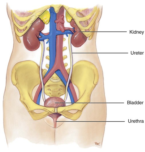
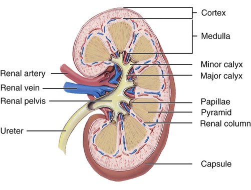
1. State six functions of the urinary system. 2. Describe the location and structural features of the kidneys. 3. Draw and label the parts of a nephron. 4. State the two parts of the juxtaglomerular apparatus. 5. Describe the location, structure, and function of the ureters, urinary bladder, and urethra. 6. List and describe the three steps in urine formation. 7. Identify the hormones that affect kidney function, and explain how they do so. 8. Explain the function of renin. 9. Describe ways in which the aging of an individual affects the urinary system. The urinary system consists of the kidneys, ureters, urinary bladder, and urethra. The kidneys produce the urine. The ureters transport the urine away from the kidneys to the urinary bladder. The urinary bladder stores the urine until it is excreted from the body. The urethra is a tubular structure that carries the urine from the urinary bladder to the outside of the body. The components of the urinary system are illustrated in Figure 15-1. Highlight on the Urinary System Hangover: Alcohol inhibits the secretion of antidiuretic hormone, so when people drink alcohol, they experience diuresis, or excessive urination. Experts believe that the dehydration caused by diuresis contributes to “hangover” symptoms. Kidney stones: Kidney stones develop when uric acid or calcium salts precipitate instead of remaining dissolved in the urine. The stones usually form in the renal pelvis, but they may also develop in the urinary bladder. If small enough, they may pass naturally with urine flow but usually cause a lot of discomfort. If kidney stones cause a serious obstruction, they may need to be surgically removed. A newer method of treatment called lithotripsy uses high-frequency sound waves to break the stone into small pieces so that it may pass naturally. The formation of stones in the urine is called urolithiasis. Nephrons: The number of nephrons does not increase after birth. Growth of the kidney occurs from enlargement of the individual nephrons. When nephrons are damaged they are not replaced. Nephroptosis: Nephroptosis, commonly referred to as a floating kidney, occurs when the kidney is no longer held in place by the renal fascia and it drops out of its normal position. This may make the kidney more vulnerable to injury if it is no longer protected by the ribs. Another danger is that the ureter may become twisted and block the flow of urine. Nephroptosis occurs more frequently in horseback riders, truck drivers, and people who ride motorcycles. Polycystic kidney disease: This inherited condition affects the tubular portion of the nephrons. Swelling or cysts develop along the tubules, and as the cysts enlarge they displace and damage functional kidney tissue. This eventually leads to a total loss of kidney function. When this occurs in both kidneys, a transplant is necessary. Uremia: When the kidneys do not function properly and fail to remove the waste products from the blood, uremia may result. Uremia is a condition in which there is a toxic level of urea in the blood. Urinary incontinence: Urinary incontinence is the inability to control urination and to retain urine in the bladder. Temporary incontinence may result when the muscles around the bladder and urethra become weakened and lose muscle tone. This is sometimes caused by stretching of the muscles during childbirth. Because these muscles help restrict the outlet of the bladder, their weakness contributes to a leakage of urine. A cough or sneeze may increase pressure within the bladder sufficiently to force urine to escape. Permanent incontinence is usually caused by damage to the central nervous system or by extensive damage to the bladder or urethra. Urinary tract infection (UTI): UTIs occur more frequently in women than in men because of differences in the urethra. In females the urethral opening is in close proximity to the anal opening, which gives intestinal bacteria easier access to the urethra. The female urethra is short, which allows any infection to spread to the urinary bladder. An infection of the urethra is called urethritis, and one of the urinary bladders is called cystitis. The macroscopic internal structure of the kidney is illustrated in Figure 15-2. The outer, reddish region is the renal cortex. The renal cortex surrounds a darker reddish-brown region called the renal medulla. The renal medulla consists of a series of renal pyramids. The renal pyramids appear striated because they contain straight tubular structures and blood vessels. The wide bases of the pyramids are adjacent to the cortex. The pointed ends of the pyramids, called renal papillae, are directed toward the center of the kidney. Portions of the renal cortex extend into the spaces between adjacent pyramids to form renal columns. The cortex and medulla make up the functional tissue of the kidney.
Urinary System
Introduction to the Urinary System
Components of the Urinary System
Kidneys
Location

Macroscopic Structure
![]()
Stay updated, free articles. Join our Telegram channel

Full access? Get Clinical Tree


