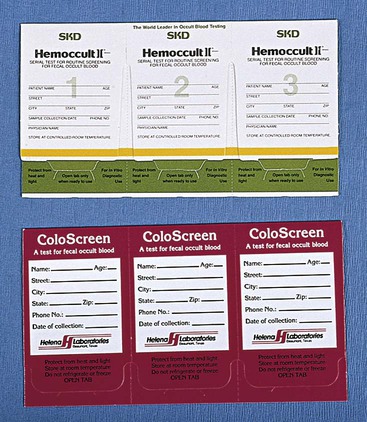
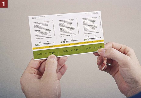
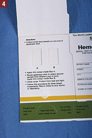
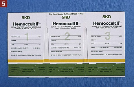
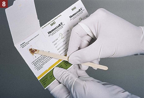
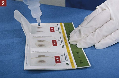
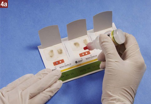
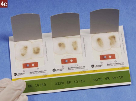
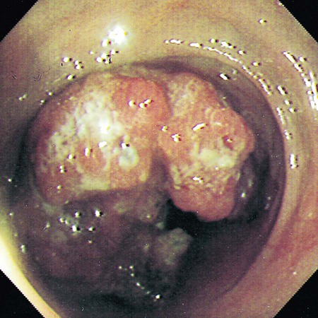
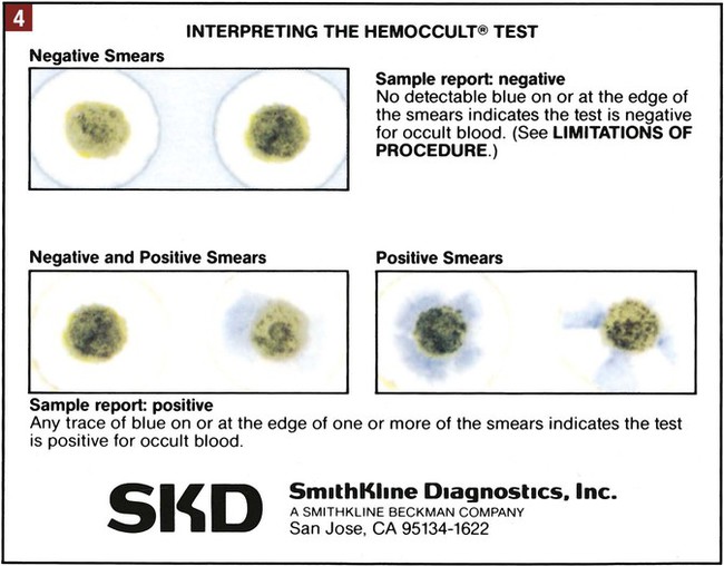
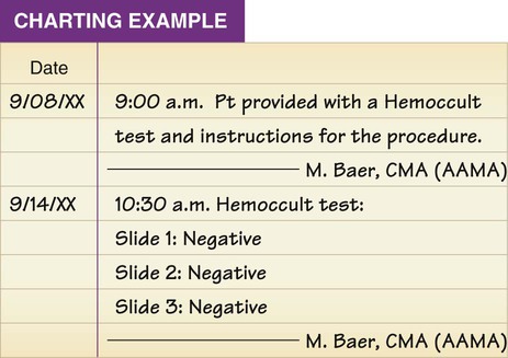
21. Explain how nuclear medicine is used to produce an image of a body part or organ. Routine screening of stool specimens for occult blood is frequently performed in the medical office. The guaiac slide test is a chemical test used to screen for fecal occult blood and is discussed in detail in this chapter. This test is commercially available with the brand names of Hemoccult, ColoScreen (Figure 28-1), and Seracult. Certain medications irritate the gastrointestinal tract, which may result in a small amount of bleeding, and thus could result in a false-positive result on the guaiac slide test. Medications that should be avoided include ibuprofen (Motrin, Advil), naproxen (Aleve), and more than one adult aspirin per day. In addition, an iron supplement may cause a false-positive result, and a vitamin C supplement (greater than 250 mg per day) can cause a false-negative result. All of these substances should be discontinued before testing. Table 28-1 lists the specific patient preparation requirements for fecal occult blood testing using the guaiac slide test. Table 28-1 Patient Preparation for the Fecal Occult Guaiac Slide Test The medical assistant should apply 1 drop of the developing solution between the positive and negative performance monitor areas on each of the three slides. The results must be read within 10 seconds after application of the developer. If the slides and developer are functioning properly, the positive area turns blue, whereas the negative area shows no color change. Failure of the expected control results to occur indicates an error, and the test results are not considered valid; possible causes include the use of outdated slides or developing solution; an error in technique; and subjection of the slides to heat, sunlight, strong fluorescent light, or volatile chemicals. Procedure 28-1 outlines the medical assistant’s responsibilities related to fecal occult blood testing using the Hemoccult guaiac slide test. Procedure 28-2 describes the development of a Hemoccult slide test. Guaiac Slide Test Instruct a patient in specimen collection for a Hemoccult guaiac slide test. 1. Procedural Step. Obtain a Hemoccult testing kit. Check the expiration date on the slides. Principle. Outdated slides can lead to inaccurate test results. 2. Procedural Step. Greet the patient and introduce yourself. Identify the patient and explain the purpose of the test. Tell the patient that the test should not be conducted during a menstrual period or when hemorrhoids are bleeding or a urinary tract infection is present. Principle. Bleeding from other (identifiable) sources causes a false-positive test result. 3. Procedural Step. Instruct the patient in proper preparation for the test. See the box Highlight on Colorectal Cancer for the specific guidelines the patient should follow. Tell the patient to begin the diet modifications 3 days before collecting the first stool specimen. Encourage the patient to adhere to the diet modifications. 4. Procedural Step. Provide the patient with the Hemoccult testing kit. The kit consists of three identical cardboard slides attached to one another; each slide contains two squares, labeled “A” and “B.” Three wooden applicator sticks and written instructions also are included in the testing kit. 5. Procedural Step. Instruct the patient on completion of the information required on the front flap of each card. This includes the patient’s name, address, phone number, and age and the date of the specimen collection. A ballpoint pen should be used to write this information. 6. Procedural Step. Provide instructions on proper care and storage of the slides. Make it clear that the slides must be stored (with the flaps in a closed position) at room temperature and protected from heat, sunlight, strong fluorescent light, and volatile chemicals. 7. Procedural Step. Instruct the patient on initiation of the test by telling him or her to begin the diet modifications and then to collect a stool specimen from the first bowel movement after the 3-day preparatory period. 8. Procedural Step. Instruct the patient on proper collection of the stool specimen: a. Fill in the sample collection date on the front flap of the first cardboard slide. b. Use a clean, dry container to collect the stool sample. The sample must be collected before it comes in contact with toilet bowl water. Allow the stool to fall into the collection container. c. Use one of the wooden applicators to obtain a specimen from one part of the stool sample. d. Open the front flap of the first cardboard slide (located on the left in the series of three). e. Spread a very thin smear of the specimen over the filter paper in the square labeled “A.” f. Using the same wooden applicator, obtain another specimen from a different area of the stool. g. Spread a thin smear of the specimen over the filter paper in the square labeled “B.” h. Close the front flap of the cardboard slide. i. Discard the wooden applicator in a waste container. Do not flush it down the toilet. j. Place the slides in a regular paper envelope to air-dry overnight. 9. Procedural Step. Instruct the patient to continue the testing period on 3 different days until all three specimens have been obtained as follows. a. Repeat Procedural Step 8 after the second bowel movement the next day. If you do not have a bowel movement on the next day, then collect the specimen on the following day. The specimens should be collected on 3 different days. Use the cardboard slide located in the middle of the series of three. b. Repeat Procedural Step 8 after the third bowel movement, using the cardboard slide located to the right in the series of three. c. Allow the completed slides to air-dry overnight in the paper envelope. 10. Procedural Step. Instruct the patient to place the cardboard slides in the envelope lined with foil, seal carefully, and return them as soon as possible to the medical office. Emphasize to the patient that only the foil-lined envelope can be used to mail the slides; a standard envelope cannot be used. Inform the patient that the slides must be returned no later than 14 days after the first specimen is collected. 11. Procedural Step. Give the patient an opportunity to ask questions; ensure that the patient understands the instructions for patient preparation and collection of the stool specimen and for storage of the slides. 12. Procedural Step. Record in the patient’s chart. Include the date and documentation that the Hemoccult test and instructions were given to the patient. Develop a Hemoccult slide test. 1. Procedural Step. Assemble the equipment. Check the expiration date on the developing solution bottle. The developing solution contains hydrogen peroxide and must be stored away from heat and light. It must be tightly capped when not in use. 2. Procedural Step. Sanitize your hands and apply gloves. Open the back flap of the cardboard slides. Apply 2 drops of the developing solution to the guaiac test paper underlying the back of each smear. 3. Procedural Step. Read the results within 60 seconds. Fecal blood loss greater than 5 mL per day results in a positive reaction, which is indicated by any trace of blue on or at the edge of the fecal smear. If no detectable color change occurs, the result is considered negative. 4. Procedural Step. Perform the quality control procedure as follows: a. Apply 1 drop of developing solution between the positive and negative control performance indicators on each of the three slides. b. Read the results within 10 seconds. c. The positive area should turn blue and the negative area should show no color change. Failure of the expected control results to occur indicates an error and that the test results are invalid. 5. Procedural Step. Properly dispose of the Hemoccult slides in a regular waste container. 6. Procedural Step. Remove gloves and sanitize your hands. Chart the results. Include the date and time, the brand name of the test (Hemoccult), and the test results for each slide (recorded as positive or negative). Highlight on Colorectal Cancer The following factors increase the risk of developing colorectal cancer: • Age. The risk of colorectal cancer increases significantly after 50 years of age and reaches a peak from ages 60 to 75 years. More than 90% of individuals diagnosed with colorectal cancer are older than 50 years of age. • Colorectal polyps. A colorectal polyp is a grapelike growth that protrudes from the inner lining (mucosa) of the colon or rectum (see Figure 28-7). Individuals with large polyps or numerous polyps are particularly at risk for colorectal cancer. Most cases of colorectal cancer arise from adenomatous polyps that gradually become malignant over many years. Colorectal polyps are fairly common in individuals older than 50 years of age. Approximately 1 in 20 colorectal polyps can become cancerous if not removed. • Personal history of colorectal cancer. Individuals who have been diagnosed previously with colorectal cancer are at higher risk for developing it in other parts of the colon and rectum, even if it was completely removed. • History of inflammatory bowel disease of long duration, such as ulcerative colitis and Crohn disease • Strong family history of colorectal cancer • Known family history of hereditary colorectal cancer syndromes such as familial adenomatous polyposis (FAP) or hereditary nonpolyposis colon cancer (HNPCC) • Other factors that have been associated with a higher incidence of colorectal cancer include smoking; heavy alcohol consumption; obesity; a diet high in fat, red meat, and processed meats (e.g., hot dogs, luncheon meats); low intake of fresh fruits and vegetables; and physical inactivity. Recommendations for early detection Tests that detect both polyps and cancer include the following: Tests that detect cancer only include these:
Specialty Examinations and Procedures
Colon Procedures, Male Reproductive Health, and Radiology and Diagnostic Imaging
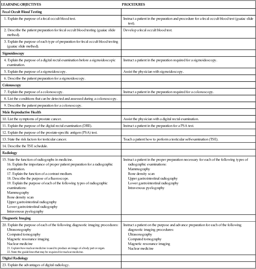
LEARNING OBJECTIVES
PROCEDURES
Fecal Occult Blood Testing
1. Explain the purpose of a fecal occult blood test.
Instruct a patient in the preparation and procedure for a fecal occult blood test (guaiac slide test).
2. Describe the patient preparation for fecal occult blood testing (guaiac slide method).
Develop a fecal occult blood test.
3. Explain the purpose of each type of preparation for fecal occult blood testing (guaiac slide method).
Sigmoidoscopy
4. Explain the purpose of a digital rectal examination before a sigmoidoscopic examination.
Instruct a patient in the preparation required for a sigmoidoscopy.
5. Explain the purpose of a sigmoidoscopy.
Assist the physician with sigmoidoscopy.
6. Describe the patient preparation for a sigmoidoscopy.
Colonoscopy
7. Explain the purpose of a colonoscopy.
Instruct a patient in the preparation required for a colonoscopy.
8. List the conditions that can be detected and assessed during a colonoscopy.
9. Describe the patient preparation for a colonoscopy.
Male Reproductive Health
10. List the symptoms of prostate cancer.
Assist the physician with a digital rectal examination.
11. Explain the purpose of the digital rectal examination (DRE).
Instruct a patient in the preparation for a PSA test.
12. Explain the purpose of the prostate-specific antigen (PSA) test.
13. State the risk factors for testicular cancer.
Teach a patient how to perform a testicular self-examination (TSE).
14. Describe the TSE schedule.
Radiology
15. State the function of radiographs in medicine.
16. Explain the importance of proper patient preparation for a radiographic examination.
17. Explain the function of a contrast medium.
18. Describe the purpose of a fluoroscope.
19. Explain the purpose of each of the following types of radiographic examinations:
Instruct a patient in the proper preparation necessary for each of the following types of radiographic examinations:
Diagnostic Imaging
20. Explain the purpose of each of the following diagnostic imaging procedures:
22. State the guidelines that may be required for nuclear medicine.
Instruct a patient on the purpose and advance preparation for each of the following diagnostic imaging procedures:
Digital Radiology
23. Explain the advantages of digital radiology.
Colon Procedures
Introduction to Colon Procedures
Fecal Occult Blood Test
Guaiac Slide Test
Patient Preparation
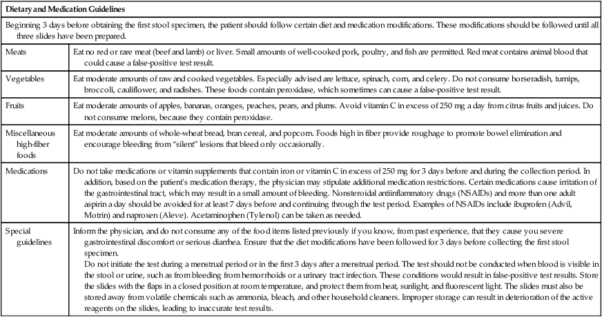
Dietary and Medication Guidelines
Beginning 3 days before obtaining the first stool specimen, the patient should follow certain diet and medication modifications. These modifications should be followed until all three slides have been prepared.
Meats
Eat no red or rare meat (beef and lamb) or liver. Small amounts of well-cooked pork, poultry, and fish are permitted. Red meat contains animal blood that could cause a false-positive test result.
Vegetables
Eat moderate amounts of raw and cooked vegetables. Especially advised are lettuce, spinach, corn, and celery. Do not consume horseradish, turnips, broccoli, cauliflower, and radishes. These foods contain peroxidase, which sometimes can cause a false-positive test result.
Fruits
Eat moderate amounts of apples, bananas, oranges, peaches, pears, and plums. Avoid vitamin C in excess of 250 mg a day from citrus fruits and juices. Do not consume melons, because they contain peroxidase.
Miscellaneous high-fiber foods
Eat moderate amounts of whole-wheat bread, bran cereal, and popcorn. Foods high in fiber provide roughage to promote bowel elimination and encourage bleeding from “silent” lesions that bleed only occasionally.
Medications
Do not take medications or vitamin supplements that contain iron or vitamin C in excess of 250 mg for 3 days before and during the collection period. In addition, based on the patient’s medication therapy, the physician may stipulate additional medication restrictions. Certain medications cause irritation of the gastrointestinal tract, which may result in a small amount of bleeding. Nonsteroidal antiinflammatory drugs (NSAIDs) and more than one adult aspirin a day should be avoided for at least 7 days before and continuing through the test period. Examples of NSAIDs include ibuprofen (Advil, Motrin) and naproxen (Aleve). Acetaminophen (Tylenol) can be taken as needed.
Special guidelines
Inform the physician, and do not consume any of the food items listed previously if you know, from past experience, that they cause you severe gastrointestinal discomfort or serious diarrhea. Ensure that the diet modifications have been followed for 3 days before collecting the first stool specimen.
Do not initiate the test during a menstrual period or in the first 3 days after a menstrual period. The test should not be conducted when blood is visible in the stool or urine, such as from bleeding from hemorrhoids or a urinary tract infection. These conditions would result in false-positive test results. Store the slides with the flaps in a closed position at room temperature, and protect them from heat, sunlight, and fluorescent light. The slides must also be stored away from volatile chemicals such as ammonia, bleach, and other household cleaners. Improper storage can result in deterioration of the active reagents on the slides, leading to inaccurate test results.
Quality Control
 Procedure 28-1 Fecal Occult Blood Testing


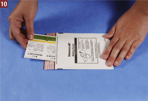
 Procedure 28-2 Developing the Hemoccult Slide Test

![]()
Stay updated, free articles. Join our Telegram channel

Full access? Get Clinical Tree