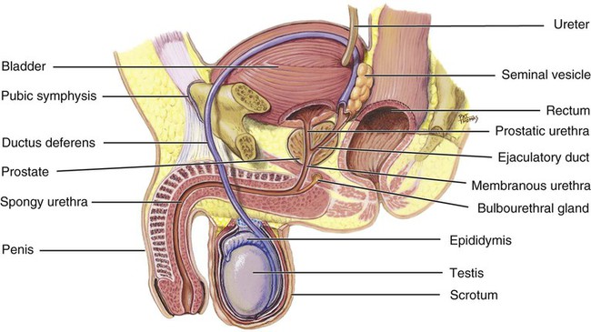
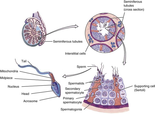
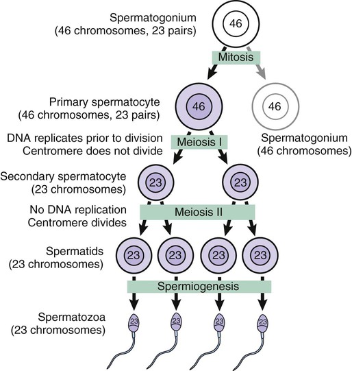
1. Distinguish between primary and secondary reproductive organs. 2. Describe the location and structure of each component of the male reproductive system. 3. Explain the process by which spermatids become mature sperm. 4. Trace the pathway of sperm from the testes to the outside of the body. 5. Outline the physiologic events in the male sexual response. 6. Describe the roles of gonadotropin-releasing hormone (GnRH), follicle-stimulating hormone (FSH), luteinizing hormone (LH), and testosterone in male reproductive functions. 7. Identify each component of the female reproductive system, including the mammary glands. 9. Describe the development of ovarian follicles. 10. Outline the physiologic events in the female sexual response. 11. Describe the roles of GnRH, FSH, LH, estrogen, and progesterone in female reproductive functions. 12. Describe what happens in each phase of the ovarian and uterine cycles, when each phase occurs, and how the cycles interact. 13. Describe ways in which the aging of an individual affects the reproductive system. • To produce egg and sperm cells • To transport and sustain these cells • To nurture the developing offspring The male reproductive system produces, sustains, and transports sperm; introduces the sperm into the female vagina; and produces hormones. Figure 16-1 illustrates the organs of the male reproductive system. Each testis is an oval structure about 5 cm long and 3 cm in diameter (Figure 16-2). A tough, white fibrous connective tissue capsule, known as the tunica albuginea (TOO-nik-ah al-byoo-JIN-ee-ah), surrounds each testis. The tunica albuginea extends inward to form septa that partition the testis into lobules. Each testis contains about 250 lobules. Each lobule contains one to four highly coiled seminiferous (seh-mye-NIFF-er-us) tubules that converge into a series of duets that exit the testes and enter the epididymis. Interstitial cells (cells of Leydig) are located between the seminiferous tubules within a lobule. Interstitial cells produce male sex hormones. Sperm are produced within the seminiferous tubules. The process of sperm formation is known as spermatogenesis (spur-mat-oh-JEN-eh-sis), which is a form of meiosis. The seminiferous tubules are packed with cells in various stages of spermatogenesis (Figure 16-3). Interspersed with these cells are large cells that extend from the periphery of the tubule to the lumen. These large cells are the supporting cells (Sertoli cells), which support and nourish the other cells. Highlight on the Reproductive System Inguinal hernia: The inguinal canal is a weak area in the abdominal wall that may rip open, resulting in an inguinal hernia. A portion of the intestine may pass through the opening into the scrotum. This is painful and potentially dangerous if the blood supply to the intestine is constricted. This condition is more common in men than in women. Inguinal hernias are frequently repaired by surgery. Undescended testicles: The condition in which the testes do not descend into the scrotum is called cryptorchidism. Crypt means “hidden” and orchid refers to the testis, so the term means “hidden testis.” Cryptorchidism results in sterility if it is not corrected before puberty because the cooler temperature of the scrotum is necessary for sperm production. Vasectomy: A vasectomy is a surgical procedure, usually accomplished through a tiny incision in the scrotum, that severs the vas deferens. A bilateral vasectomy results in sterility because it interrupts the pathway of the sperm to the outside of the body. Enlarged prostate gland: Benign prostatic hyperplasia is a common condition in older men. In this condition, the prostate enlarges and compresses the urethra, making urination difficult. This situation results in urine retention in the bladder, which makes the individual more susceptible to urinary tract infections. Prostate cancer: Cancer of the prostate is a common cancer in men. It usually starts in one of the secretory glands, and as it continues it produces a lump on the surface of the prostate. In many cases by the time the lump can be palpated through the wall of the rectum, the cancer has metastasized to other areas of the body. It is hoped that using blood-screening techniques (e.g., prostate-specific antigen [PSA]) in addition to rectal palpation will result in earlier detection of the tumor so that treatment can begin before metastasis occurs. Circumcision: Circumcision is the surgical removal of the prepuce of the penis. Sometimes this is done to correct phimosis, a condition in which the prepuce is too tight and obstructs urine flow. In certain cultures, circumcision is performed as a religious rite or an ethnic custom. For others, it is a matter of family preference. The medical benefits of circumcision are a subject of debate in the medical community. Some believe it is practical for hygienic reasons. Evidence indicates that circumcision may reduce the risk of penile cancer. Impotence: Impotence is the inability to achieve an erection. Psychological stresses are often blamed for impotence, but other causes can lead to the difficulty. Impotence may result from an abnormality of the erectile tissue or failure of the parasympathetic reflexes that produce an erection. Drugs and alcohol may cause temporary impotence because they can interfere with the nerve and blood vessel actions that are necessary for an erection. Ovarian cancer: Carcinomas of the ovary account for more deaths than those of cervical and uterine cancers together. Because there are no screening tests and few symptoms in the early stages, ovarian carcinomas are usually in an advanced stage when they are discovered. Surgery, radiation therapy, and chemotherapy are used as therapeutic measures. Tubal ligation: Tubal ligation is a surgical procedure in which the uterine tubes are burned or severed and tied off. This is a permanent method of birth control because sperm are unable to reach the egg for fertilization. The technique involves making a small incision in the abdomen and inserting a small tube through which the ligation instruments can be introduced. Ectopic pregnancy: An ectopic pregnancy occurs when a fertilized egg implants in some site other than the uterus. Ectopic pregnancies may occur in the uterine tubes. Because the tubes are not equipped to sustain and nourish the developing embryo, miscarriages often occur. The uterine tubes are unable to expand like the uterus and may rupture, with subsequent hemorrhage. Surgery may be indicated to remove the implant and to preserve the uterine tube before rupture occurs. Mittelschmerz: Mittelschmerz is a term to describe one-sided lower abdominal pain, which may switch sides from one month to another, at or around the time of ovulation. The pain is usually described as sharp or cramping and lasts from 24 to 48 hours. There is no known prevention, and treatment consists of analgesics. Fibrocystic disease: Fibrocystic disease is a common benign condition of the breast. Small sacs of tissue and fluid develop in the breast tissue, and the patient notices lumps in the breast often associated with premenstrual tenderness. Mammography and surgical biopsy may be indicated to differentiate between fibrocystic disease and carcinoma of the breast. Each primary spermatocyte goes through the first meiotic division (meiosis I) to produce two secondary spermatocytes. In the second meiotic division (meiosis II), each secondary spermatocyte divides to produce two spermatids. As a result of the two meiotic divisions, each primary spermatocyte produces four spermatids (Figure 16-4). During spermatogenesis there are two cellular divisions but only one replication of DNA, so each spermatid has 23 chromosomes (haploid), one from each pair in the original primary spermatocyte. Each successive stage in spermatogenesis is pushed toward the center of the tubule. This results in the more immature cells being at the periphery, and the more differentiated cells are nearer the center (see Figure 16-3). Spermatogenesis (and oogenesis in the female) differs from mitosis (review Chapter 5) because the resulting cells have only half the number of chromosomes as the original cell. When the sperm cell nucleus unites with an egg cell nucleus, the full number of chromosomes is restored. If sperm and egg cells were produced by mitosis, then each successive generation would have twice the number of chromosomes as the preceding one. The final step in the development of sperm is called spermiogenesis (spur-mee-oh-JEN-eh-sis). In this process, the spermatids formed from spermatogenesis become mature spermatozoa, or sperm. The mature sperm cell has a head, midpiece, and tail (see Figure 16-3). The head contains the 23 chromosomes surrounded by a nuclear membrane. The tip of the head is covered by an acrosome (AK-roh-sohm). The acrosome contains enzymes that help the sperm penetrate the female gamete. The midpiece contains mitochondria that provide adenosine triphosphate (ATP). The tail, also called the locomotor region, is a typical flagellum for locomotion. The sperm are released into the lumen of the seminiferous tubule, where they leave the testes and enter the epididymis. In the epididymis the sperm undergo final maturation and become capable of fertilizing a female gamete. Sperm leave the testes through a series of ducts that enter the epididymis (ep-ih-DID-ih-mis) (see Figure 16-2). The epididymis is a long tube that is tightly coiled to form a comma-shaped organ. When the sperm leave the testes, they are immature and incapable of fertilizing ova. They complete their maturation process and become fertile as they move through the epididymis. Mature sperm are stored in the lower portion of the epididymis. The ductus deferens (also called vas deferens) is a tube that is continuous with the epididymis (see Figure 16-2). The ductus deferens enters the abdominopelvic cavity, then descends along the posterior wall of the bladder toward the prostate gland (see Figure 16-1). Sperm are stored in the ductus deferens, near the epididymis, and peristaltic movements propel the sperm through the tube.
Reproductive System
Introduction to the Reproductive System
Male Reproductive System
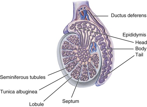
Testes
Structure
Spermatogenesis

Duct System
Epididymis
Ductus Deferens
![]()
Stay updated, free articles. Join our Telegram channel

Full access? Get Clinical Tree


Reproductive System
Get Clinical Tree app for offline access
