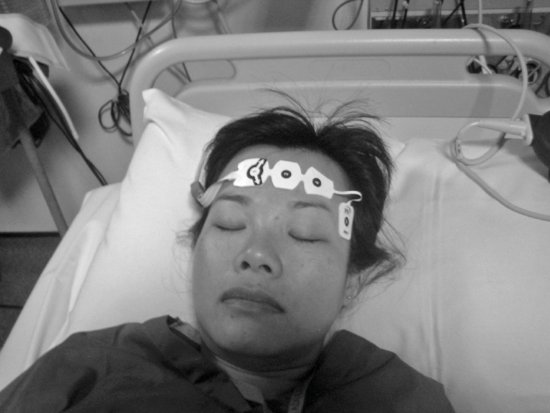
An infusion of propofol 100 mg/h has been commenced, with the intention to gradually reduce the dosage over the next 24 hours. The plan is to wake Betty as quickly as possible to allow for neurological assessment, once her condition has stabilised.
Reader activities
Having read this scenario, consider the following:
- Why does Betty need sedation?
- What criteria should be used to select the type of sedation and the dosage to be administered?
- What information is required for monitoring the effects of sedation?
- What is best practice for appropriate management of Betty’s sedation therapy?
- What assessments would you undertake to ensure appropriate use of sedation?
Pathophysiology and pharmacology
Pain and agitation are common in critically ill patients. This is, in part, due to activation of the stress response causing neurohormonal elevation of plasma catecholamines (Blanchard 2002), and sympathetic over-activity, with an associated increase in heart rate, myocardial oxygen consumption and respiratory rate (Ferguson and Mehta 2002). As well as untreated or intractable pain, the stress response can be attributed to a vast array of other factors including the critical care environment; medications; invasive procedures; uncomfortable positioning; an inability to communicate; fear; sleep deprivation and the vast array of monitoring and technology with which they are surrounded. The resulting anxiety and agitation can lead to dyspnoea, patient ventilator asynchrony, elevated blood pressure and heart rate and possible aggressive behaviour and PTSD. Pain, delirium, anxiety and agitation can manifest with similar signs but each may have different causes requiring different management.
Sedation is important for Betty to ensure comfort from both a psychological and physiological perspective. She currently appears mildly hypertensive possibly due to anxiety and/or pain. Sedation may be used to help control this stress response and reduce her oxygen requirements, particularly as she is already hypoxaemic, possibly due to sepsis evidenced by her pyrexia and mild metabolic acidaemia. It can also be used to reduce any patient-ventilator dys-synchrony (Ferguson and Mehta 2002). Betty needs to be awoken quickly for neurological assessment purposes. It is currently unclear if she has sustained a neurological injury. This could also be compromising her blood gases and haemodynamic status. Short-term sedation which allows for neurological assessment is therefore desirable. However, continued sedation may be necessary to facilitate other interventions and ongoing management.
In order to optimise sedation management, it is essential that nurses understand the basic pharmacological and clinical uses of the most commonly used sedative agents.
Sedatives
Benzodiazepines
Benzodiazepines (lorazepam, midazolam, diazepam) work by enhancing the effects of GABA (γ-aminobutyric acid), a potent inhibitory neurotransmitter, making neurons resistant to excitation and producing sedatory and amnesic effects (Pun and Dunn 2007). They also have anticonvulsant effects. However, they do not have analgesic properties and should therefore be used in conjunction with an appropriate analgesic agent (see Chapter 10).
The use of lorazepam has recently been reported to be an independent risk factor for the development of delirium in critically ill patients, particularly as the dosage increases (Pandharipande et al. 2006). Generally, midazolam is the preferred benzodiazepine for use (Jacobi et al. 2002) as it has a rapid onset (2–5 minutes) and has a short half-life. The intravenous (IV) route is the most reliable and effective because of absorption and administration difficulties associated with oral and/or intramuscular administration.
Midazolam may produce adverse effects, including respiratory and cardiovascular depression, physical dependence and agitation as well as unpredictable awakening (with prolonged sedation) (Nasraway 2001; Fullwood and Sargent 2010). These adverse effects result from either an accumulation of the drug or its active metabolites (Hassan et al. 1998; Jacobi et al. 2002). When associated with alcohol, used in an elderly patient such as Betty, or in patients with cirrhosis of the liver, it can also depress respiration.
Respiratory depression and hypotension are dose-dependent. Hypotension occurs primarily in hypovolaemic patients and is potentiated by the associated use of opioids (Blanchard 2002), infection and hypoxaemia. Despite the relatively short half-life, extensive re-distribution can cause prolonged sedation. Recovery time is proportional to duration; therefore, midazolam infusions generally should not exceed 48 hours (Jacobi et al. 2002).
Midazolam would not be the most appropriate agent of choice for Betty initially due to the desire to wake and neurologically assess. However, if agitation were to become problematic then small IV boluses may be considered. It is also likely that Betty may not wean easily from ventilation due to her poor physical state and respiratory failure. She may therefore require benzodiazepines as part of her ongoing management.
Propofol
Propofol (Diprovan®) a sedative–hypnotic agent has a very rapid onset (1–2 minutes) and a short half-life, making it attractive to use for Betty. How propofol works is not completely understood (Fullwood and Sargent 2010), but it is believed to enhance GABA’s affinity for its receptors, much like the benzodiazepines. Propofol concentration in plasma falls quickly once the drug is discontinued.
Propofol is a useful drug for patients with neurologic injury. It decreases intracranial pressure, cerebral blood flow and cerebral metabolism (Rhoney and Parker 2001; Jacobi et al. 2002). For Betty this drug could be considered to be the safest option because currently the reason for her low GCS is unknown.
Vigilance is important when using prolonged or high doses of propofol (more than 48 hours at doses higher than 5 mg/kg/h), particularly in patients with acute neurological or inflammatory illnesses (Jacobi et al. 2002) such as Betty. Caution is also required in cases of hypovolaemia or poor cardiovascular system function due to its vasodilator and potent negative inotropic properties, which can potentially cause large decreases in blood pressure. In addition, high-dose propofol use has been linked to ‘Propofol infusion syndrome’, a potentially fatal syndrome characterised by cardiac failure, rhabdomyolysis (the breakdown of muscle fibres, resulting in the release of muscle fibre content (myoglobin) into the bloodstream), severe metabolic acidosis and renal failure (Ostermann et al. 2000; Jacobi et al. 2002; Vasile et al. 2003; Marik 2004) and painful peripheral administration (Ostermann et al. 2000; Jacobi et al. 2002).
This drug is prepared in a lipid emulsion, so long-term or high dose infusion may lead to elevated triglyceride levels. Serum triglyceride levels should be monitored and the emulsion considered a calorific source (Ostermann et al. 2000; Jacobi et al. 2002). Additionally, because the lipid emulsion can promote bacterial growth, strict aseptic technique and a dedicated IV catheter are also essential to help prevent the development of further sepsis in Betty.
Propofol is metabolised at least partially by the liver to inactive metabolites and excreted by the kidneys. However, the presence of renal or hepatic dysfunction does not significantly affect clearance (Sang Ko and Gwak 2008). It can also accumulate in peripheral tissues, prolonging its effects.
Neuromuscular blocking (paralysing) agents
These drugs block transmission at the neuromuscular junction causing paralysis of the affected skeletal muscles. Neuromuscular blocking agents, for example, suxamethonium, atracurium, vecuronium or pancuronium are useful to facilitate mechanical ventilation but can also be harmful. These agents should not be avoided in Betty as neurological assessment, and the ability to monitor for any seizure activity would be compromised.
If patients are adequately sedated, neuromuscular blockade is usually un-necessary. It should be used as a last resort for patients resisting or fighting the ventilator or those who are at risk of harm. It should not be used as a form of chemical restraint.
Tests and investigations
Assessment of sedation
A comprehensive assessment of Betty’s sedation needs and level of sedation is required. However, the nurse must first rule out or treat any pain that Betty has as this could be influencing any agitation (see Chapters 10 and 13 for more information on pain and agitation). Although Betty should not be agitated, nervous or experiencing pain, at the same time she should not be over-sedated as this can lead to complications of its own. This balance can be difficult to achieve.
The ability to assess sedation levels may be affected by patient variables such as age, language (including aphasia), disease pathology and pain (Weinert et al. 2001). Older patients like Betty can also appear adequately sedated, but actually be under-sedated. Disruptions to communication can further decrease the reliability of any sedation assessment. For example, in the non-communicative patient it may be difficult to differentiate whether movement is a symptom of pain or inadequate sedation (Frazier et al. 2002).
Because of the limitations of the subjective assessment tools available, over- and under-sedation remain major challenges to critical care nurses. In general, there appears to be a tendency to over-sedate. This, can lead to avoidable prolongation of mechanical ventilation and ICU and hospital stay (Kress et al. 2000). Excessive sedation can cause an increased risk of venous thrombosis, decreased intestinal motility, hypotension and reduced tissue oxygen extraction capabilities. Cardiopulmonary dysfunction including restlessness, tachypnoea, irritability and hypertension may also occur secondarily to subsequent withdrawal. Equally, under-sedation may instigate psychological consequences, leading to extreme agitation, potentially associated with the accidental removal of the artificial airway or invasive lines and monitoring equipment. Evidence from follow-up of former critically ill patients suggests that unpleasant experiences may have potentially serious implications for psychological and emotional recovery (Jones et al. 2001), including the development of delirium see chapter 13 for further details on delirium. Physiological insults include myocardial ischemia, hypoxia and increased myocardial oxygen consumption (White et al. 2001), resulting in catecholamine release (tachycardia) and increased irritability, prolonging recovery and overall hospital stay. Many of these problems can, in part, be related to difficulty in accurately judging patients’ medication requirements (Egerod 2002).
Titration of sedative agents continues to be reliant upon methods of assessment that are influenced by a variety of subjective factors including family presence and preconditioned social, personal and professional norms of the nurse performing the assessment. Various sedation scoring tools have been developed in order to facilitate the assessment process. However, many units continue to rely on nursing staff to assess sedation rather than routinely employing any scoring system (Soliman et al. 2001). Even when scoring systems are in place, some nurses continue to avoid their use as part of their assessment. Additionally, there is, on occasion, disagreement amongst multi-disciplinary team members about the goals of sedation, which potentially originates from miscommunication (Slomka et al. 2000).
Standardised sedation-assessment scales provide health care professionals with a common language. They can be used to judge the level of sedation through indicators such as movement and response to physical or verbal stimuli. A variety of such scales exist, although not all have been validated. These include the Ramsay sedation scale (Ramsay et al. 1974), Richmond agitation–sedation scale (Sessler et al. 2002), motor activity assessment scale (Devlin et al. 1999), the Vancouver interactive and calmness scale (de Lemos et al. 2000) and the Nursing Instrument for the Communication of Sedation (NICS) (Mirski et al. 2010).
The Ramsay scale (Ramsay et al. 1974) is still one of the most widely used tools for evaluating sedation in the United Kingdom. Sedation levels range from one (patient awake and anxious, agitated or both) to six (patient asleep and unresponsive to both light touch and loud noise). Despite its popularity, limitations of the Ramsay scoring system have resulted in some units adapting the tool, and one author comments that nurses perceive no advantages in its use (Elliott et al. 2006). Behavioural responses cannot be used to classify patients who are quietly disorientated and distressed (Elliott et al. 2006). In addition, the tool incorporates three very different constructs (agitation, anxiety and conscious level) in the same scale making classification difficult. It has also been criticised as not adequately reflecting states of consciousness in the brain-injured patient (Hansen-Flaschen et al. 1994), an important consideration for Betty. However, the assessment of consciousness and the assessment of sedation, although interlinked, should not be considered interchangeable elements of assessment. Thus, although assessment of Betty’s conscious level is difficult whilst receiving sedation, an attempt to assess her neurology should be made using a recognised tool designed for such purpose (see Chapter 12 for more information on neurological issues). Further, as might be a criticism of many sedation assessment tools, observation is limited to a short, discrete period of time and does not account for changes in response that may occur between assessments. Therefore, despite the plethora of tools available, there exists little practical difference between the majority.
Objective sedation assessment tools may prove beneficial as an adjunct to subjective scales, particularly in patients receiving neuromuscular blockade. Bispectral index (BIS) monitoring (see Figures 11.1 and Figures 11.2) has been advocated to be a reliable form of sedation assessment and monitoring (Olson et al. 2004). However, this research was conducted in a neurological critical care unit with staff adept in EEG monitoring. They may be less reliable in general ICUs, with staff less familiar with such monitoring. Although BIS is used in some general units, there is currently a lack of adequate research to support its use in such areas and some studies suggest that BIS is not reliable for routine monitoring (Frenzel et al. 2002; Riess et al. 2002).
Stay updated, free articles. Join our Telegram channel

Full access? Get Clinical Tree


