Chapter 19 Rheumatology
Insider’s Guide to Rheumatology for the USMLE Step 1
When it comes to rheumatology on the USMLE, practice makes perfect. You are highly likely to encounter “textbook” presentations of the rheumatologic and bone conditions you must learn for Step 1, so the more cases you read before your examination, the better you will get at spotting the unique features that distinguish one disease from another. We have specifically designed this chapter to include high-yield cases that closely resemble the clinical presentations you should expect to see on boards. As you go through these cases, pay special attention to buzzwords and laboratory values (including autoantibodies) that will clue you into the correct diagnosis.
Basic concepts
1 What is a “diarthrodial” joint?
A diarthrodial joint is a joint in which bones meet in cartilage-covered surfaces. They are designed for mobility, such as that seen in the shoulder, hip, and interphalangeal joints, where substantial degrees of movement are possible. The diarthrodial joint is the most common type of joint found in the body.
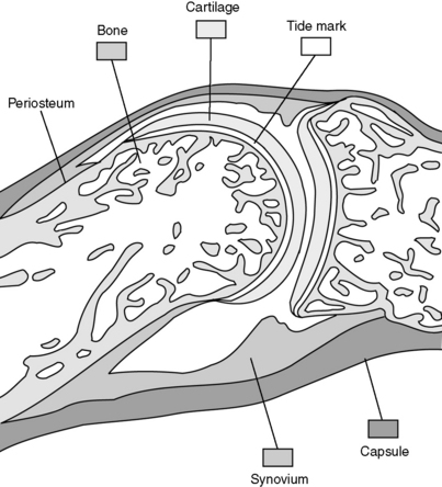
2 What are the components of a diarthrodial joint and which sites within a diarthrodial joint are vulnerable to disease?
These joints are composed of articulating bones covered by cartilage (usually hyaline cartilage). A fibrous capsule surrounds and protects the joint. The diarthrodial joint cavity is lined with a synovial membrane, which produces synovial fluid to lubricate the joint space. Ligamentous connections provide support and typically allow for a large amount of movement. Each of these components of the joint may be involved in disease processes (Fig. 19-1).
3 What are synarthrodial and amphiarthrodial joints?
Synarthrodial joints exist where bones meet by fibrous connections without a joint space. These joints prevent motion between bones. The suture lines in the skull are an example of synarthroses. Amphiarthrodial joints consist of bones bound by fibrocartilage, which allows some limited degree of movement. The joints between vertebrae and the pubic symphysis are examples of amphiarthroses.
Diarthrodial joints are the most common sort of joint and allow a large degree of motion between bones (examples: knees, hips).
The diarthrodial joint is made up of bone, cartilage, a joint space lined with synovium, synovial fluid, a surrounding capsule, and ligamentous insertions.
Synarthrodial joints consist of bones bound together by fibrous tissue and are nearly immobile (example: sutures in skull).
Amphiarthrodial joints consist of bones bound together by cartilage and are slightly more mobile than synarthrodial joints (example: pubic symphysis).
Case 19-1
A 55-year-old obese woman presents with several months of bilateral knee pain of several months’ duration. The pain is exacerbated by activity and decreases with rest. She experiences approximately 10 to 15 minutes of morning stiffness each day but otherwise denies constitutional complaints such as fever, anorexia, weight loss, or fatigue.
1 What is the differential diagnosis?
The differential diagnosis for symmetrical joint pain is broad and includes osteoarthritis, rheumatoid arthritis, and spondyloarthropathies such as ankylosing spondylitis or psoriasis, crystal arthropathy such as gout or pseudogout, and septic arthritis. In a middle-aged obese woman with involvement of weight-bearing joints, osteoarthritis seems likely.
Case 19-1 continued:
The patient is moderately obese. None of her joints are warm, swollen, or tender. Aside from some crepitus with passive movement, examination of her knees is normal. The proximal and distal interphalangeal joints in both hands are enlarged but nontender. Plain film of the right knee shows narrowing of the medial compartment joint space (Fig. 19-2).
2 What is the likely diagnosis?
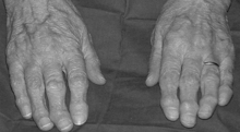
Osteoarthritis (also known as OA, osteoarthrosis, degenerative joint disease, hypertrophic arthritis) is likely and is the most common joint disease worldwide. OA is characterized by loss of articular cartilage, which results in damage to the underlying bone. This process results primarily in pain (especially in weight-bearing joints), as well as stiffness and loss of joint mobility. The process is noninflammatory, so there is no ankylosis (fusion) of the joint. Loss of the smooth articulating surface accounts for the finding of crepitus when the joint is moved. Pain is typically worse with use of the joint and decreases with rest. Reactive bone formation resulting in osteophytes (bone spurs) also occurs at the joint margins and may cause slight elevations in serum alkaline phosphatase. Joints typically affected include the proximal and distal interphalangeal joints (Bouchard’s and Heberden’s nodes, respectively), knees, and hips. Figure 19-3 shows both Heberden’s and Bouchard’s nodes. Recall that rheumatoid arthritis typically does not affect the distal interphalangeal joints.

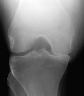
Figure 19-2 Plain film of the knee for patient in Case 19-1.
(From Harris ED, Budd RC, Genovese MC, et al: Kelley’s Textbook of Rheumatology, 7th ed. Philadelphia, WB Saunders, 2005.)
3 Is the pathogenesis of this condition primarily related to degeneration of bone, cartilage, or synovial membrane?
Osteoarthritis is characterized by degeneration of articular cartilage and is often associated with “overuse” or trauma to the joint. Chondrocytes produce the type II cartilage that makes up the articular cartilage, and altered chondrocyte function has been demonstrated to occur in osteoarthritis. When articular cartilage is not maintained properly, the bones in the diarthrodial joint may come into direct contact with one another. The wear and tear that bones experience in this situation leads to abnormal bone proliferation, with the formation of osteophytes (bone spurs).
Rheumatoid arthritis differs in that the primary site of damage is the synovium, via an autoimmune mechanism. Unlike rheumatoid arthritis, osteoarthritis is not characterized by systemic inflammation or autoimmune phenomena. Rheumatoid joints often appear red and swollen (synovitis), whereas joints affected by osteoarthritis typically do not.
4 What is the anatomic source of the joint pain in osteoarthritis?
Although cartilage is the primary site of injury in this disease, there is no neural input to cartilage and therefore no pain transmission from it. The pain of osteoarthritis actually comes from the periosteum (dense fibrous tissue) surrounding the bone. The periosteum is highly innervated and is damaged when the cartilage has worn away to the point that bone is rubbing on bone.
Note: Joint cartilage is also completely avascular, which explains why injured cartilage will not heal.
5 What are some risk factors associated with developing osteoarthritis?
Obesity, occupation (repetitive motions), intense physical activity, joint trauma, and muscle weakness (likely from joint instability) play a role. Notice that all of these factors increase the mechanical forces to which the joint cartilage is exposed.
Gender, hormones, and genetics are involved as well. Women are more likely to suffer from osteoarthritis than men, and elderly populations are affected by this disease much more often than young people. There are certain forms of osteoarthritis that appear to be heritable.
Obesity is the strongest modifiable risk factor.
Note: Osteoarthritis can be classified as primary (idiopathic), the most common, or secondary, with an underlying cause (e.g., trauma, obesity, Paget disease, metabolic disorders). Diseases that involve the systemic deposition of certain compounds, such as hemochromatosis (iron), Wilson’s disease (copper), and the crystal arthropathies, may be causes of secondary osteoarthritis.
6 Would you expect the erythrocyte sedimentation rate (ESR) or C-reactive protein (CRP) to be elevated in this patient?
No. An elevated ESR or CRP is a nonspecific indicator of a systemic inflammatory process. Because osteoarthritis is a local degenerative disease, these measurements would probably not be elevated in this patient (note that unlike rheumatoid arthritis, osteoarthritis does not result in systemic symptoms). In addition, although severe joint degeneration caused by OA may lead to inflammation, this inflammatory response would be confined to the joint space.
Note: Although osteoarthritis is classically considered a noninflammatory disease, it is interesting to note that pharmacologic treatment frequently involves the use of nonsteroidal anti-inflammatory drugs (NSAIDs) and intra-articular injections of steroids, which clearly act by reducing inflammation.
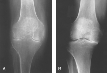
7 How do the findings on x-ray studies generally differ between osteoarthritis and rheumatoid arthritis?
The characteristic radiologic finding in osteoarthritis is joint space narrowing, as in this patient, due to the loss of cartilage between the bones. There may also be evidence of bony proliferation, such as increased density of the bones abutting the joint (subchondral sclerosis or eburnation) and presence of osteophytes. Chondrocalcinosis (calcium in the articular cartilage) and subchondral cysts may also be evident.
In rheumatoid arthritis, on the other hand, there are often marginal erosions of bone and osteoporotic changes (demineralization), and the joint space is typically normal. However, if the rheumatoid arthritis is severe enough, the inflammatory process may eventually destroy articular cartilage also and narrow the joint space.
Figure 19-4A demonstrates nearly complete loss of the lateral and medial joint spaces in rheumatoid arthritis, whereas Figure 19-4B demonstrates loss of only the medial joint space with subchondral sclerosis (increased density) of the underlying bone in OA.

Figure 19-4 Radiographs of the knees in the two most common forms of arthritis: rheumatoid arthritis and osteoarthritis. A, Severe involvement in rheumatoid arthritis, with almost complete symmetrical loss of joint space in both the medial and lateral compartments, with little subchondral sclerosis or osteophyte formation. B, Typical osteoarthritis, with severe, near-total loss of joint space of one compartment and a normal or actually increased joint space of the other compartment.
(From Goldman L, Ausiello D: Cecil Textbook of Medicine, 22nd ed. Philadelphia, WB Saunders, 2004.)
8 Nonsteroidal anti-inflammatory drugs and cyclooxygenase-2 inhibitors are both possible treatments for this patient. What is similar about their mechanisms of action, and what is different?
Regarding similarities, both classes of drug inhibit prostaglandin synthesis by inhibiting cyclooxygenase (COX), and by doing so cause relief of pain. All NSAIDs reversibly inhibit COX, except aspirin, which irreversibly inhibits COX. The COX-2 inhibitors (rofecoxib, celecoxib) selectively inhibit a form of COX that is induced in inflammatory cells, but not the constitutively expressed COX-1 that is produced for various normal body functions.
9 What is the principal therapeutic advantage of the cyclooxygenase-2 inhibitors? What can be given to this patient with a nonsteroidal anti-inflammatory drug to prevent their principal side effect?
By not inhibiting gastrointestinal (GI) prostaglandin synthesis, COX-2 inhibitors are supposed to cause less gastric ulceration, a very common problem with the NSAIDs. Drugs such as misoprostol, a synthetic prostaglandin analog, can be given orally with NSAIDs to reduce the risk of gastric ulceration and bleeding. However, more commonly proton pump inhibitors such as omeprazole are given to reduce the GI side effects of NSAIDs.
Note: NSAID use can also precipitate renal failure in patients with borderline renal function due to reduced synthesis of vasodilatory prostaglandins that help maintain renal perfusion.
10 Why are cyclooxygenase-2 inhibitors considered dangerous in some patients?
COX-2 inhibitors do not inhibit COX-1 in platelets. They therefore leave platelets capable of aggregating and forming thrombi. Additionally, COX-2 inhibitors do inhibit the production of prostaglandins in endothelial cells, potentially creating a prothrombotic state and promoting vasoconstriction. It is therefore believed that COX-2 inhibitors may pose a cardiovascular threat to certain patients at risk for stroke or myocardial infarction (MI), which is why Vioxx (rofecoxib) was withdrawn from the market in 2004.
11 Is acetaminophen a reasonable option for treating joint pain in this patient?
Yes. Actually, acetaminophen should be first-line therapy. Acetaminophen primarily works in the central nervous system (CNS) by raising the pain threshold. Its primary use is therefore as an analgesic. Because acetaminophen is not an NSAID and does not inhibit peripheral prostaglandin synthesis to a significant extent, it does not have the potential GI or renal side effects of the NSAIDs.
12 What sort of analgesic would you prescribe for a patient with preexisting renal disease? Or liver disease? Or pregnancy?
NSAIDs are renally excreted, and they decrease perfusion to the kidneys. They would therefore be relatively contraindicated in a patient with kidney disease, and acetaminophen is a better choice for this patient. Acetaminophen, however, is metabolized primarily by the liver (remember that patients who overdose on acetaminophen develop liver failure). A patient with cirrhosis or other liver disease would be better served by an NSAID than by large doses of acetaminophen. In a pregnant patient, NSAIDs should be avoided, particularly in the third trimester. The fetal ductus arteriosus is kept open by prostaglandins, and NSAIDs cause it to close prematurely in utero. However, in the case of a patent ductus arteriosus that persists after birth, indomethacin can be given to promote closure.
13 Quick review: Look only at the left column in Table 19-1 and try to list the class of drug, mechanism of action, and major side effects for each drug listed
Osteoarthritis is the most common joint disease worldwide.
Although there may be local joint inflammation, it is considered a noninflammatory condition without systemic (constitutional) symptoms.
The disease process is centered in the cartilage and leads to destruction of bone.
Pain is typically worse with activity.
Obesity, gender, age, and trauma all contribute to its development.
Radiographs show joint space narrowing, subchondral sclerosis, and osteophytes.
Acetaminophen, cyclooxygenase (COX)-2 inhibitors, and nonsteroidal anti-inflammatory drugs (NSAIDs) are the mainstay of pharmacotherapy.
Case 19-2
A 52-year-old woman presents with a chief complaint of polyarticular joint pain for several months. Her pain is worst in the morning and lessens with activity over the course of the day. She typically experiences 1 hour of stiffness after she wakes up. She also reports malaise, anorexia, night sweats, and a persistent low-grade fever. Her symptoms seem to come and go at will.
1 What is the differential diagnosis?
The differential diagnosis for polyarticular arthritis includes osteoarthritis, rheumatoid arthritis, septic arthritis (e.g., disseminated gonococcal infection), reactive arthritis (e.g., in response to infections, previously known as Reiter’s syndrome), systemic lupus erythematosus (SLE), hepatitis, and paraneoplastic syndromes. Although the differential here is broad, her morning stiffness and constitutional complaints are suggestive of an inflammatory arthropathy such as rheumatoid arthritis.
Case 19-2 continued:
Upon examination, she has symmetrical involvement of the metacarpophalangeal (MCP) and proximal interphalangeal joints, with sparing of the distal interphalangeals (Fig. 19-5). Involved joints in her hands are swollen, warm, and tender to palpation. She has moderate-sized bilateral knee effusions. Laboratory tests show elevated ESR and CRP and positive rheumatoid factor (RF). She is diagnosed with a chronic autoimmune disease and prescribed methotrexate, an NSAID, and prednisone.
2 What is the diagnosis?
This patient presents with classic signs of rheumatoid arthritis, which is an autoimmune disease of synovial joints. The principal pathologic process in rheumatoid arthritis is synovitis—inflammation of the synovial membrane—which leads to destruction of the joint. Because rheumatoid arthritis is a systemic inflammatory condition, patients often present with constitutional symptoms such as fever, weight loss, and fatigue as well as joint pain. Note that joint involvement in rheumatoid arthritis is usually symmetrical.
3 What are the criteria for the diagnosis of rheumatoid arthritis?
Four of the following seven criteria must be met for the diagnosis to be made:
1. Morning stiffness lasting at least 1 hour
2. Arthritis of three or more joints
4. Symmetrical joint involvement
This patient has morning stiffness, symmetrical arthritis of more than three joints including the hand, and positive RF. She therefore meets the criteria for the definitive diagnosis of rheumatoid arthritis.
4 What sort of damage occurs in the joints of patients with rheumatoid arthritis?
The initial site of damage is the synovium lining the joint space, which becomes the center of an inflammatory process that will involve the entire joint. Lymphocytes, macrophages, osteoclasts, and fibroblasts are all involved and ultimately lead to destruction of the cartilage and bone of the joint. Pannus formation occurs as synovial tissue is aberrantly stimulated to proliferate, and bony erosions develop as the inflammatory process continues. Joints will be painful, show signs of inflammation, and eventually lose normal architecture and mobility.
5 What is the significance of rheumatoid factor in this disease?
RF is an autoantibody (IgM) directed against the Fc region of IgG. This autoantibody forms immune complexes (type III hypersensitivity reaction), which deposit throughout the body and are implicated in the extra-articular manifestations of rheumatoid arthritis. RF is present in roughly 80% of rheumatoid arthritis patients, but it is not highly specific for the condition; RF can also appear in lupus, tuberculosis, Sjögren’s syndrome, and other disorders. It is therefore not necessary for the diagnosis of rheumatoid arthritis, but it is useful for determining prognosis. Virtually 100% of patients with extra-articular manifestations have a positive RF.
Note: The current best serologic test for the diagnosis of rheumatoid arthritis is the anti-CCP (citrullinated cyclic peptide) antibody. Its sensitivity is the same as that for RF, but it is more specific for rheumatoid arthritis and is a better predictor of disease progression.
6 What is the epidemiology of rheumatoid arthritis?
As is the case with most autoimmune conditions, rheumatoid arthritis affects women more often than men, and its prevalence increases with age. However, there is no ethnic background or geographic distribution associated with the disease. Genetic susceptibility has been demonstrated, as the disease is associated with certain major histocompatibility complex (MHC) class II proteins expressed by antigen-presenting cells. The HLA-DR4 haplotype confers risk for rheumatoid arthritis, and a specific HLA-DR epitope is shared by many people with rheumatoid arthritis. However, these HLA genes do not tell the whole story, and many genes are likely to be responsible for the development of the disease.
7 What is the typical treatment for rheumatoid arthritis?
Three classes of medications are used to manage rheumatoid arthritis: analgesics to treat pain, corticosteroids to suppress inflammation, and disease-modifying antirheumatic drugs (DMARDs) to limit progression of the disease. NSAIDs are commonly used as analgesics and have the added benefit of dampening inflammation. Corticosteroids can be injected directly into affected joints and may be used to manage acute flares of rheumatoid arthritis. DMARDs include a variety of drugs such as methotrexate, sulfasalazine, gold, and tumor necrosis factor (TNF) inhibitors. Early use of DMARDs is desirable, as these drugs halt progression of irreversible joint damage.
8 What would be learned from aspiration of this patient’s knee?
Probably not much would be revealed by aspiration. The synovial fluid should have an inflammatory composition with 2000 to 50,000 white blood cells (WBCs)/mm3, with the cells being predominantly polymorphocnuclear neutrophils (PMNs). If crystals or bacteria were present, or the WBC count was not in this range, one would need to consider other diagnoses on the differential.
Note: Rheumatoid arthritis may also show decreased C3 protein in the synovium but increased C3 levels in the serum.
Case 19-2 continued:
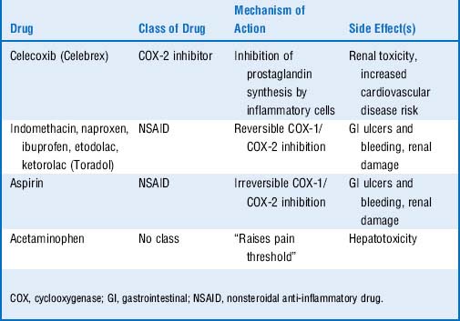
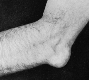
Your rheumatoid arthritis patient is lost to follow-up but then returns to your office years later. She complains of diffuse arthralgias, weight loss, and shortness of breath. Her hands appear as shown in Figure 19-6, and subcutaneous nodules are present at the olecranon bilaterally. She has decreased breath sounds at the lung bases, and chest x-ray film shows bilateral pleural effusions and several pulmonary nodules.
9 What are the characteristic deformities in the hands in advanced rheumatoid arthritis?
Destruction of the metacarpophalangeal joints leads to ulnar deviation (shown in Fig. 19-6) as the distal portions of the digits shift toward the ulna. Boutonnière and swan-neck deformities consist of contractions of the fingers at the proximal interphalangeal and distal interphalangeal joints. Remember that the distal interphalangeal joints are not typically involved with rheumatoid arthritis, though they may be forced into flexion/extension due to involvement of the proximal interphalangeal joints and the tendons of the hand.

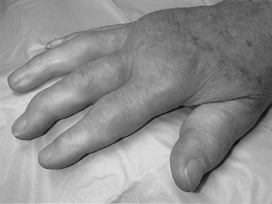
Figure 19-5 Appearance of the proximal interphalangeal and metacarpophalangeal joints of the patient in Case 19-2.
(From Goldman L, Ausiello D: Cecil Textbook of Medicine, 22nd ed. Philadelphia, WB Saunders, 2004.)
10 What are the extra-articular manifestations of rheumatoid arthritis?
Because rheumatoid arthritis is a systemic inflammatory disease, it is not limited to the joints; immune complexes deposit in the vasculature and may affect nearly any organ system. Rheumatoid nodules typically form in the subcutaneous tissue or along tendon sheaths. Pericarditis, pulmonary nodules, interstitial fibrosis, episcleritis, and effusions in the pleural and pericardial space may all be seen in rheumatoid patients. Carpal tunnel syndrome can result from median nerve compression. A normocytic normochromic anemia (anemia of chronic disease) is also common.
Note: Felty’s syndrome is the combination of seropositive (RF+) rheumatoid arthritis, granulocytopenia, and splenomegaly.
Summary Box: Rheumatoid Arthritis
Rheumatoid arthritis is a systemic inflammatory disease caused in part by immune complex deposition in the joints and potentially various other tissues.
Gender, age, and human leukocyte antigens (HLAs) have been associated.
The disease process is centered in the synovium of the joint.
Joints are typically warm, erythematous, swollen, and tender (synovitis).
Distal interphalangeal joints are typically spared.
Pain is worse in the morning, classically lasting more than an hour.
Rheumatoid factor is an autoantibody, the presence of which confers a poor prognosis.
Extra-articular manifestations include rheumatoid nodules, pericarditis, pleural effusions, pulmonary nodules, pulmonary fibrosis, carpal tunnel syndrome, ocular manifestations, and anemia of chronic disease.
Treatment includes nonsteroidal anti-inflammatory drugs (NSAIDs), glucocorticoids, and disease-modifying antirheumatic drugs (DMARDs). In recent years there has been a strong push to start DMARDs at the time of initial diagnosis.
Case 19-3
A 47-year-old man presents with excruciatingly pain in the right knee that began 24 hours earlier. He had been to a banquet the night before and woke up with a red, swollen knee. He tried taking aspirin, but his pain only worsened. He has a history of hypertension treated with hydrochlorothiazide, and his body mass index (BMI) is 32.
1 What is your differential diagnosis?
The differential diagnosis for an acutely inflamed joint includes septic arthritis, cellulitis, gout, pseudogout, and osteomyelitis.
2 How can crystal and septic arthritis be differentiated?
Examination of joint fluid is of paramount importance. Both conditions will cause a high WBC and PMN percentage in the fluid, but Gram stain and culture must be done to look for infectious causes. Septic arthritis may cause more systemic illness, with symptoms such as fever, malaise, lymphadenopathy, and skin lesions. Organisms responsible for septic arthritis include Staphylococcus aureus, Streptococcus species, and Neisseria gonorrhoeae. Rapid diagnosis and treatment are crucial in septic arthritis.
Case 19-3 continued:
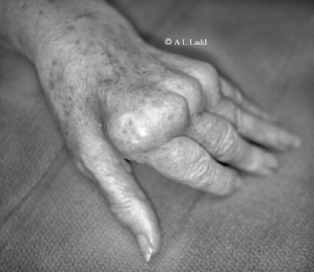
On examination, the right knee is warm, erythematous, swollen, and tender to touch. The patient is afebrile, and the remainder of his examination in unremarkable. You aspirate cloudy yellow fluid from the joint and find 24,000 WBCs, 70% PMNs, and needle-shaped crystals that are strongly negatively birefringent under polarized light (Fig. 19-7).
3 What is the likely diagnosis?
Gout is an inflammatory arthritis caused by the intra-articular deposition of uric acid crystals. Although this patient’s presentation is consistent with the other diseases listed for the differential diagnosis, the crystals seen here are pathognomonic for gout.
The term to associate with gout is negative birefringence, which means that when the crystals are examined under polarized light, they appear yellow when oriented parallel to the direction of slow light vibration, and blue when oriented perpendicularly.
4 What is the pathophysiology of this condition?
Uric acid is the end product of purine nucleotide metabolism and is excreted from the body by the kidneys. High levels of serum uric acid may occur with either overproduction or underexcretion of uric acid. The vast majority of the time (>90% of cases), gout is caused by underexcretion. In either situation, uric acid is deposited in synovial fluid in the form of monosodium urate crystals, which are phagocytosed and induce a local inflammatory response. Free uric acid crystals also activate synovial cells, leukocytes, and complement proteins including C5a, which recruits large numbers of PMNs into the joint space.
5 Do most people with hyperuricemia develop gout?
No. Most people with hyperuricemia do not have gout, and many gout patients do not have high serum levels of urate. The risk of developing gout becomes substantial only with quite high levels of uric acid (>9 mg/dL vs. normal level of ~5 mg/dL). Other risk factors for gout are a purine-rich diet (e.g., seafood, certain meats), male gender age (fifth decade), obesity, hypertension, alcohol intake, renal disease, family history of gout, use of certain drugs, and certain genetic conditions.
6 What is Lesch-Nyhan syndrome and why might it predispose to gout?
Lesch-Nyhan syndrome consists of mental retardation, spasticity, choreoathetosis, aggressive behavior, and self-mutilation, as well as gouty arthritis. It is due to an X-linked defect in hypoxanthine phosphoribosyltransferase (HGPRT), an enzyme involved in the purine salvage pathway. Absence of HGPRT leads to overproduction of uric acid with resultant hyperuricemia.
7 Are there any complications of gout besides the monoarticular inflammatory arthritis?
The classic location for gouty arthritis is in the metatarsophalangeal joint of the great toe, which is known as podagra. In patients with chronic gout, monosodium urate crystals can also deposit in subcutaneous tissues, forming nodules called tophi. Tophi are often found in the helix of the ear and over the elbow. Figure 19-8 shows olecranon bursitis in chronic tophaceous gout. Uric acid calculi may form in the renal pelvis or ureters, leading to obstruction of the urinary tract. Crystals may also cause damage to the renal interstitial tissue, a condition termed gouty interstitial nephropathy.

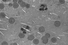
Figure 19-7 Microscopic appearance of synovial fluid under polarized light aspirated from patient in Case 19-3.
(From McPherson RA, Pincus MR: Henry’s Clinical Diagnosis and Management by Laboratory Methods, 21st ed. Philadelphia, WB Saunders, 2006.)
8 Are there any medications that can precipitate attacks of gout?
Drugs that decrease the renal excretion of uric acid may contribute to gout. Such offenders include low-dose salicylates, thiazide diuretics, and furosemide. Our patient may have been predisposed to developing gout because of his hydrochlorothiazide use, and the aspirin he took decreased his renal excretion of uric acid even further. Other precipitants include alcoholic beverages and purine-rich foods (meats and seafood).
Note: Although low-dose aspirin (<2 g/day) decreases renal excretion of uric acid, high-dose aspirin actually increases renal clearance.
9 What other treatment options exist for an acute gout attack?
Acute attacks can be managed with NSAIDs (typically indomethacin), glucocorticoids, and colchicine, all of which dampen the inflammatory response and inhibit the continued phagocytosis of monosodium urate crystals.
Note: Colchicine works by binding to the molecule tubulin and inhibiting the polymerization of microtubules, thereby inhibiting mitosis in inflammatory cells. By inhibiting microtubule function, it also serves to limit the mobility of inflammatory cells. A worrisome side effect of colchicine is bone marrow suppression. It also commonly causes intestinal upset and diarrhea.
10 Who needs chronic treatment for gout, and what does this treatment consist of?
Patients with recurrent bouts of gouty arthritis, radiographic evidence of joint damage, and extra-articular manifestations such as tophi and urate kidney stones merit chronic treatment. Note that asymptomatic hyperuricemia is not reason for treatment. Probenecid is used for most chronic gout patients (underexcretors), as it serves to increase renal excretion of uric acid. Allopurinol, an inhibitor of the uric acid-synthesizing enzyme xanthine oxidase, can be used to treat overproducers. For boards, patients are designated underexcretors or overproducers using a 24-hour urinary uric acid excretion test, although in clinical practice this is rarely done.
Note: Patients undergoing treatment for hematologic malignancies are at risk for tumor lysis syndrome. As malignant cells are killed, their DNA is degraded into purine and pyrimidine nucleotides. Allopurinol is often given before radiation therapy or chemotherapy to prevent the production of uric acid through purine catabolism.
Note: At the time of this writing, febuxostat (Uloric) has recently been approved as an alternative to allopurinol. While febuxostat has been shown to be more effective than allopurinol at lowering serum urate concentration, it has not been shown to be more clinically efficacious at preventing gouty flares.

Stay updated, free articles. Join our Telegram channel

Full access? Get Clinical Tree

