Respiratory Disorders


Respiratory Disorders


Respiratory System


imagehttp://evolve.elsevier.com/Price/pediatric/

The respiratory system consists of the nose, pharynx, and larynx (upper respiratory tract) and the trachea, bronchi, and lungs (lower respiratory tract). The respiratory tract continually changes during the first 12 years of a child’s life. There are several anatomic differences that predispose children to respiratory difficulties (Box 11-1). This makes respiratory problems common during childhood. Most problems are mild and can be managed at home. Other conditions can be chronic and have an impact on the growth and development of the child. Respiratory distress and failure are the common factors for cardiac arrest in children. Respiratory diseases account for 25% of hospitalizations for children younger than 15 years of age. Pneumonia accounts for 31%, bronchiolitis and bronchitis for 25%, and asthma for 25% of the respiratory diseases (CDC, 2007).


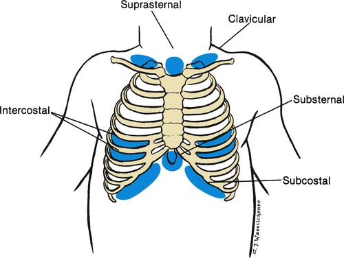
Children with respiratory difficulties have common signs and symptoms that can progress to respiratory failure if not recognized. Assessment findings of the child’s respiratory system will vary depending on the age of the child. The nurse must recognize any change in the child’s breathing status. Signs and symptoms may include fever, anorexia, vomiting, diarrhea, abdominal pain, nasal blockage, nasal drainage, cough, sore throat, retractions, and abnormal respiratory sounds. Respiratory assessment should be comprehensive and frequent as the status can change rapidly. The assessment should include the guidelines shown in Box 11-2. Retractions can indicate respiratory distress. The severity of respiratory distress can be assessed using the depth and location of the retractions (Figure 11-1).


A variety of diagnostic tests can assist in monitoring respiratory function. Chest x-rays assist in identifying foreign objects or abnormalities in lung tissue. Pulse oximetry is a noninvasive method for measuring oxygen saturation. Pulmonary function test (PFT) and spirometry measure the vital and expiratory capacity.


Sudden Infant Death Syndrome


Sudden infant death syndrome (SIDS) is defined clinically as the sudden, unexpected death of an apparently healthy infant younger than 1 year of age for which a routine autopsy fails to identify the cause. It is also referred to as crib death or cot death. Although precise data are not available, it is estimated that in the United States, SIDS kills about 3000 infants per year. In industrialized countries, SIDS is one of the leading causes of death in early infancy; the peak incidence is between 2 and 4 months of age. It is more common in low–birth weight babies, in boys, in families with crowded living conditions, and during the winter months. Autopsy may reveal slight respiratory infection or otitis media, petechiae over the pleura, and pulmonary edema. Two clinical features of the disease remain constant: (1) death occurs during sleep, and (2) the infant does not cry or make other sounds of distress. In some cases, the baby is found in one corner of the crib with blood-tinged froth coming from the nose.


Theories concerning the cause of SIDS are numerous. Although there appears to be an increased incidence among siblings, no genetic pattern has been determined. The risk for SIDS is increased in twins.


Many theories concerning cause (e.g., suffocation, aspiration allergy, and hormone deficiency) have been disproved. The exact cause is not known. Some researchers propose that crib death results from an interruption of some basic function in the central or autonomic nervous system that causes apnea. Carotid bodies located in the neck and involved in the control of breathing have been found to be abnormal in victims of SIDS. Current opinion holds that SIDS has more than one cause.


The death rate has continued to decrease, and the focus is on decreasing risk factors that contribute to SIDS. The Back to Sleep program has produced positive results. Co-sleeping has been identified as an increased risk of SIDS even if mothers do not smoke or if they breastfeed. Health care providers should encourage a separate crib or bassinet for sleeping.


imageHealth Promotion


Guidelines for Prevention of SIDS


• Always place infant on his or her back to sleep, and do NOT use side-lying position (see Box 11-3).


• Use a firm sleep surface with a safety-approved crib mattress.


• Keep soft objects and loose bedding out of infant’s sleep area.


• Avoid overheating, keeping head uncovered.


• Do not smoke during pregnancy or near babies.


• Avoid co-sleeping, but keep infant’s sleep area close. Keep infant’s bedroom door open.


• Avoid home respiratory or cardiac monitors used to reduce SIDS risk.


• Avoid devices that claim to maintain sleep position to reduce SIDS.


• Consider offering a clean, dry pacifier (controversial).


• Provide tummy time during awake periods.


• Stress that all care providers for infant follow the guidelines (AAP, 2005).


Babies with infantile apnea (also called near-miss infants) and subsequent siblings of babies with SIDS are often monitored at home until they are past the age of danger. Monitors can be leased. Parents are provided with ongoing education and support during this period. Parents are taught cardiopulmonary resuscitation before being discharged.


In dealing with grieving parents after the death of their infant, the nurse must convey some important facts: that the baby died of a disease entity called SIDS, that this disease currently cannot be predicted or prevented, and that they are not responsible for the child’s death. Grieving parents need time to say good-bye to their child. They should be encouraged to hold and rock their infant, shed tears, and assist in burial preparations. This process, not common in the past, is conducive to the resolution of grief (see also Chapter 22). One mother who was denied this experience stated that 5 years later, while visiting a florist’s shop, she noticed a heart-shaped wreath intended for an infant. She unexpectedly burst into tears and wept.


Parents of a child who dies of SIDS experience a great deal of guilt and are catapulted into a totally unexpected bereavement, requiring numerous explanations to relatives and friends. Often needless blame has been placed on one parent by the other or by relatives. The family babysitter and physician may also be targets of attack. Emergency department personnel need to be especially sensitive and supportive during this crisis. There have been occurrences of SIDS for which parents have been charged with child abuse and have even been jailed because of lack of public knowledge about the disease.


Sudden infant death syndrome can occur in the hospital, and many nurses and physicians have personal experience of the suffering that losing a child to SIDS can cause. Group therapy with other parents of SIDS victims is recommended. Two nationally supported organizations are the Compassionate Friends, Inc., and the National Sudden Infant Death Syndrome Foundation. These groups have local chapters in most states.


Bronchopulmonary Dysplasia


Bronchopulmonary dysplasia (BPD) is a chronic lung disease that occurs in newborns that are premature or have pulmonary disorders that require mechanical ventilator support with high positive pressure and oxygen. The lung tissue is immature and unable to withstand tissue damage resulting from the required oxygen supplement. The resulting fibrosis and alteration in lung compliance may last from several months to years. Improvements in treatment of low–birth weight preterm infants have increased the incidence of this disorder, and continue to be the primary issue for infants younger than 27 weeks of age (Belcastro, 2004). Newer mechanical ventilators are being used for low birth weight neonates in hopes of decreasing BPD.


Signs and Symptoms


The symptoms of BPD are directly related to the pathophysiology of the disease. Tachypnea, dyspnea, and wheezing can be a result of airway obstruction and increased airway resistance. Increased work of breathing can cause retractions and use of accessory muscles. The infants may display activity intolerance during feedings. They may be irritable and difficult to comfort. Cyanosis may develop during crying spells. Infants who needed intubation for a long period of time may have subglottic stenosis and inspiratory stridor develop. All of the symptoms can be associated with the chronic hypoxia state. The diagnosis is made on the basis of abnormal radiographic findings, signs of respiratory distress, oxygen dependency after 28 days of age, and a history of required mechanical ventilation during the first week of life.


Treatment and Nursing Care


The treatment for infants with BPD is to provide adequate oxygenation and prevent progression of the disease process. Treatment includes oxygen, drug therapy, and nutritional support. Surfactant continues to be included in the course of medical treatment (Askins and Diehl-Jones, 2009). Infants may continue to need oxygen after hospital discharge. These infants do not tolerate excessive or even normal amounts of fluids. They may have problems develop with accumulation of fluids in the lungs that require the use of diuretics. The use of these drugs requires the monitoring of electrolytes and edema. Oral electrolyte supplements may be given. Bronchodilators (albuterol) and steroids may promote improved lung function. These infants are at risk for respiratory infections and should be given RSV-immune globulin (RespiGam) or palivizumab (Synagis) during the RSV season.


Infants with BPD are at high risk for growth failure, and nutrition is an important issue (VanRiper, 2010). They have higher metabolic needs, and providing adequate nutrition without causing respiratory distress can be difficult. Nursing care should be organized to provide periods of rest. Small, frequent feedings and nutritional supplements may be used. The environment should include measures to decrease stimulation.


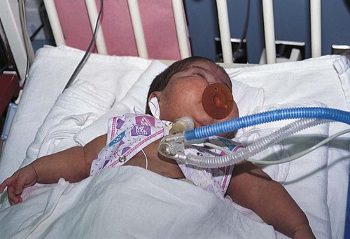
Parents may be extremely anxious caring for a child with BPD. All equipment and procedures should be explained in simple terms. Children with tracheostomies can be cared for in the home setting, and home care teaching of the equipment is needed (Figure 11-2). Review care of the child with tracheostomy in Chapter 3. Extended hospitalization can interfere with the development of the normal parent-child relationship and with the normal development of the infant. Parental participation in the infant’s care should be encouraged. The family’s ability to cope and care for a child with a chronic illness needs to be evaluated as home discharge plans are developed. An adequate period of education may be necessary for the parents to become comfortable with the care required for their child. Families should be referred to social services to assist in providing additional support and to assist in helping the parent gain access to available community services. Parental support groups can be beneficial in providing additional assistance with coping skills necessary for caring for a child with complex care.


Otitis Media


Otitis media (ot, ear; itis, inflammation of; media, middle) is an inflammation of the middle ear. The middle ear is a tiny cavity in the temporal bone. Its entrance is guarded by the sensitive tympanic membrane, or eardrum, which transmits sound waves through the oval window to the inner ear. The inner ear contains the organs of hearing and balance. The middle ear opens into air spaces, or sinuses, in the mastoid process of the temporal bone. It is also connected to the throat by a channel called the eustachian tube. These structures—the mastoid sinuses, the middle ear, and the eustachian tube—are lined with mucous membranes. As a result, an infection of the throat can easily spread to the middle ear and mastoid. The eustachian tube also protects the middle ear from nasopharyngeal secretions and provides drainage of middle ear secretions into the nasopharynx and equalizes air pressure between the middle ear and the outside atmosphere. These protective functions are diminished when the tubes are blocked. Unequalized air within the ear creates a negative pressure that allows organisms to be swept up into the tube if it opens.


Otitis media may be the result of an upper respiratory tract infection, caused by a variety of organisms. The most common cause of these infections is viral. Bacteria cause the rest of otitis media cases. Of the bacterial cases, 40% to 50% are caused by Streptococcus pneumoniae, which is increasingly demonstrating resistance to penicillin. Other common bacteria that cause otitis media are Haemophilus influenzae and Moraxella catarrhalis. With the use of H. influenzae type b vaccine (Hib) as a routine immunization, the number of cases of otitis media caused by this organism has decreased. The addition of the seven-valent S. pneumoniae conjugate vaccine to the immunization schedule should also decrease the incidence rate of otitis media caused by this organism (Morris, 2009). Infants are more prone to ear infections because the eustachian tube is shorter, wider, and straighter than in older children and adults. Because babies lie flat for long periods, microorganisms have easy access from the eustachian tube to the middle ear. This is thought by some investigators to be a contributing factor.


There are two types of otitis media. The acute disease is suppurative or purulent otitis media (AOM). It is most commonly caused by S. pneumoniae and H. influenzae. The second type is called serous or nonsuppurative otitis media with effusion (OME). The cause is unknown, but it often occurs after an acute episode. OME is the most common cause of hearing loss and hearing impairment in children.


Signs and Symptoms


The symptoms of acute otitis media (AOM) are pain in the ear (often severe), irritability, and interference with hearing. Sucking or chewing has a tendency to increase the pain. Fever, which may run as high as 40° C (104° F), headache, and vomiting may also accompany the illness as may diarrhea. The nurse may suspect an earache in the infant who rubs the ear frequently or pulls at it. The infant may also roll the head from side to side and cry piercingly. The older child can point to the place that is tender. OME is the result of chronic otitis media. Children may be asymptomatic but may report a feeling of fullness or popping in the ears.


If an abscess forms, the eardrum may rupture as a result and pus may drain from the ear. When this happens, the pressure is relieved and the child is more comfortable.


Complications of an ear infection include hearing loss, mastoiditis, chronic otitis media, and meningitis. These complications are rare with modern treatment. Prevention lies in the prompt treatment of respiratory infections or infected tonsils and adenoids.


Treatment and Nursing Care


The professional who examines the ears first observes their appearance and general hygiene. The lymph nodes about the ear are observed for swelling or tenderness. The child’s head is adequately stabilized to prevent injury to the ear canal from sudden, unexpected movement. Excess cerumen or wax in the ear, which may obstruct visibility, is carefully removed. The examiner ensures that no foreign bodies are lodged in the outer canal before inserting the otoscope. To straighten the canal and improve viewing, the ear is pulled down and back in infants and small children. The ear is pulled up and back in older children and adults. The physician may also perform a pneumatic otoscopic examination. The ear speculum is used to seal the ear canal, and air is expressed into the canal. The movement or lack of movement of the tympanic membrane is indicative of the degree of fluid behind the membrane. This examination has proven useful in determining the degree of the condition.


New treatment guidelines were released in 2004 by the American Academy of Pediatrics (AAP) (Box 11-4). Treatment included an observation option, pain management, and antimicrobial treatment. Development of resistant strains of bacteria and misuse of antimicrobials assisted in the development of new guidelines. When antimicrobials are used, amoxicillin remains the drug of choice, but recurrent infections may require other antimicrobials. Parents are taught to give the entire dose of the antimicrobial even though the child may appear well. Parents will need to understand the new course of treatment using the guidelines and their role in the management of AOM.


Pain control may be needed for the child with AOM. Pain control is achieved using acetaminophen or ibuprofen as prescribed. Antihistamines and decongestants are not effective and may have side effects. Eardrops (Auralgan) may be prescribed to control pain. Warm or cold compresses may be applied to the ear. The child can be placed on the affected side with the ear on top of a hot water bottle (temperature of the water 115° F, or 46° C) or on a heating pad on the low setting. Children should be placed upright to decrease pain. If the ear is draining, the outer canal can be cleaned with sterile water or hydrogen peroxide. Parents should be instructed not to use cotton swabs in the ears.


Nurses need to be aware that environmental factors have been identified that can contribute to the risk for ear infections. Daycare outside the home, parental smoking, and pacifier use has been shown to increase the risk for recurrent otitis media. Breastfeeding for at least 6 months has reduced the risk for AOM. These factors should be discussed with parents.


For children with recurrent AOM or chronic OME, tympanostomy tubes may be effective. The physician performs the procedure by completing a myringotomy (myringo, eardrum; otomy, incision) and inserts a tiny tube into the eardrum. These tubes require surgical placement and special care by the parents. Eventually, the tubes fall out spontaneously.


Bronchiolitis


Bronchiolitis is an inflammation of the small airways. It occurs most often during late autumn through late spring and in children younger than 2 years of age. Bronchiolitis is usually caused by a viral infection. The most common causative organism is the respiratory syncytial virus (RSV). Children are usually exposed through other family members who have symptoms of an upper respiratory tract infection. Children who are at risk for respiratory distress have chronic lung diseases such as bronchopulmonary dysplasia (BPD) or cystic fibrosis (CF).


Inflammation of the bronchioles is associated with obstruction that is caused by edema and accumulation of mucus. There may be partial or complete obstruction. The alveoli are usually not affected. Normal gas exchange in the lung is affected. This leads to hypoxemia.


Treatment


Mild cases of bronchiolitis can be managed at home. Treatment at home includes increasing the intake of fluids and increasing the humidity in the air. Also useful are antipyretics to control fever. The parents or caregiver should be instructed to bring the child back for reevaluation if any signs of increased respiratory distress occur or if the child’s condition worsens.


Indications for hospitalization include an infant younger than 6 months of age, sleeping respiratory rates of 50 to 60 per minute or higher, hypoxemia, apnea, or the inability to tolerate oral feeding.


When the child is hospitalized, intravenous fluids are started to hydrate the child and thin the secretions. The child is placed in an atmosphere of humidified oxygen (mist tent, croupette, or nasal cannula/mask). The goal is to keep oxygen levels at 92% or better with a pulse oximeter. With severe bronchiolitis, the physician may use a bronchodilator and a corticosteroid, but these remain controversial (Zorc, 2010). Antimicrobials may also be used for small or severely ill infants because these infants may be susceptible to a secondary bacterial infection. Fever is controlled with antipyretics. A laboratory study of a nasopharyngeal washing should be done to determine whether the causative organism is RSV. As a precautionary measure for the safety and concern of other children, the infant is placed in contact isolation until RSV has been ruled out.


When the causative organism is RSV, no medications can effectively treat the disease. Ribavirin, antimicrobials, antihistamines, and oral decongestants have been identified as being ineffective for treatment (Zorc, 2010). Medical attention has recently focused on active and passive immunizations. RSV-immune globulin (RespiGam) and palivizumab (Synagis) have been approved for use with children at high risk. Palivizumab may be preferred because of ease of administration (intramuscular); lack of interference with mumps, measles, and rubella (MMR) vaccine and varicella vaccine; and lack of complications associated with intravenous immune globulin (RespiGam). Monthly administration during RSV season (October to May) is recommended (AAP, 2009).


Nursing Care


Nursing diagnoses for the infant with bronchiolitis include the following:


The child with bronchiolitis is monitored closely for signs and symptoms of increasing respiratory distress. Breath sounds, skin color, depth and rate of respirations, and vital signs are assessed. Changes in alertness and increased anxiety can be signs of impending distress. Continuous or intermittent pulse oximetry may be used to monitor the infant’s oxygen level.


Intravenous fluids are monitored in the acutely ill child. As the child improves, oral fluids are increased and frequent small meals are offered. The child is on intake and output recording, and daily weights are taken. The fontanel and the child’s skin turgor are also assessed as indicators of hydration status.


Formula-fed infants may have thickened feeding to improve swallowing dysfunction and to prevent aspiration. Breastfed infants should have more frequent feeding with shorter times. This assists in decreasing the workload of the infant and conserves energy. Nasal secretions should be removed with a bulb syringe before feedings (Allen, 2006).


The child in a mist tent should have the gown and linens changed if they become damp. Also, moisture buildup should be removed from the tubing and the sides of the tent (see Chapter 3 for detailed care of the child in a mist tent). For home care, cold air humidifiers can be used but must be cleaned properly to prevent bacteria or fungal growth.


As always, parents are encouraged to stay with the child. This may be even more important because the child may already be anxious because of respiratory distress. Parents should understand the importance of the child staying in the tent. They should be included in the care and diversional activities for the child.


Preventing the spread of infection is also important. If an infant has RSV, then contact isolation is recommended. RSV is primarily spread by large droplets and fomites. RSV can survive on hands for almost an hour and on hard surfaces up to 24 hours. Health care–associated infections (HAIs) or nosocomial infections can be a serious nursing issue. Hand hygiene is extremely important in all issues of infection. All caregivers, including parents, need to know and apply measures to prevent the spread of infection.


Support of the parents is significant. Most of the infants who are in severe or critical condition are usually young infants or those who have an underlying disease. Their parents may lack confidence when it comes to the care of the infant and need to be supported and reassured in their actions. It can be frightening for them to see their infant so ill. If the infant is admitted to a critical care area, the support of the parents is crucial. Explanations should be given in terms the parents can understand. The family needs information from the physician or the nurse concerning the infant’s condition, medications, treatments, and procedures. Plans for all of these issues and discharge information can aid the parents in coping with the situation. Family, friends, and clergy can be a great support for the parents.


Cystic Fibrosis


Cystic fibrosis (CF) is a genetic disorder that results in a multisystem disease involving the cell membrane and the electrolyte and water system of the cell. This disease affects many parts of the body but particularly the lungs and pancreas. It occurs in about 1 in 3000 live births. CF is an inherited congenital disorder. The condition is believed to be inherited as a Mendelian recessive trait from both parents. The parents, who are carriers of this disease, do not show any symptoms. CF disease results when the two genes for the disease combine during conception. CF affects both genders equally. The survival rate of the children has increased, and many are living into adulthood. Better antimicrobial control of pulmonary infection both at home and during hospitalization and increased numbers of CF centers have contributed to this success.


The gene associated with CF was identified in 1989, and it is now possible to identify healthy individuals who carry the trait. Chromosome 7 is the location of the gene responsible for CF. Sodium and chloride at the cell membrane are controlled by this gene. With a defect in the CF transmembrane regulator (CFTR) protein, secretions become thick and pasty. There are many possible mutations, which helps to explain the various degrees of involvement of the systems: respiratory and GI.


With the ability to identify the CF gene, diagnosis can be assisted with genetic analysis. Caution is noted because of the possibilities of false-positive and false-negative results. Genetic analysis can be used in conjunction with other diagnostic criteria such as the sweat test. New aggressive approaches in treatment have resulted in increasing the life expectancy from less than 10 years to 40 years.


Signs and Symptoms


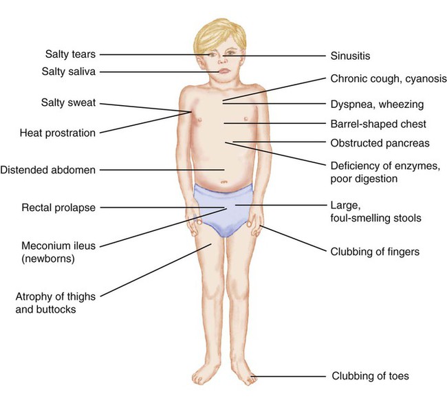
The major symptoms of CF are manifested in the respiratory tract and the GI tract. The first symptom may be seen in the newborn infant who has a meconium ileus. This condition is seen in approximately 10% to 20% of children who are born with CF. An overview of the manifestations of CF is shown in Figure 11-3.


Lung Involvement. 

Cystic fibrosis is considered the most serious lung problem in children in the United States. The air passages of the lungs become clogged with mucus. There is widespread obstruction of the bronchioles. It is hard for the child to breathe; expiration is especially difficult. More and more air becomes trapped in the lungs (obstructive emphysema), and small areas of collapse (atelectasis) may occur. Eventually, the chest assumes a barrel shape, with increased diameter across the front and back. The right ventricle of the heart, which supplies the lungs, may become strained and enlarged. Clubbing of the fingers and toes, indicating a chronic lack of oxygen, may be present. Staphylococcus and Pseudomonas infections can easily occur in the lungs, which provide a suitable medium for these organisms to grow. This causes more thickening of the abnormal secretions, irritates and damages lung tissues, and further increases lung obstruction.


The time of onset of this disease varies. Symptoms may appear weeks, months, or years after birth. In general, the earlier the onset of the disease symptoms, the more severe course of the disease. Symptoms range from mild to severe. Any or all symptoms may be present in varying degrees of severity in one individual. A chronic cough develops that may produce vomiting. Dyspnea, wheezing, and cyanosis may occur. The child is irritable and tires easily. Gradually, there is a change in physical appearance. Chest radiographs reveal widespread infection. Evidence of obstructive emphysema, atelectasis, and fibrosis of lung tissue may also be present. The prognosis for survival depends on the extent of lung damage. However, this is only part of the picture because CF also affects the pancreas and sweat glands.


Pancreatic Involvement. 

The pancreas lies behind the stomach. Some of its cells secrete pancreatic enzymes that drain from the pancreatic duct into the duodenum at the same area in which bile enters. Changes occurring in the pancreas are the result of obstruction by thickened secretions that block the flow of pancreatic digestive enzymes. Consequently, foodstuffs, particularly fats and proteins, are not properly used by the body.


In infants, the stools may be loose. Gradually, because of impaired digestion and food absorption, the feces of the child become large, fatty, and foul-smelling. They are usually light in color. In spite of having a good appetite, the infant does not gain weight and may look undernourished. The abdomen becomes distended, and the buttocks and thighs atrophy as fat disappears from the main deposit sites. Laboratory test results show a deficiency in pancreatic enzymes (trypsin, lipase, and amylase).


An oral pancreatic extract such as Pancrease is given to the child with each meal and snack to replace the pancreatic enzymes the child’s body cannot produce. This medication is considered specific for the disease because it aids in the digestion and absorption of food, thus improving the condition of the stools. If the child is ill and not eating, the medication is withheld.


A presenting condition known as meconium ileus develops after birth when the intestine of the newborn becomes obstructed with abnormally thick meconium while in utero. This is caused by the absence of pancreatic enzymes that normally digest proteins in the meconium. The abnormal, puttylike stool sticks to the walls of the intestine, causing blockage. Vomiting, abdominal distention, and absence of stools lead to the suspicion of intestinal obstruction. Radiographs confirm the diagnosis. The condition is treated surgically. The death rate is high, but the prognosis is more favorable when the obstruction is detected early. Most infants who survive manifest CF. Fortunately, meconium ileus is rare because the pancreatic enzyme deficiency is seldom complete. Nevertheless, the nurse assigned to the nursery must constantly be on guard for suspicious symptoms.


Sweat Glands. 

The sweat, tears, and saliva of the child with CF become abnormally salty from an increase in sodium and chloride levels. There is also an increase in the potassium level of sweat glands. The normal amount of chloride in sweat is 1 to 60 mEq/L. Higher concentrations are considered specific for the disease. The analysis of sweat is a major aid in the diagnosis of the condition. The sweat test, with pilocarpine iontophoresis, is the best diagnostic study. A dilute solution of pilocarpine is applied to the arm, and a weak electrical current is used to stimulate sweating. A positive test should be repeated for confirmation. Because large amounts of salt are lost through perspiration, the child must be observed for heat prostration. Liberal amounts of salt should be given with food, and extra fluids and salt should be provided during hot weather. Infants do not have a lot of sweat; therefore obtaining enough sweat for an accurate test may be difficult. Mothers often report that when they kiss their infants they taste salty.


Complications


Cystic fibrosis is often responsible for rectal prolapse in infants and children. This is partly from poor muscle tone in the rectal area and excessive leanness of the buttocks of the child.


As the disease progresses, the liver may become hard, nodular, and enlarged. There may be edema of the extremities. The retina of the eye may hemorrhage, there may be damage to the eye from swelling, and inflammation in part of the optic nerve may occur. Cor pulmonale (cor, heart; pulmon, lung), heart strain from improper lung function, is frequently a cause of death. Osteoporosis (osteo, bone, pore; osis, disease) may occur. When it is caused by CF, the bones become porous because of poor utilization of fat-soluble vitamin D, which is necessary for proper calcium metabolism. There is a deficiency in vitamin A also because the child is unable to absorb the fats from which this vitamin is obtained.


With the extended life expectancy, about 20% or higher of the children develop hyperglycemia and cystic fibrosis–related diabetes (CFRD). Blood sugar control becomes an important objective. With malabsorption and pancreatic insufficiency issues, control becomes challenging. Oral hypoglycemia drugs and insulin may be used. As they become adults, these children can develop the vascular problems seen with the long-term effects of diabetes.


Treatment and Nursing Care


Cystic fibrosis is a chronic condition and must be monitored and maintained daily. The family providers of care need support, as does the child. The care in a regional CF center, where all disciplines are located in one facility, can be extremely helpful because it allows the family to go to one location rather than traveling to several different clinics for care. The CF team can work with the family and the primary care physician to meet the needs of the child and family.


Respiratory Relief. 

Most new approaches in treatment are focused on the lung. The targeted outcomes are improved airway clearance, thinning of secretions, treatment of infections, and reduction of inflammation. Antimicrobials may be given as a preventive measure against respiratory infection; however, this treatment is subject to controversy. Full dosages of antimicrobials are given in an acute infection. The physician determines the particular antimicrobial to be used on the basis of the results of throat and sputum cultures. The route may be oral or intravenous. Intravenous medication may be given via heparin lock or, in some cases, a Broviac catheter or implanted port. This can be used successfully in both inpatient and outpatient settings. The child’s respiratory status can be monitored through the pulmonary function test, which indicates the lung’s capacity.


Intermittent aerosol therapy is administered to provide medication to the lower respiratory tract and to promote evacuation of secretions. DNase, an enzyme, is administered by inhalation and results in decreasing the viscosity of the sputum. An inhaled antimicrobial, tobramycin (TOBI), is being used as a maintenance prophylactic with chronic Pseudomonas aeruginosa to help suppress bacterial growth. Bronchodilators such as Albuterol are used to increase the width of the bronchi, allowing free passage of air into the lungs.


Postural drainage, chest clapping, and breathing exercises are also important. These are performed by the respiratory therapist during hospitalization. When postural drainage and chest clapping are done properly, the secretions in the chest are moved up and out. During latent periods or in mild cases, the child may not raise sputum. This should be explained to the parents so that they do not discontinue this valuable procedure when the child goes home. Instructions may need to be repeated frequently to encourage full cooperation of the parents and child. These procedures should be done after nebulization and at least 1 hour after eating. General exercise is good for the child because it stimulates coughing. Somersaults, headstands, and wheelbarrow play within the child’s endurance are therapeutic.


Preventing respiratory tract infections is important. The child should be isolated from individuals and personnel who may harbor infections. The child must be given the necessary immunizations against childhood diseases. Appropriate boosters should be given so that the immunity obtained is kept up to date.


Diet. 

Adequate nutrition is essential. The diet should be high in calories, as much as 50% more than normal. There should be increased protein and moderate amounts of fat in conjunction with pancreatic extracts. Simple sugars are easy to digest, and banana products are particularly good. Fruits, cottage cheese, vegetables, and lean meats, which are high in protein and low in fat and starches, are recommended. With the improvement of nutritional enzymes, many of these children can tolerate normal to higher amounts of fat in their diet. Enzymes can be adjusted if there is an increased amount of fat in the stool. Extra salt may be provided with pretzels and salted breadsticks and crackers. As the disease progresses, some children will benefit from nighttime feedings with a nasogastric tube or gastrostomy tube (G-button). Older children can add supplemental drinks such as Boost or Ensure to increase their calorie input.


Supplements of vitamins A, D, and E in a water-miscible base are given each day in double the recommended dose. Vitamin K may also be given when indicated. Salt tablets may be given to the older child during hot weather. Forcing fluids may be ordered because larger amounts of fluid are lost in the stools. The nurse may be asked to weigh the child daily.


The nurse feeding the infant with CF must be calm and unhurried. The infant may cough, have difficulty breathing, and vomit. Careful burping is necessary to avoid abdominal distention. In general, the appetite is good. Older children need small amounts of food served attractively and frequently. Food piled high on a child’s tray is discouraging. The amount of a meal eaten should be compared to what the child normally ingests rather than the amount left on the tray. This practice will decrease a child’s appetite being reported as poor. The nurse records the fluid intake at the end of the meal. The child’s reaction to new foods and any variations in stools resulting from the food are noted. The food refused and the type, character, and amount of vomiting, if any, are also noted.


Stay updated, free articles. Join our Telegram channel

Dec 22, 2016 | Posted by in NURSING | Comments Off on Respiratory Disorders

Full access? Get Clinical Tree

Get Clinical Tree app for offline access