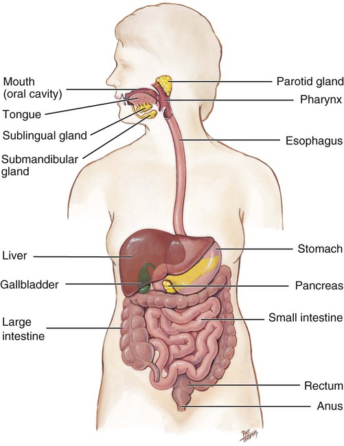
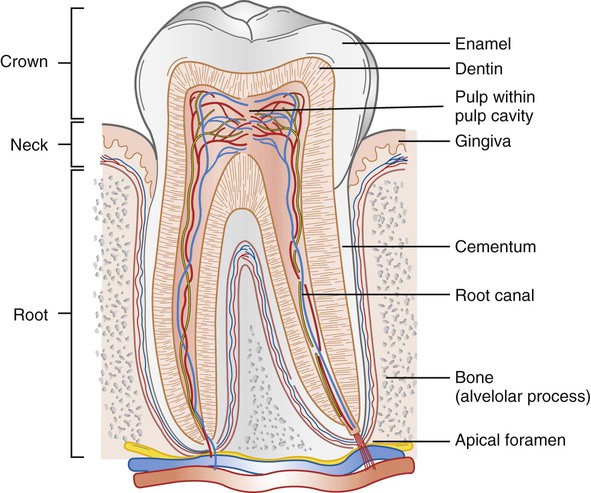
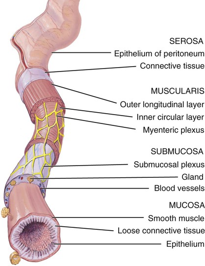
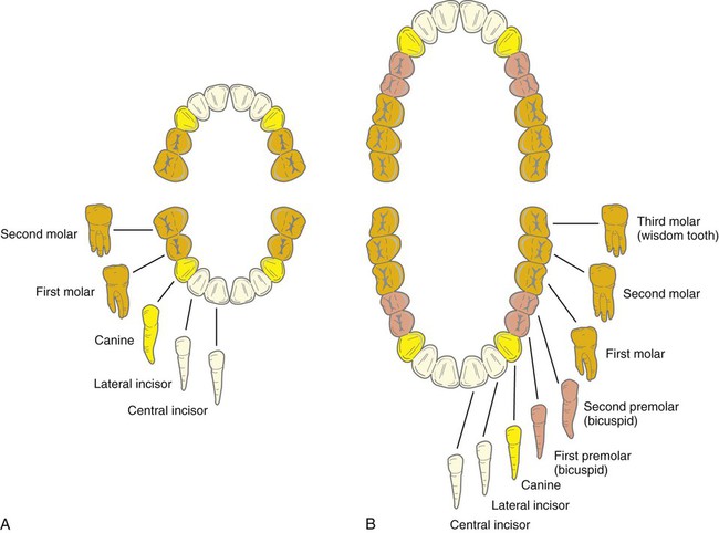
1. Identify the components of the digestive tract and the accessory organs. 2. List six functions of the digestive system. 3. Describe the general histology of the four layers in the digestive tract wall. 4. Describe the features and function of the oral cavity, teeth, pharynx, and esophagus. 5. List and describe the location of the three salivary glands. 6. Explain the function of saliva. 7. Describe the structure and features of the stomach and its role in digestion. 8. Describe the structure and features of the small intestine and its role in digestion and absorption. 9. Describe the structure, features, and function of the large intestine. 10. Describe the structure and function of the liver, gallbladder, and pancreas. 11. Explain how substances are absorbed into the body through the small intestine. 12. Describe ways in which the aging of an individual affects the digestive system. The digestive system includes the digestive tract and its accessory organs (Figure 14-1). The function of the digestive system is to process food into molecules that can be absorbed and used by the cells of the body. Food is broken down, bit by bit, until the molecules are small enough to be absorbed and the waste products are eliminated. The digestive tract (also called the alimentary canal or gastrointestinal [GI] tract) consists of a long, continuous tube that extends from the mouth to the anus. It includes the mouth, pharynx, esophagus, stomach, small intestine, and large intestine. The tongue and teeth are accessory structures located in the mouth. The salivary glands, liver, gallbladder, and pancreas are not part of the digestive tract but are major accessory organs that have a role in digestion. These secrete fluids into the digestive tract. Food undergoes three types of processes in the body: • Ingestion—The first activity of the digestive system is to take in food. This process is called ingestion. Ingestion has to take place before anything else can happen. • Mechanical digestion—The large pieces of food that are ingested have to be broken into smaller particles that can be acted on by various enzymes. This is called mechanical digestion. Mechanical digestion begins in the mouth with chewing, or mastication (mas-tih-KAY-shun), and continues with churning and mixing actions in the stomach. • Chemical digestion—The complex molecules of carbohydrates, proteins, and fats are transformed by chemical digestion into smaller molecules that can be absorbed and used by the cells. Chemical digestion uses water to break down the complex molecules. This process is known as hydrolysis. Digestive enzymes speed up the hydrolysis process, which is otherwise slow. • Movements—After ingestion and mastication, the food particles move from the mouth into the pharynx, and then into the esophagus. This movement is called deglutition (dee-gloo-TISH-un), or swallowing. Mixing movements occur in the stomach as a result of smooth muscle contraction. These repetitive contractions mix the food particles with enzymes and other fluids. The movements that propel the food particles through the digestive tract are called peristalsis. These are rhythmic waves of contractions that move the food particles through the various regions in which mechanical and chemical digestion takes place. • Absorption—The simple molecules that are produced from chemical digestion pass through the lining of the small intestine into the blood. This process is called absorption. • Elimination—The food molecules that cannot be digested need to be eliminated from the body. The removal of indigestible wastes through the anus, in the form of feces, is defecation (def-eh-KAY-shun). The wall of the digestive tract has four layers or tunics (Figure 14-2): Highlight on the Digestive System Cold sores: Cold sores, or fever blisters, are small fluid-filled blisters that itch and are painful, usually appearing around the lips and in the mouth. They are caused by recurring infections with the herpes simplex virus. After the initial infection, the virus remains dormant in a cutaneous nerve until it is activated by stress, fever, or ultraviolet radiation. Cleft palate: Cleft palate is a condition in which the bones in the hard palate do not fuse completely during prenatal development. This leaves an opening between the nasal and oral cavities. An infant with this problem has difficulty creating enough suction for proper feeding. Cleft palate can usually be corrected surgically. Tongue-tied: A person with a short lingual frenulum is said to be “tongue-tied.” The movement of the tongue is abnormally limited, which causes difficulties in speech. Surgically cutting the frenulum corrects this problem. Wisdom teeth: The third molars are the last teeth to erupt. These are sometimes called “wisdom teeth” because they usually erupt between the ages of 17 and 25 years, when one is supposed to be wise. These teeth may remain embedded in the jawbone. If this happens, they are said to be impacted. In some cases, wisdom teeth are absent altogether. Gingivitis: Gingivitis is an inflammation of the gingiva, or gum. The gums become sore and red and may bleed. This condition is reversible if it is not neglected and if corrective action is taken. Periodontal disease results when gingivitis is neglected and bacteria invade the bone around the tooth. This is a major cause of tooth loss in adults. Cavities: Caries, or dental cavities, are caused by the demineralization of the teeth resulting from the action of bacteria that live in the mouth. The bacteria metabolize sugars in the mouth, producing acids that dissolve the calcium salts of the tooth. If the bacteria reach the pulp cavity, it is necessary to perform a root canal procedure. In this procedure, the pulp cavity with its nerve is destroyed, and the cavity is completely filled with a solid filling material. Mumps: Mumps is a viral infection of the parotid glands. The infection causes inflammation in the gland, which makes opening the mouth and chewing difficult. If the disease occurs in postadolescent males, the infection may spread to the testes, which in severe cases may result in sterility. Bad breath: Halitosis, commonly called “bad breath,” results from an overabundance of bacteria in the mouth. In some cases it may be caused by poor oral hygiene. In others, it may be caused by a disease process that reduces the secretion of saliva for cleansing the mouth and moving food particles to the pharynx for swallowing. As a result, some food particles remain in the mouth and decompose, which provides a growth medium for the bacteria. Hiatal hernia: A hiatal hernia occurs when a portion of the stomach protrudes into the thoracic cavity through a weakened area of the diaphragm. Frequently it develops when a small region of the fundus balloons backward through the esophageal hiatus. Symptoms of this condition include pain in the upper abdomen and “heartburn” caused by the reflux of stomach acid into the esophagus, especially when the person is lying down. Vomiting: Vomiting is the forceful ejection of the stomach contents through the mouth. It can be initiated by extreme stretching of the stomach or by the presence of irritants such as bacterial toxins, alcohol, spicy foods, and certain drugs. The vomiting action is a coordinated reflex controlled by the vomiting center of the medulla oblongata. Lactose intolerance: Lactose intolerance is caused by a deficiency of the intestinal enzyme lactase, which acts on lactose, a sugar found in milk. When people with lactose intolerance drink milk, this sugar is not digested properly. Bacterial action on the undigested sugar causes gas and a bloated feeling. The undigested lactose also prevents absorption of water from the small intestine, which leads to diarrhea. The solution to this problem is to avoid milk and milk products. Appendicitis: Appendicitis is an inflammation that sometimes occurs when infectious material becomes trapped inside the appendix. If the inflamed appendix ruptures and releases the infectious contents into the abdominal cavity, the peritoneum may become involved, resulting in a potentially life-threatening inflammation of the peritoneum, called peritonitis. Treatment for appendicitis is usually the surgical removal of the appendix. Cirrhosis: Cirrhosis is a chronic liver disease that may develop as a result of chronic alcoholism or severe hepatitis. The hepatic cells of the liver are destroyed and replaced with fibrous connective tissue such that the liver no longer functions properly. One consequence of cirrhosis is the buildup of bilirubin in the blood because it is not properly incorporated into the bile and excreted. The word cirrhosis means “orange-colored condition,” which refers to the discoloration of the liver in this disease. Gallstones: Gallstones are formed in the gallbladder when cholesterol precipitates from the bile and hardens into stones because there is a lack of bile salts. Problems develop when the stones leave the gallbladder and lodge in the bile duct. This obstructs the flow of bile into the small intestine and interferes with fat absorption. Surgery may be required to remove the gallstones. The muscular layer (labeled muscularis in Figure 14-2) consists of two layers of smooth muscle. The inner circular layer has fibers arranged in a circular manner around the circumference of the tube. When these muscles contract, the diameter of the tube is decreased. In the outer longitudinal layer the fibers run lengthwise along the long axis of the tube. When these fibers contract, their length decreases and the tube shortens. A network of autonomic nerve fibers, called the myenteric (mye-en-TAIR-ik) plexus, exists between the circular and longitudinal muscle layers. The myenteric plexus, along with the submucosal plexus, is important for controlling the movements and secretions of the digestive tract. In general, parasympathetic impulses stimulate movement and secretion in the GI tract and sympathetic impulses inhibit these activities. The largest and most movable organ in the oral cavity is the tongue. Most of the tongue consists of skeletal muscle. The major attachment for the tongue is the posterior region, or root, which is anchored to the hyoid bone. The anterior portion is relatively free but is connected to the floor of the mouth, in the midline, by a membranous fold of tissue called the lingual frenulum. The dorsal surface of the tongue is covered by tiny projections called papillae. The papillae provide friction for manipulating food in the mouth, and they also contain the taste buds (see Chapter 10). The lingual tonsils are embedded in the posterior surface of the tongue. The lingual tonsils provide defense against bacteria that enter the mouth. Two different sets of teeth develop in the mouth. The first set begins to appear at approximately 6 months of age and continues to develop until about Different teeth are shaped to handle food in different ways. The incisors are chisel-shaped and have sharp edges for biting food. Cuspids (canines) are cone-shaped and have points for grasping and tearing food. Bicuspids (premolars) and molars have flat surfaces with rounded projections for crushing and grinding. Note the location of each type of tooth in Figure 14-3. Although the different types of teeth have different shapes, each tooth has three parts: The central core of a tooth is the pulp cavity. It contains the pulp, which consists of connective tissue, blood vessels, and nerves. In the root, the pulp cavity is called the root canal. Nerves and blood vessels enter the root through an apical foramen. The pulp cavity is surrounded by dentin, which forms the bulk of the tooth. Dentin is a living cellular substance similar to bone. In the root, the dentin is surrounded by a thin layer of calcified connective tissue called cementum, which attaches the root to the periodontal ligaments. The ligaments have fibers that firmly anchor the root in the alveolar process. Enamel surrounds the dentin in the crown of the tooth. Enamel is the hardest substance in the body. Figure 14-4 shows a longitudinal section of a tooth and illustrates the major features.
Digestive System
Introduction to the Digestive System
Functions of the Digestive System
General Structure of the Digestive Tract

Muscular Layer
Components of the Digestive Tract
Mouth
Tongue
Teeth
 years of age. This set is known as the primary or deciduous teeth. The primary teeth contain 10 teeth in each jaw for a total of 20 teeth. Figure 14-3, A illustrates the types of primary teeth. Starting at 6 years of age, the primary teeth begin to fall out and are replaced by the secondary or permanent teeth. This set contains 16 teeth in each jaw for a total of 32 teeth. These teeth are illustrated in Figure 14-3, B.
![]()
Stay updated, free articles. Join our Telegram channel

Full access? Get Clinical Tree


Digestive System
Get Clinical Tree app for offline access
