Nursing Management: Musculoskeletal Trauma and Orthopedic Surgery

Chapter 63


Nursing Management


Musculoskeletal Trauma and Orthopedic Surgery


Damien Zsiros and Mary Wollan


Reviewed by Julie Darby, RN, MSN, Assistant Professor, Baptist College of Health Sciences, Memphis, Tennessee.


Musculoskeletal problems resulting from trauma, along with common orthopedic surgical procedures, are discussed in this chapter. The nurse’s role in prevention of complications and promotion of function in patients with fractures and orthopedic surgery is emphasized.


The most common cause of musculoskeletal injuries is a traumatic event resulting in fracture, dislocation, and/or soft tissue injuries. Although most of these injuries are not fatal, the cost in terms of pain, disability, medical expense, and lost wages is enormous. For all age-groups, accidents are exceeded only by heart disease, cancer, chronic lower respiratory tract diseases, and strokes as a cause of death.1 Accidental injuries (e.g., motor vehicle collisions, drowning, burns) are the leading cause of death in young adults in the United States.


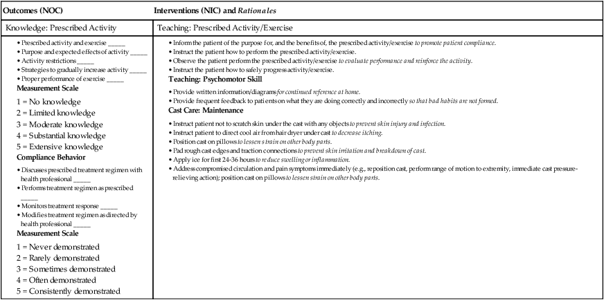
image eNursing Care Plan 63-1   Patient With a Fracture


Patient Goals


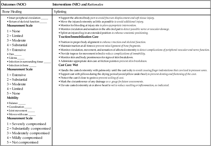
Outcomes (NOC) Interventions (NIC) and Rationales
Bone Healing Splinting


image


Patient Goal


Experiences no peripheral neurovascular dysfunction


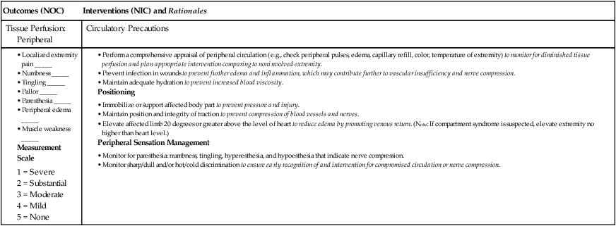
Outcomes (NOC) Interventions (NIC) and Rationales
Tissue Perfusion: Peripheral Circulatory Precautions


image


Patient Goal


Reports satisfaction with pain relief measures


Patient Goals


Outcomes (NOC) Interventions (NIC) and Rationales
Knowledge: Prescribed Activity Teaching: Prescribed Activity/Exercise


image


*Nursing diagnoses listed in order of priority.


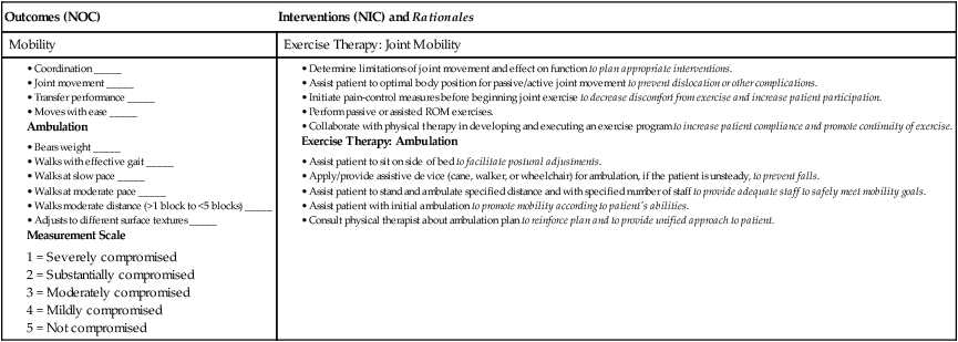
image eNursing Care Plan 63-2   Patient Having Orthopedic Surgery*


Patient Goals


Outcomes (NOC) Interventions (NIC) and Rationales
Mobility Exercise Therapy: Joint Mobility


image


Patient Goal


Reports satisfactory relief of pain


Patient Goals


ROM, Range of motion.


*This NCP is appropriate for a patient with an open reduction with internal fixation (ORIF) or joint replacement surgery.


**Nursing diagnoses listed in order of priority.


It is important to teach the public about the basic principles of safety and accident prevention. The morbidity associated with accidents can be significantly reduced if people are aware of environmental hazards, use appropriate safety equipment, and apply safety and traffic rules. In the occupational and industrial setting, teach employees and employers about the use of proper safety equipment and avoidance of hazardous working situations.


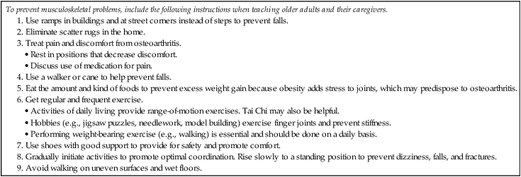
Ways to prevent common musculoskeletal problems in the older adult are listed in Table 63-1.


Soft Tissue Injuries


Soft tissue injuries, which include sprains, strains, dislocations, and subluxations, are usually caused by trauma. The increasing number of people involved in a fitness program or participating in sports has contributed to the increased incidence of soft tissue injuries. Common sports-related injuries are summarized in Table 63-2. The most common sports injuries that result in a visit to the emergency department for younger patients are bruises, sprains and strains, tendinitis, and fractures.2


TABLE 63-2


SPORTS-RELATED INJURIES


Injury Description Treatment
Impingement syndrome Entrapment of soft tissue structures under coracoacromial arch of shoulder. NSAIDs. Rest until symptoms decrease and then gradual ROM and strengthening exercises.
Rotator cuff tear Tear within muscle or tendinoligamentous structures around shoulder. If minor tear: Rest, NSAIDs, and gradual mobilization with ROM and strengthening exercises.
If major tear: Surgical repair.
Shin splints Inflammation along anterior aspect of calf from periostitis caused by improper shoes, overuse, or running on hard pavement. Rest, ice, NSAIDs, proper shoes. Gradual increase in activity.
If pain persists, x-ray to rule out stress fracture of tibia.
Tendinitis Inflammation of tendon as a result of overuse or incorrect use. Rest, ice, NSAIDs. Gradual return to sport activity. Protective brace (orthosis) may be necessary if symptoms recur.
Ligament injury Tearing or stretching of ligament. Usually occurs as a result of inversion, eversion, shearing, or torque applied to a joint.
Characterized by sudden pain, swelling, and instability.
Rest, ice, elevation of extremity if possible, NSAIDs. Protection of affected extremity by use of brace. If symptoms persist, surgical repair may be necessary.
Meniscus injury Injury to fibrocartilage of knee characterized by popping, clicking, tearing sensation, effusion, and/or swelling. Rest, ice, elevation of extremity if possible, NSAIDs. Gradual return to regular activities.
If symptoms persist, MRI to diagnose meniscus injury and possible arthroscopic surgery.
Anterior cruciate ligament tear Traumatic tearing of ligament by deceleration forces together with pivoting or odd positions of the knee or leg. Physical therapy with rehabilitation, knee brace.
If knee instability or continued injuries, reconstructive surgery may be done.


Sprains and Strains


Sprains and strains are common injuries from abnormal stretching or twisting forces that may occur during vigorous activities. These injuries tend to occur around joints and in the spinal musculature.


A sprain is an injury to the ligamentous structures surrounding a joint, usually caused by a wrenching or twisting motion. Most sprains occur in the ankle, wrist, and knee joints.3 A sprain is classified according to the degree of ligament damage. A first-degree (mild) sprain involves tears in only a few fibers, resulting in mild tenderness and minimal swelling. A second-degree (moderate) sprain is partial disruption of the involved tissue with more swelling and tenderness. A third-degree (severe) sprain is complete tearing of the ligament in association with moderate to severe swelling. A gap in the muscle may be apparent or palpated through the skin if the muscle is torn. Because areas around joints are rich in nerve endings, the injury can be extremely painful.


A strain is an excessive stretching of a muscle, its fascial sheath, or a tendon. Most strains occur in the large muscle groups, including the lower back, calf, and hamstrings. Strains may also be classified as first degree (mild or slightly pulled muscle), second degree (moderate or moderately torn muscle), and third degree (severely torn or ruptured muscle).


The clinical manifestations of sprains and strains are similar and include pain, edema, decreased function, and contusion. Pain aggravated by continued use is common. Edema develops in the injured area because of the local inflammatory response.


Mild sprains and strains are usually self-limiting, with full function returning within 3 to 6 weeks. X-rays of the affected part may be taken to rule out a fracture. A severe sprain can result in a concomitant avulsion fracture, in which the ligament pulls loose a fragment of bone. Alternatively, the joint structure may become unstable and result in subluxation or dislocation. At the time of injury, hemarthrosis (bleeding into a joint space or cavity) or disruption of the synovial lining may occur. Severe strains may require surgical repair of the muscle, tendon, or surrounding fascia.


Nursing Management Sprains and Strains


Nursing Implementation


Health Promotion.


Warming up muscles before exercising and vigorous activity, followed by stretching, may significantly reduce the risk of sprains and strains. Strength, balance, and endurance exercises are also important. Strengthening exercises that involve working against resistance build up muscle strength and bone density. Balance exercises, which may overlap with some strengthening exercises, help to prevent falling. Endurance exercises should start at a low level of effort and progress gradually to a moderate level.4 Exercise instructions for these types of physical activity are available at www.weboflife.ksc.nasa.gov/exerciseandaging.


Acute Intervention.


If an injury occurs, the immediate care focuses on (1) stopping the activity and limiting movement, (2) applying ice compresses to the injured area, (3) compressing the involved extremity, (4) elevating the extremity, and (5) providing analgesia as necessary (Table 63-3).


RICE (Rest, Ice, Compression, Elevation) may decrease local inflammation and pain for most musculoskeletal injuries. Movement should be restricted and the extremity rested as soon as pain is felt. Unless the injury is severe, prolonged rest is usually not indicated.


Cold (cryotherapy) in several forms can be used to produce hypothermia in the involved part. The cold induces physiologic changes in soft tissue, including vasoconstriction and a reduction in the transmission and perception of nerve pain impulses. In addition to pain relief, these changes reduce muscle spasms, inflammation, and edema. Cold is most useful when applied immediately after the injury has occurred. Ice applications should not exceed 20 to 30 minutes per application, and ice should not be applied directly to the skin.


An elastic compression bandage can be wrapped around the injured part. To prevent edema and encourage fluid return, wrap the bandage starting distally (at the point farthest from the midline of the body) and progress proximally (toward the midline of the body). The bandage is too tight if numbness is felt below the area of compression or there is additional pain or swelling beyond the edge of the bandage. The bandage can be left in place for 30 minutes and then removed for 15 minutes. However, some elastic wraps are left on during training, athletic, and occupational activities.


The injured part should be elevated above the heart level to help mobilize excess fluid from the area and prevent further edema. The injured part should be elevated even during sleep. Mild analgesics and nonsteroidal antiinflammatory drugs (NSAIDs) may be necessary to manage patient discomfort.


After the acute phase (usually 24 to 48 hours), warm, moist heat may be applied to the affected part to reduce swelling and provide comfort. Heat applications should not exceed 20 to 30 minutes, allowing a “cool-down” time between applications. Encourage the patient to use the limb, provided that the joint is protected by means of casting, bracing, taping, or splinting. Movement of the joint maintains nutrition to the cartilage, and muscle contraction improves circulation and resolution of the contusion and swelling.


Dislocation and Subluxation


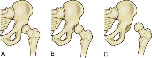
A dislocation is a severe injury of the ligamentous structures that surround a joint. Dislocation results in the complete displacement or separation of the articular surfaces of the joint. A subluxation is a partial or incomplete displacement of the joint surface. The clinical manifestations of a subluxation are similar to those of a dislocation but are less severe.


Dislocations characteristically result from forces transmitted to the joint that disrupt the soft tissue support structures surrounding it. The joints most frequently dislocated in the upper extremity include the thumb, elbow, and shoulder. In the lower extremity, the hip is vulnerable to dislocation as a result of severe trauma, often associated with motor vehicle collisions (Fig. 63-1). The patella may dislocate because of a sharp blow to the kneecap or after a sudden twisting inward motion while the planted foot is pointed outward.5


The most obvious clinical manifestation of a dislocation is deformity. For example, if a hip is dislocated in a posterior (or backward) direction, the limb can be shorter and is often internally rotated on the affected side. Additional manifestations include local pain, tenderness, loss of function of the injured part, and swelling of the soft tissues in the joint region. The major complications of a dislocated joint are open joint injuries, intraarticular fractures, avascular necrosis (bone cell death as a result of inadequate blood supply), and damage to adjacent neurovascular tissue.


X-ray studies are performed to determine the extent of displacement of the involved structures. The joint may also be aspirated to assess for hemarthrosis or fat cells. Fat cells in the aspirate indicate a probable intraarticular (within the joint) fracture.


Nursing and Collaborative Management Dislocation


A dislocation requires prompt attention and is often considered an orthopedic emergency. It may be associated with significant vascular injury. The longer the joint remains unreduced, the greater the possibility of avascular necrosis. The hip joint is particularly susceptible to avascular necrosis. Compartment syndrome (discussed on p. 1522) may also occur after a dislocation. Neurovascular assessment is critical (see pp. 15171518).


The first goal of management is to realign the dislocated portion of the joint in its original anatomic position. This can be accomplished by a closed reduction, which may be performed under local or general anesthesia or IV conscious sedation. Anesthesia is often necessary to relax the muscle so that the bones can be manipulated. In some situations, surgical open reduction may be necessary. After reduction, the extremity is usually immobilized by bracing, splinting, taping, or using a sling to allow the torn ligaments and capsular tissue time to heal.


Nursing management of subluxation or dislocation is directed toward relief of pain and support and protection of the injured joint. After the joint has been reduced and immobilized, motion is usually restricted. A carefully monitored rehabilitation program can prevent fracture instability and joint dysfunction. Gentle range-of-motion (ROM) exercises may be recommended if the joint is stable and well supported. An exercise program slowly restores the joint to its original ROM without causing another dislocation. The patient should gradually return to normal activities.


A patient who has dislocated a joint may be at greater risk for repeated dislocations because of loose ligaments. Activity restrictions may be imposed on the affected joint to decrease the risk of repeated dislocations.


Repetitive Strain Injury


Repetitive strain injury (RSI) and cumulative trauma disorder are terms used to describe injuries resulting from prolonged force or repetitive movements and awkward postures. RSI is also referred to as repetitive trauma disorder, nontraumatic musculoskeletal injury, overuse syndrome (sports medicine), regional musculoskeletal disorder, and work-related musculoskeletal disorder. Repeated movements strain the tendons, ligaments, and muscles, causing tiny tears that become inflamed. The exact cause of these disorders is unknown. There are no specific diagnostic tests, and diagnosis is often difficult.


Persons at risk for RSI include musicians, dancers, butchers, grocery clerks, vibratory tool workers, and those who frequently use a computer mouse and keyboard. Competitive athletes and poorly trained athletes may also develop RSI. Swimming, overhead throwing (e.g., baseball), weight lifting, gymnastics, tennis, skiing, and kicking sports (e.g., soccer) require repetitive motion, and overtraining compounds the effects of RSI.


In addition to repetitive movements, other factors related to RSI include poor posture and positioning, poor workspace ergonomics, badly designed workplace equipment (e.g., computer keyboard), and repetitive lifting of heavy objects without sufficient muscle rest. The result may be inflammation, swelling, and pain in the muscles, tendons, and nerves of the neck, spine, shoulder, forearm, and hand. Symptoms of RSI include pain, weakness, numbness, or impairment of motor function. RSI can be prevented through education and ergonomics (the science that promotes efficiency and safety in the interaction of humans and their work environment). Ergonomic considerations for persons who work at a desk and use a computer include keeping the hips and knees flexed to 90 degrees with the feet flat, keeping the wrist straight to type, having the top of the computer monitor even with the forehead, and taking at least hourly stretch breaks. Once RSI is diagnosed, treatment consists of identification of the precipitating activity; modification of equipment or activity; pain management, including heat or cold application and NSAIDs; rest; physical therapy for strengthening and conditioning exercises; and lifestyle changes.

Stay updated, free articles. Join our Telegram channel

Nov 17, 2016 | Posted by in NURSING | Comments Off on Nursing Management: Musculoskeletal Trauma and Orthopedic Surgery

Full access? Get Clinical Tree

Get Clinical Tree app for offline access