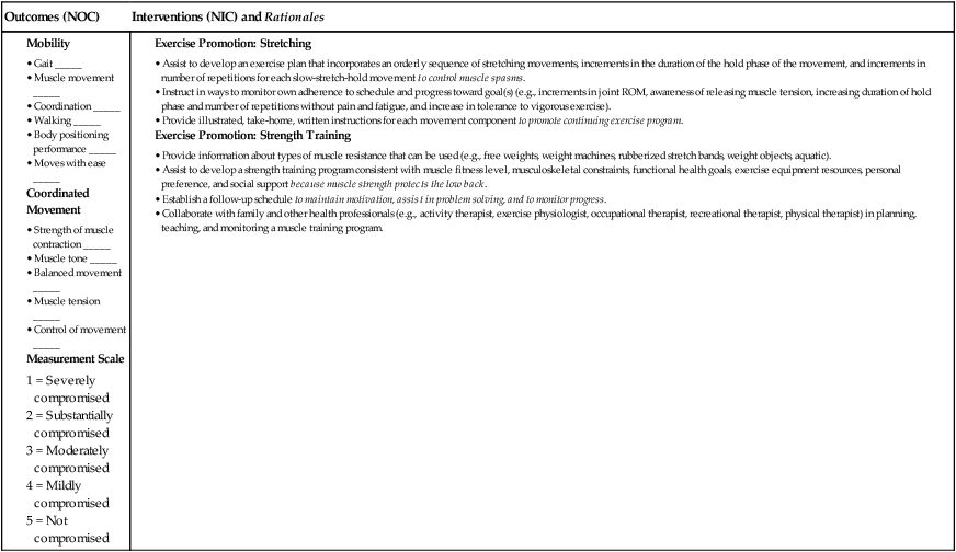
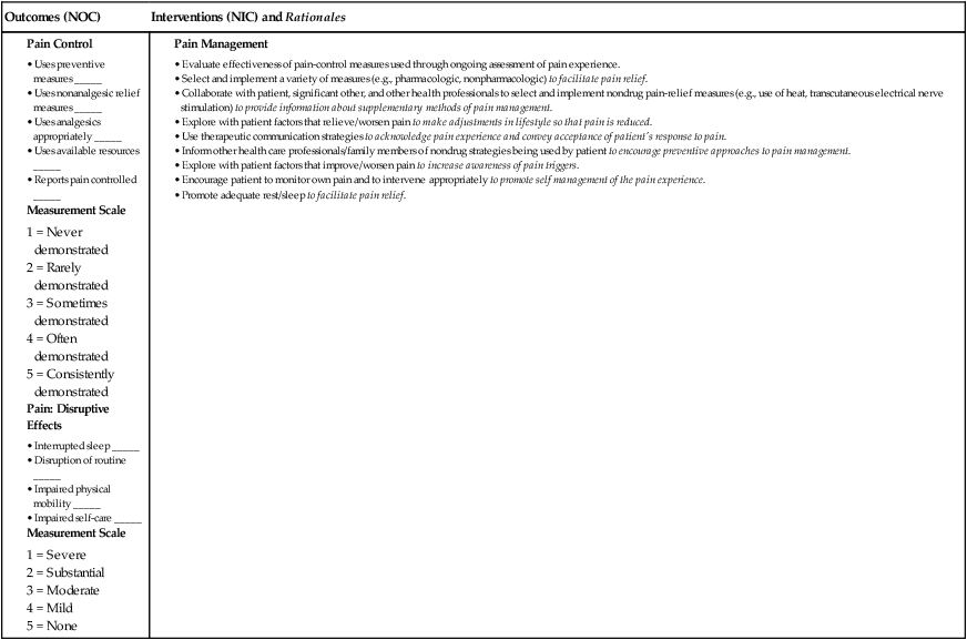
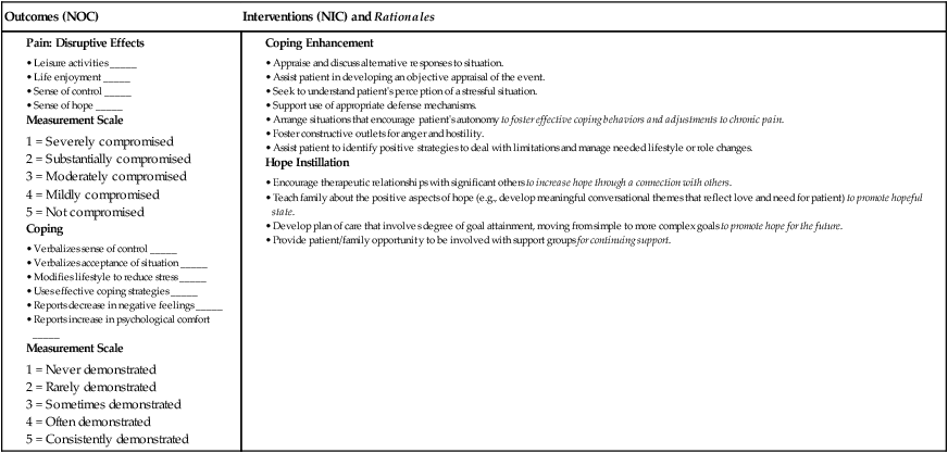
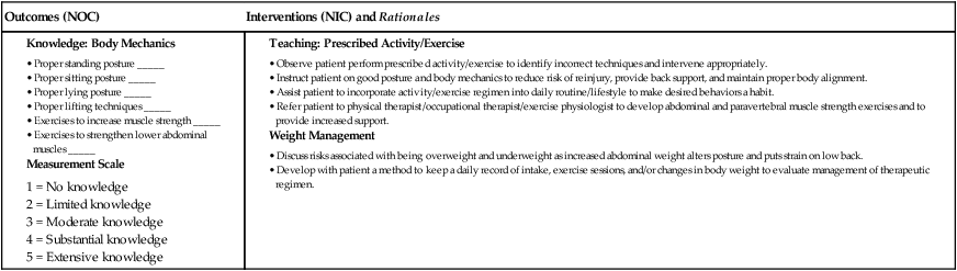
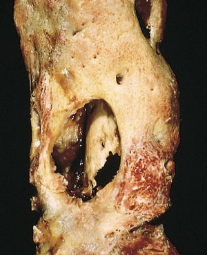
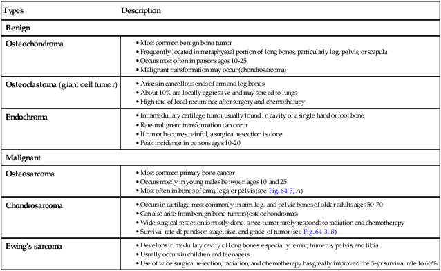
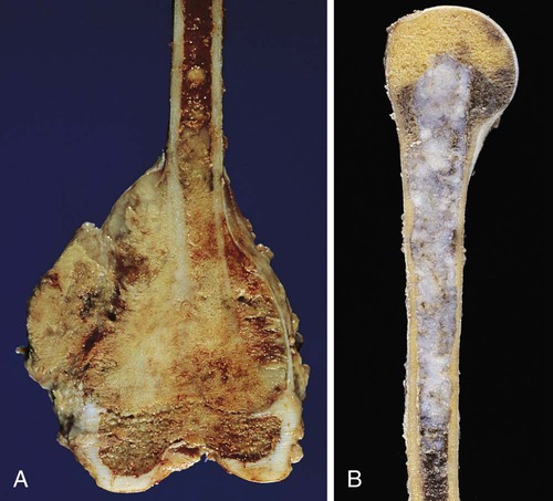
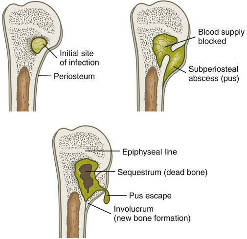
Nursing Management: Musculoskeletal Problems

Chapter 64


Nursing Management


Musculoskeletal Problems


Jerry Harvey


Reviewed by Tammy C. Roman, RN, EdD, CNE, Assistant Professor of Nursing, St. John Fisher College, Wegmans School of Nursing, Rochester, New York; and Dianne Travers Gustafson, RN, PhD, Associate Professor, Creighton University School of Nursing, Omaha, Nebraska.


Acute and chronic musculoskeletal problems are a common source of pain and disability. A variety of problems unrelated to trauma that affect the musculoskeletal system are presented in this chapter, including osteomyelitis, bone cancer, muscular dystrophy, foot disorders, and metabolic bone diseases. Management of the patient with both acute and chronic low back pain is addressed, and spinal surgery is discussed as an intervention for a herniated disc. Throughout the discussion of all of these problems, the nurse’s role in prevention of injury and maintenance of mobility is emphasized.


Osteomyelitis


Etiology and Pathophysiology


Osteomyelitis is a severe infection of the bone, bone marrow, and surrounding soft tissue. Although Staphylococcus aureus is a common cause of infection, a variety of microorganisms may cause osteomyelitis1 (Table 64-1).


The infecting microorganisms can invade by indirect or direct entry. The indirect entry (hematogenous) of microorganisms most frequently affects growing bone in boys younger than 12 years old, and is associated with their higher incidence of blunt trauma. Adults with vascular insufficiency disorders (e.g., diabetes mellitus) and genitourinary and respiratory tract infections are at higher risk for a primary infection to spread via the blood to the bone. The pelvis, tibia, and vertebrae, which are vascular-rich sites of bone, are the most common sites of infection.


image 
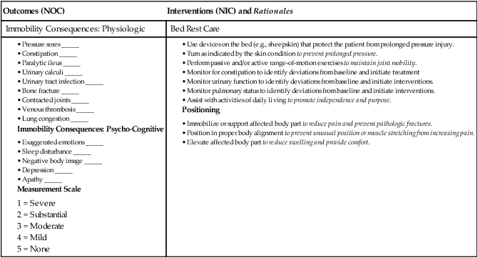
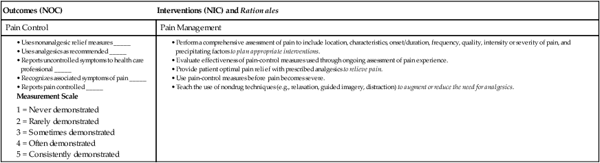
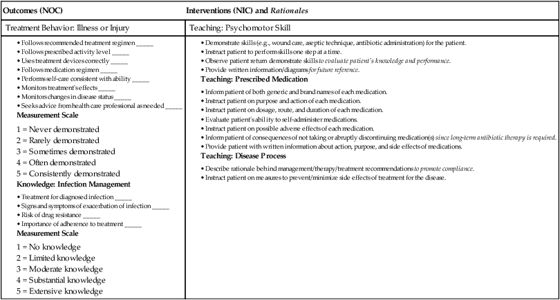
eNursing Care Plan 64-1   Patient With Osteomyelitis


Patient Goals


Outcomes (NOC) Interventions (NIC) and Rationales
Immobility Consequences: Physiologic Bed Rest Care


image


Patient Goals


Patient Goals


Outcomes (NOC) Interventions (NIC) and Rationales
Treatment Behavior: Illness or Injury Teaching: Psychomotor Skill


image


*Nursing diagnoses listed in order of priority.


image 
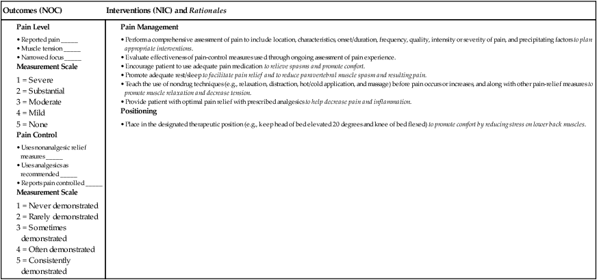
eNursing Care Plan 64-2


Patient With Low Back Pain: Acute Management


Patient Goal


Reports satisfactory pain relief with pain <4 on 10-point scale


Outcomes (NOC) Interventions (NIC) and Rationales


Pain Management


• Perform a comprehensive assessment of pain to include location, characteristics, onset/duration, frequency, quality, intensity or severity of pain, and precipitating factors to plan appropriate interventions.


• Evaluate effectiveness of pain-control measures used through ongoing assessment of pain experience.


• Encourage patient to use adequate pain medication to relieve spasms and promote comfort.


• Promote adequate rest/sleep to facilitate pain relief and to reduce paravertebral muscle spasm and resulting pain.


• Teach the use of nondrug techniques (e.g., relaxation, distraction, hot/cold application, and massage) before pain occurs or increases, and along with other pain-relief measures to promote muscle relaxation and decrease tension.


• Provide patient with optimal pain relief with prescribed analgesics to help decrease pain and inflammation.


image


Patient Goals


Outcomes (NOC) Interventions (NIC) and Rationales


Exercise Promotion: Strength Training


• Provide information about types of muscle resistance that can be used (e.g., free weights, weight machines, rubberized stretch bands, weight objects, aquatic).


• Assist to develop a strength training program consistent with muscle fitness level, musculoskeletal constraints, functional health goals, exercise equipment resources, personal preference, and social support because muscle strength protects the low back.


• Establish a follow-up schedule to maintain motivation, assist in problem solving, and to monitor progress.


• Collaborate with family and other health professionals (e.g., activity therapist, exercise physiologist, occupational therapist, recreational therapist, physical therapist) in planning, teaching, and monitoring a muscle training program.


image


Patient With Low Back Pain: Chronic Management


Patient Goal


Reports ability to control pain at an acceptable level with the use of preventive and nonpharmacologic measures


Outcomes (NOC) Interventions (NIC) and Rationales


Pain Management


• Evaluate effectiveness of pain-control measures used through ongoing assessment of pain experience.


• Select and implement a variety of measures (e.g., pharmacologic, nonpharmacologic) to facilitate pain relief.


• Collaborate with patient, significant other, and other health professionals to select and implement nondrug pain-relief measures (e.g., use of heat, transcutaneous electrical nerve stimulation) to provide information about supplementary methods of pain management.


• Explore with patient factors that relieve/worsen pain to make adjustments in lifestyle so that pain is reduced.


• Use therapeutic communication strategies to acknowledge pain experience and convey acceptance of patient’s response to pain.


• Inform other health care professionals/family members of nondrug strategies being used by patient to encourage preventive approaches to pain management.


• Explore with patient factors that improve/worsen pain to increase awareness of pain triggers.


• Encourage patient to monitor own pain and to intervene appropriately to promote self management of the pain experience.


• Promote adequate rest/sleep to facilitate pain relief.


image


Patient Goals


Outcomes (NOC) Interventions (NIC) and Rationales


image


Patient Goal


Integrates a program of appropriate posture, body mechanics, exercises, and weight management into daily routine


*Nursing diagnoses listed in order of priority.


Direct entry osteomyelitis can occur at any age when there is an open wound (e.g., penetrating wounds, fractures) and microorganisms gain entry to the body. Osteomyelitis may also occur in the presence of a foreign body such as an implant or an orthopedic prosthetic device (e.g., plate, total joint prosthesis).


After gaining entry into the blood, the microorganisms grow, resulting in an increase in pressure because of the nonexpanding nature of most bone. This increasing pressure eventually leads to ischemia and vascular compromise of the periosteum. The infection spreads through the bone cortex and marrow cavity, ultimately resulting in cortical devascularization and necrosis.


Once ischemia occurs, the bone dies. The area of devitalized bone eventually separates from the surrounding living bone, forming sequestra. The part of the periosteum that continues to have a blood supply forms new bone called involucrum (Fig. 64-1). It is difficult for blood-borne antibiotics or white blood cells (WBCs) to reach the sequestrum. A sequestrum may become a reservoir for microorganisms that spread to other sites, including the lungs and brain. If the sequestrum does not resolve on its own or is debrided surgically, a sinus tract may develop, resulting in chronic, purulent cutaneous drainage.


Clinical Manifestations and Complications


Acute osteomyelitis refers to the initial infection or an infection of less than 1 month in duration. The clinical manifestations of acute osteomyelitis are both local and systemic. Local manifestations include constant bone pain unrelieved by rest that worsens with activity; swelling, tenderness, and warmth at the infection site; and restricted movement of the affected part. Systemic manifestations include fever, night sweats, chills, restlessness, nausea, and malaise. Later signs include drainage from cutaneous sinus tracts or the fracture site.


Chronic osteomyelitis refers to a bone infection that persists for longer than 1 month or an infection that has failed to respond to the initial course of antibiotic therapy. Chronic osteomyelitis is either a continuous, persistent problem (a result of inadequate acute treatment) or a process of exacerbations and remissions (Fig. 64-2). Systemic signs may be diminished, with local signs of infection more common, including constant bone pain and swelling and warmth at the infection site. Over time, granulation tissue turns to scar tissue. This avascular scar tissue provides an ideal site for continued microorganism growth that cannot be penetrated by antibiotics.


Long-term and mostly rare complications of osteomyelitis include septicemia, septic arthritis, pathologic fractures, and amyloidosis.


Diagnostic Studies


A bone or soft tissue biopsy is the definitive way to determine the causative microorganism. The patient’s blood and wound cultures are frequently positive for microorganisms.2 An elevated WBC count and erythrocyte sedimentation rate (ESR) may also be found. X-ray signs suggestive of osteomyelitis usually do not appear until 10 days to weeks after the initial clinical symptoms, by which time the disease will have progressed. Radionuclide bone scans (gallium and indium) are helpful in diagnosis and are usually positive in the area of infection. Magnetic resonance imaging (MRI) and computed tomography (CT) scans may be used to help identify the extent of the infection.3


Collaborative Care


Vigorous and prolonged IV antibiotic therapy is the treatment of choice for acute osteomyelitis, as long as bone ischemia has not yet occurred. Cultures or a bone biopsy should be done if possible before initiating drug therapy. If antibiotic therapy is delayed, surgical debridement and decompression are often necessary.


Patients are often discharged to home care or a skilled nursing facility with IV antibiotics delivered via a central venous catheter or peripherally inserted central catheter. IV antibiotic therapy may be started in the hospital and continued at home for 4 to 6 weeks or as long as 3 to 6 months. A variety of antibiotics may be prescribed depending on the microorganism. These drugs include penicillin, nafcillin (Nafcil), neomycin, vancomycin, cephalexin (Keflex), cefazolin (Ancef), cefoxitin (Mefoxin), gentamicin (Garamycin), and tobramycin (Nebcin).


In adults with chronic osteomyelitis, oral therapy with a fluoroquinolone (ciprofloxacin [Cipro]) for 6 to 8 weeks may be prescribed instead of IV antibiotics. Oral antibiotic therapy may also be given after acute IV therapy is completed to ensure resolution of the infection. The patient’s response to drug therapy is monitored through bone scans and ESR tests.


Treatment of chronic osteomyelitis includes surgical removal of the poorly vascularized tissue and dead bone and the extended use of antibiotics.5 Antibiotic-impregnated polymethyl methacrylate bead chains may also be implanted at this time to help combat the infection. After debridement of the devitalized and infected tissue, the wound may be closed and a suction irrigation system inserted. Intermittent or constant irrigation of the affected bone with antibiotics may also be initiated. Protection of the limb or the surgical site with casts or braces is often done. Negative-pressure wound therapy (vacuum-assisted wound closure) may be used (see pp. 181182 in Chapter 12).


Hyperbaric oxygen with 100% oxygen may be given as an adjunct therapy in refractory cases of chronic osteomyelitis. This therapy is thought to stimulate circulation and healing in the infected tissue (see pp. 182183 in Chapter 12). Orthopedic prosthetic devices, if a source of chronic infection, must be removed. Muscle flaps or skin grafting provides wound coverage over the dead space (cavity) in the bone. Bone grafts may help to restore blood flow. Amputation of the extremity may be indicated when bone destruction is extensive and to save the patient’s life or to improve quality of life.


Nursing Management Osteomyelitis
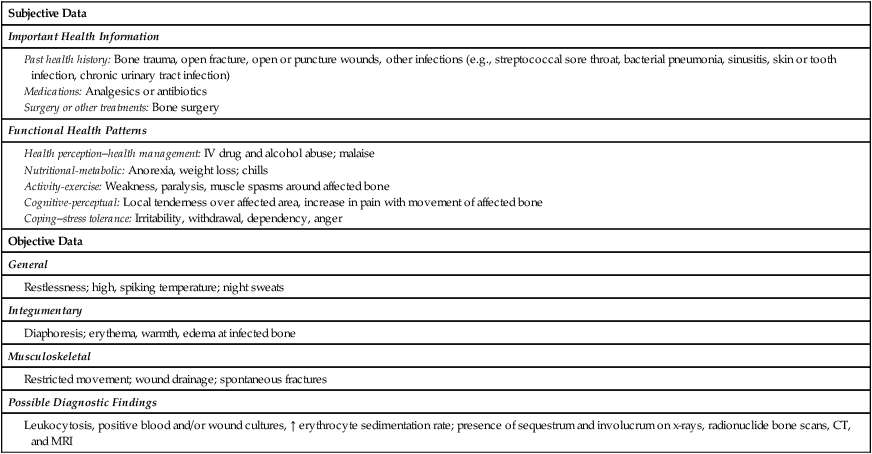

Nursing Assessment


Subjective and objective data that should be obtained from an individual with osteomyelitis are presented in Table 64-2.


Nursing Implementation


Health Promotion.


The control of infections already in the body (e.g., urinary, respiratory tract, deep pressure ulcers) is important in preventing osteomyelitis. Individuals who are especially at risk for osteomyelitis are those who are immunocompromised, have orthopedic prosthetic devices, or have vascular insufficiencies. Instruct these patients regarding the local and systemic manifestations of osteomyelitis. Also make family members aware of their role in monitoring the patient’s health. Instruct patients to immediately report symptoms of bone pain, fever, swelling, and restricted limb movement to the health care provider so treatment can be started.


Acute Intervention.


Some immobilization of the affected limb (e.g., splint, traction) is usually indicated to decrease pain. Carefully handle the involved limb and avoid excessive manipulation, which increases pain and may cause a pathologic fracture. Assess the patient’s pain. Minor to severe pain may be experienced with muscle spasms. Nonsteroidal antiinflammatory drugs (NSAIDs), opioid analgesics, and muscle relaxants may be prescribed to provide patient comfort. Encourage nondrug approaches to pain management (e.g., guided imagery, relaxation breathing) (see Chapters 7 and 9).


Dressings are used to absorb the exudate from draining wounds and to debride devitalized tissue from the wound site. Types of dressings include dry, sterile dressings; dressings saturated in saline or antibiotic solution; and wet-to-dry dressings. Handle soiled dressings carefully to prevent cross-contamination of the wound or spread of the infection to other patients. Sterile technique is essential when changing the dressing.


The patient is frequently on bed rest in the early stages of the acute infection. Good body alignment and frequent position changes prevent complications associated with immobility and promote comfort. Flexion contracture of the lower extremity is a common complication of osteomyelitis because the patient frequently positions the affected extremity in a flexed position to promote comfort. Footdrop can develop quickly if the foot is not correctly supported in a neutral position by a splint or if a splint applies excessive pressure, which can injure the peroneal nerve.


Teach the patient the potential adverse and toxic reactions associated with prolonged and high-dose antibiotic therapy (e.g., tobramycin, neomycin). These reactions include hearing deficit, nephrotoxicity, and neurotoxicity. With cephalosporins (e.g., cefazolin) these reactions include hives, severe or watery diarrhea, blood in stools, and throat or mouth sores.


Tendon rupture (especially the Achilles tendon) can occur with use of the fluoroquinolones (e.g., ciprofloxacin, levofloxacin [Levaquin]). Peak and trough blood levels of most antibiotics should be monitored to avoid adverse effects. Lengthy antibiotic therapy can also result in an overgrowth of Candida albicans and Clostridium difficile in the genitourinary and gastrointestinal (GI) tract, especially in immunosuppressed and older patients. Instruct the patient to report any changes in the oral cavity such as whitish yellow, curdlike lesions or changes in the genitourinary cavity such as any perianal itching or diarrhea.


The patient, caregiver, and family may be anxious and discouraged because of the serious nature of osteomyelitis, the uncertainty of the outcome, and the cost and lengthy course of treatment. Continued psychologic and emotional support is an integral part of nursing management. A nursing care plan for the patient with osteomyelitis (eNursing Care Plan 64-1) is available on the website for this chapter.


Bone Tumors


Primary bone tumors, both benign and malignant, are relatively rare in adults. They account for only about 3% of all tumors. Metastatic bone cancer in which the cancer has spread from another site is a more common problem.


Benign Bone Tumors


Benign bone tumors are more common than primary malignant tumors. The main types of benign bone tumors are osteochondroma, osteoclastoma, and endochroma (Table 64-3). These types of tumors are often removed by surgery.


TABLE 64-3


TYPES OF PRIMARY BONE TUMORS


Types Description
Benign
Osteochondroma
Osteoclastoma (giant cell tumor)
Endochroma
Malignant
Osteosarcoma
Chondrosarcoma
Ewing’s sarcoma


image


Osteochondroma


Osteochondroma is the most common primary benign bone tumor. It is characterized by an overgrowth of cartilage and bone near the end of the bone at the growth plate. It is more commonly found in the long bones of the leg, pelvis, or scapula.


Clinical manifestations include a painless, hard, immobile mass; lower-than-normal height for age; soreness of muscles in close proximity to the tumor; one leg or arm longer than the other; and pressure or irritation with exercise. Patients may also be asymptomatic. Diagnosis is confirmed using x-ray, CT scan, and MRI.


No treatment is necessary for asymptomatic osteochondroma. If the tumor is causing pain or neurologic symptoms because of compression, surgical resection is usually done. Patients should have regular screening examinations for early detection of malignant transformation.


Malignant Bone Tumors


A sarcoma is a malignant tumor that can develop in bone, muscle, fat, nerve, or cartilage. The most common types of malignant bone tumors are osteosarcoma, chondrosarcoma, and Ewing’s sarcoma (see Table 64-3). Annually about 2900 new cases of bone (and joint) cancer occur in the United States, with an estimated 1400 deaths.6 Primary malignant tumors occur most often during childhood and young adulthood. They are characterized by their rapid metastasis and bone destruction.


Osteosarcoma


Osteosarcoma is a primary malignant bone tumor that is extremely aggressive and rapidly metastasizes to distant sites. It usually occurs in the metaphyseal region of the long bones of extremities, particularly in the regions of the distal femur, proximal tibia, and proximal humerus, as well as the pelvis (Fig. 64-3, A). Osteosarcoma is the most common malignant bone tumor affecting children and young adults. It can also occur, but not as commonly, in older adults. It is most often associated with Paget’s disease and prior radiation.


Clinical manifestations of osteosarcoma are usually associated with the gradual onset of pain and swelling, especially around the knee. A minor injury does not cause the neoplasm but may bring the preexisting condition to medical attention. Metastasis is present in 10% to 20% of individuals when they are diagnosed with osteosarcoma.


Diagnosis is confirmed from tissue biopsy, elevation of serum alkaline phosphatase and calcium levels, x-ray, computed tomography (CT) or positron emission tomography (PET) scans, and magnetic resonance imaging (MRI).


Preoperative (neoadjuvant) chemotherapy may be used in the treatment of osteosarcoma to decrease tumor size before surgery. Limb salvage procedures are usually considered when there is a clear (no cancer present) 6- to 7-cm margin surrounding the lesion. Limb salvage is usually contraindicated if there is major neurovascular involvement, pathologic fracture, infection, or extensive muscle involvement.


The use of adjunct chemotherapy after amputation or limb salvage has increased the 5-year survival rate to 70% in people without metastasis. Chemotherapy includes methotrexate, doxorubicin (Adriamycin), cisplatin (Platinol), cyclophosphamide (Cytoxan), etoposide (VePesid), bleomycin (Blenoxane), dactinomycin (Cosmegen), and ifosfamide (Ifex).7

Only gold members can continue reading. Log In or Register to continue

Stay updated, free articles. Join our Telegram channel

Nov 17, 2016 | Posted by in NURSING | Comments Off on Nursing Management: Musculoskeletal Problems

Full access? Get Clinical Tree

Get Clinical Tree app for offline access