Nursing Assessment: Reproductive System

Chapter 51


Nursing Assessment


Reproductive System


Catherine (Kate) Lein


Reviewed by Brenda Pavill, RN, PhD, FNP, Associate Professor–Nursing, Misericordia University, Dallas, Pennsylvania.


Structures and Functions of Male and Female Reproductive Systems


The reproductive system of both males and females consists of primary (or essential) organs and secondary (or accessory) organs. The primary reproductive organs are referred to as gonads. The female gonads are the ovaries; the male gonads are the testes. The main responsibility of the gonads is secretion of hormones and production of gametes (ova and sperm). Secondary (or accessory) organs are responsible for (1) transporting and nourishing the ova and sperm and (2) preserving and protecting the fertilized eggs.


Male Reproductive System


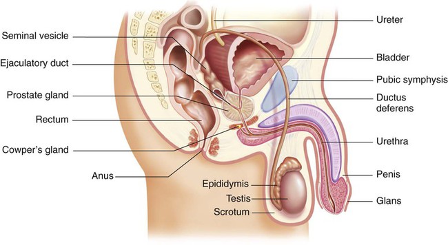
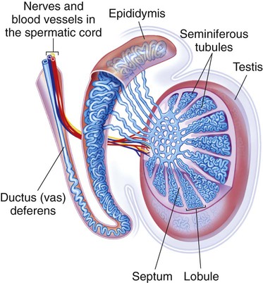
The three primary roles of the male reproductive system are (1) production and transportation of sperm, (2) deposition of sperm in the female reproductive tract, and (3) secretion of hormones. The primary reproductive organs in the male are the testes. Secondary reproductive organs include ducts (epididymis, ductus deferens, ejaculatory duct, and urethra), sex glands (prostate gland, Cowper’s glands, and seminal vesicles), and the external genitalia (scrotum and penis)1 (Fig. 51-1).


Ducts.

Sperm formed in the seminiferous tubules move through a series of ducts. These ducts transport the sperm from the testes to the outside of the body. As sperm exit the testes, they enter and pass through the epididymis, ductus deferens, ejaculatory duct, and urethra.


The epididymis is a comma-shaped structure located on the posterosuperior aspect of each testis within the scrotum (see Figs. 51-1 and 51-2). It is a very long, tightly coiled structure that measures about 20 ft in length.1 The epididymis transports the sperm as they mature. Sperm exit the epididymis through a long, thick tube known as the ductus deferens.


The ductus deferens (also known as the vas deferens) is continuous with the epididymis within the scrotal sac. It travels upward through the scrotum and continues through the inguinal ring into the abdominal cavity. The spermatic cord is composed of a connective tissue sheath that encloses the ductus deferens, arteries, veins, nerves, and lymph vessels as it ascends up through the inguinal canal (see Fig. 51-2). In the abdominal cavity, the ductus deferens travels up, over, and behind the bladder. Posterior to the bladder the ductus deferens joins the seminal vesicle to form the ejaculatory duct (see Fig. 51-1).


The ejaculatory duct passes downward through the prostate gland, connecting with the urethra. The urethra extends from the bladder, through the prostate, and ends in a slitlike opening (the meatus) on the ventral side of the glans, the tip of the penis. During the process of ejaculation, sperm travels through the urethra and out of the penis.


Glands.

The seminal vesicles, the prostate gland, and Cowper’s (bulbourethral) glands are the accessory glands of the male reproductive system. These glands produce and secrete seminal fluid (semen), which surrounds the sperm and forms the ejaculate.


The seminal vesicles lie posterior to the bladder and between the rectum and bladder. The ducts of the seminal vesicles fuse with the ductus deferens to form the ejaculatory ducts that enter the prostate gland. The prostate gland lies beneath the bladder. Its posterior surface is in contact with the rectal wall. The prostate normally measures 0.8 in (2 cm) wide and 1.2 in (3 cm) long and is divided into the right and left lateral lobes and an anteroposterior median lobe. Cowper’s glands lie on each side of the urethra and slightly posterior to it, just below the prostate. The ducts of these glands enter directly into the urethra.


Secretions from the seminal vesicles, prostate, and Cowper’s glands make up most of the fluid in the ejaculate. These various secretions serve as a medium for the transport of sperm and create an alkaline, nutritious environment that promotes sperm motility and survival.


Female Reproductive System


The three primary roles of the female reproductive system are (1) production of ova (eggs), (2) secretion of hormones, and (3) protection and facilitation of the development of the fetus in a pregnant female. Like the male, the female has primary and secondary reproductive organs. The primary reproductive organs in the female are the paired ovaries. Secondary reproductive organs include the ducts (fallopian tubes), uterus, vagina, sex glands (Bartholin’s glands and breasts), and external genitalia (vulva).


Pelvic Organs


Ovaries.

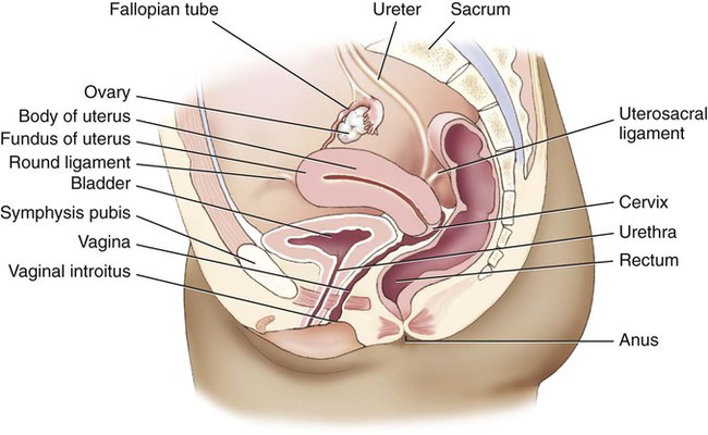
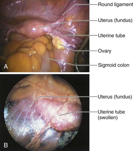
The ovaries are located on either side of the uterus, just behind and below the fallopian (uterine) tubes (Fig. 51-3). The almond-shaped ovaries are firm and solid, approximately 0.6 in (1.5 cm) wide and 1.2 in (3 cm) long. Their functions include ovulation and secretion of the two major reproductive hormones, estrogen and progesterone.


The outer zone of the ovary contains follicles with germ cells, or oocytes. Each follicle contains a primordial (immature) oocyte surrounded by granulosa and theca cells. These two layers protect and nourish the oocyte until the follicle reaches maturity and ovulation occurs. However, not all follicles reach maturity. In a process termed atresia, most of the primordial follicles become smaller and are reabsorbed by the body. Thus the number of follicles declines from 2 million to 4 million at birth to approximately 300,000 to 400,000 at menarche. Fewer than 500 oocytes are actually released by ovulation during the reproductive years of the normal healthy woman.


Fallopian Tubes.

Normally, each month during a woman’s reproductive years, one ovarian follicle reaches maturity, and the ovum is ovulated, or expelled, from the ovary through the stimulus of the gonadotropic hormones, follicle-stimulating hormone (FSH) and luteinizing hormone (LH). The ovum then travels up a fallopian tube where fertilization by sperm may occur, if sperm are present. An ovum can be fertilized up to 72 hours after its release.


The distal ends of the fallopian tubes consist of fingerlike projections called fimbriae that “massage” the ovaries at ovulation to help extract the mature ovum. The tubes, which average 4.8 in (12 cm) in length, extend from the fimbriae to the superior lateral borders of the uterus. Fertilization usually takes place within the outer one third of the fallopian tubes.


Uterus.

The uterus is a pear-shaped, hollow, muscular organ (see Fig. 51-3). It is located between the bladder and the rectum. In the mature nulliparous (never pregnant) woman, the uterus is approximately 2.4 to 3.2 in (6 to 8 cm) long and 1.6 in (4 cm) wide. The uterine walls consist of an outer serosal layer, the perimetrium; a middle muscular layer, the myometrium; and an inner mucosal layer, the endometrium.


The uterus consists of the fundus, body (or corpus), and cervix (see Fig. 51-3). The body makes up about 80% of the uterus and connects with the cervix at the isthmus, or neck. The cervix is the lower portion of the uterus that projects into the anterior wall of the vaginal canal. It makes up about 15% to 20% of the uterus in the nulliparous female. The cervix consists of the ectocervix, the outer portion that protrudes into the vagina, and the endocervix, the canal in the opening of the cervix. The ectocervix is covered with squamous epithelial cells, which give it a smooth, pinkish appearance. The endocervix contains a lining of columnar epithelial cells, which give it a rough, reddened appearance. The junction at which the two types of epithelial cells meet is termed the squamocolumnar junction and contains the optimal types of cells needed for an accurate Papanicolaou (Pap) test to screen for malignancies.


The cervical canal is 0.8 to 1.6 in (2 to 4 cm) long and is relatively tightly closed. However, the cervix allows sperm to enter the uterus and also allows menses to be expelled. The columnar epithelium, under hormonal influence, provides elasticity during labor for the cervix to stretch to allow passage of a fetus during the birth process. The entrance of sperm into the uterus is facilitated by mucus produced by the cervix under the influence of estrogen. Under normal conditions, the cervical mucus becomes watery, stretchy, and more abundant at ovulation. The postovulatory cervical mucus, under the influence of progesterone, is thick and inhibits sperm passage.


External Genitalia.

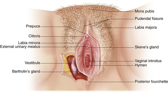
The external portion of the female reproductive system (Fig. 51-4), commonly called the vulva, consists of the mons pubis, labia majora, labia minora, clitoris, urethral meatus, Skene’s glands, vaginal introitus (opening), and Bartholin’s glands.


The mons pubis is a fatty layer lying over the pubic bone. It is covered with coarse hair that lies in a triangular pattern. (The male hair pattern is diamond shaped.) The labia majora are folds of adipose tissue that form the outer borders of the vulva. The hairless labia minora form the borders of the vaginal orifice and extend anteriorly to enclose the clitoris.


The vestibule is a boat-shaped fossa between the labia minora, extending from the clitoris at the anterior end to the vaginal opening at the posterior end. The perineum is the area between the vagina and the anus. The vaginal introitus is surrounded by thin membranous tissue called the hymen. In the adult woman the hymen usually appears as folds or hymenal tags and separates the external genitalia from the vagina. At the posterior aspect of the vagina, a tense band of mucous membrane connecting the posterior ends of the labia minora is referred to as the posterior fourchette.


The clitoris is erectile tissue that becomes engorged during sexual excitation. It lies anterior to the urethral meatus and the vaginal orifice and is usually covered by the prepuce. Clitoral stimulation is an important part of sexual activity for many women.


Ducts of the Skene’s glands lie alongside the urinary meatus and are thought to help lubricate the urinary meatus.2 The Bartholin’s glands, located at the posterior and lateral aspects of the vaginal orifice, secrete a thin, mucoid material believed to contribute slightly to lubrication during sexual intercourse. These glands are not usually palpable unless sebaceous-like cysts form or they are swollen in the presence of an infection, such as a sexually transmitted infection (STI).


Breasts.

The breasts are a secondary sex characteristic that develops during puberty in response to estrogen and progesterone. Cyclic hormonal changes lead to regular changes in breast tissue to prepare it for lactation when fertilization and pregnancy occur.


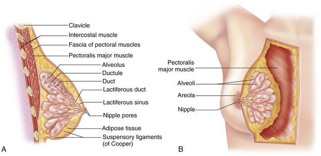
The breasts extend from the second to the sixth ribs, with the tail reaching the axilla (Fig. 51-5). The fully mature breast is dome shaped and contains a pigmented center termed the areola. The areolar region contains Montgomery’s tubercles, which are similar to sebaceous glands and assist in lubricating the nipple. During lactation, the alveoli secrete milk. The milk then flows into a ductal system and is transported to the lactiferous sinuses. The nipple contains 15 to 20 tiny openings through which the milk flows during breastfeeding. The fibrous and fatty tissue that supports and separates the channels of the mammary duct system is primarily responsible for the varying sizes and shapes of the breasts in different individuals.


The breast has a rich lymphatic network that drains into axillary and clavicular channels (see Fig. 52-5). Superficial lymph nodes are located in the axilla and are accessible to examination. This system is often responsible for the metastasis of a malignant tumor from the breast to other parts of the body.


Neuroendocrine Regulation of Reproductive System

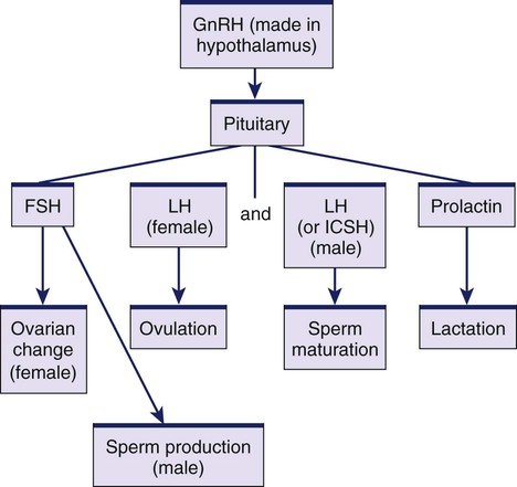
The hypothalamus, pituitary gland, and gonads secrete numerous hormones (Fig. 51-6). (Endocrine hormones are discussed in Chapter 48.) These hormones regulate the processes of ovulation, spermatogenesis (formation of sperm), and fertilization and the formation and function of the secondary sex characteristics. The hypothalamus secretes gonadotropin-releasing hormone (GnRH), which stimulates the anterior pituitary gland to secrete its hormones, including FSH and LH. LH in males is sometimes called interstitial cell–stimulating hormone (ICSH). The gonadal hormones are estrogen, progesterone, and testosterone.


In women, FSH production by the anterior pituitary stimulates the growth and maturity of the ovarian follicles necessary for ovulation.3 The mature follicle produces estrogen, which in turn suppresses the release of FSH. Another hormone, inhibin, is also secreted by the ovarian follicle and inhibits both GnRH and FSH secretion. In men, FSH stimulates the seminiferous tubules to produce sperm.


LH contributes to the ovulatory process because it causes follicles to complete maturation and undergo ovulation. It also affects the development of a ruptured follicle (area where ovum exited during ovulation), which turns into a corpus luteum from which progesterone is secreted. Progesterone maintains the rich vascular state of the uterus (secretory phase) in preparation for fertilization and implantation. In men, LH (or ICSH) triggers testosterone production by the interstitial cells of the testes and thus is essential for the full maturation of sperm.


Prolactin has no known function in men. In women, prolactin stimulates the development and growth of the mammary glands. During lactation, it initiates and maintains milk production.


The gonadal hormones, estrogen and progesterone, are produced by the ovaries in women. Small amounts of an estrogen precursor are also produced in the adrenal cortices. Estrogen is essential to the development and maintenance of the secondary sex characteristics, the proliferative phase of the menstrual cycle immediately after menstruation, and the uterine changes essential to pregnancy. The role and importance of estrogen in men are not well understood. In men, estrogen is produced predominantly in the adrenal cortex.


Progesterone plays a major role in the menstrual cycle but most specifically in the secretory phase. Like estrogen, progesterone is involved in the bodily changes associated with pregnancy. Adequate progesterone is necessary to maintain an implanted egg.


In men, the major gonadal hormone is testosterone, which is produced by the testes. Testosterone is responsible for the development and maintenance of secondary sex characteristics, as well as for adequate spermatogenesis. In women, androgens are produced in small amounts by the adrenal glands and ovaries.


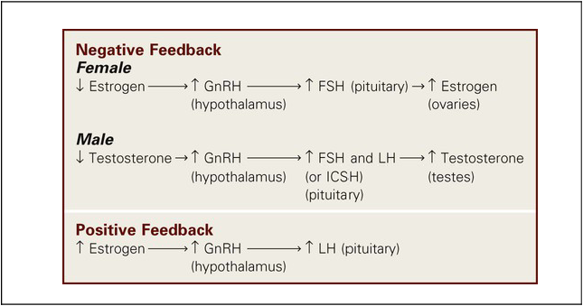
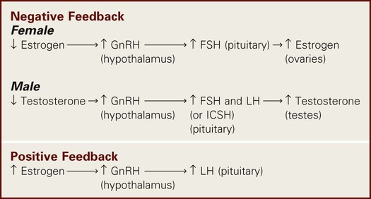
The circulating levels of gonadal hormones are controlled primarily by a negative feedback process. Receptors within the hypothalamus and pituitary are sensitive to the circulating blood levels of the hormones (Table 51-1). Increased levels of hormones stimulate a hypothalamic response to decrease the high circulating levels. Likewise, low circulating levels provoke a hypothalamic response that increases the low circulating levels. For example, if the circulating level of testosterone in men is low, the hypothalamus is stimulated to secrete GnRH. This triggers the anterior pituitary to secrete greater amounts of FSH and LH, which in turn causes an increase in the production of testosterone. The high level of testosterone signals a decrease in the production of GnRH and thus of FSH and LH.


In women, however, there is a slight variation. The circulating levels are controlled through a combination of both a negative and a positive feedback system. A negative feedback control mechanism exists similar to that described above. When circulating estrogen levels are low, the hypothalamus is stimulated to increase its production of GnRH. GnRH stimulates the pituitary to secrete greater amounts of FSH and LH, resulting in higher levels of estrogen production by the ovaries. Reciprocally higher levels of circulating estrogen result in a decreasing secretion of GnRH and thus a decrease in the secretion of FSH by the pituitary.


Women also have a positive feedback control mechanism. Thus with increasing levels of circulating estrogen, a greater level of GnRH is produced, resulting in an increased level of LH from the pituitary. Likewise, lowered levels of estrogen result in a lowered level of LH.


Menstrual Cycle


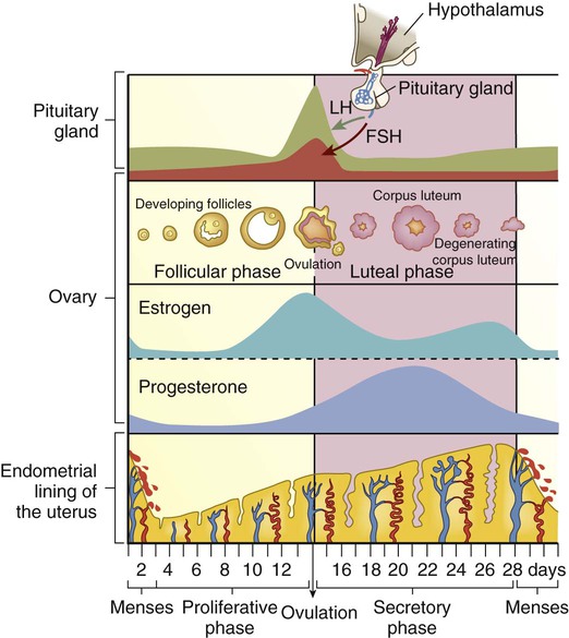
The major functions of the ovaries are ovulation and the secretion of hormones. These functions are accomplished during the normal menstrual cycle, a monthly process mediated by the hormonal activity of the hypothalamus, pituitary gland, and ovaries. Menstruation occurs during each month in which an egg is not fertilized (Fig. 51-7). The endometrial cycle is divided into three phases labeled in relation to uterine and ovarian changes: (1) the proliferative, or follicular, phase; (2) the secretory, or luteal, phase; and (3) the menstrual, or ischemic, phase. The length of the menstrual cycle ranges from 21 to 35 days, with an average of 28 days.4


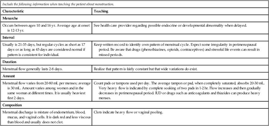
The menstrual cycle begins on the first day of menstruation, which usually lasts 4 to 6 days. Table 51-2 includes characteristics of the menstrual cycle and related teaching. During menstruation, estrogen and progesterone levels are low, but FSH levels begin to increase. During the follicular phase, a single follicle matures fully under the stimulation of FSH. (The mechanism that ensures that usually only one follicle reaches maturity is not known.) The mature follicle stimulates estrogen production, causing a negative feedback with resulting decreased FSH secretion.


TABLE 51-2


PATIENT TEACHING GUIDE
Characteristics of Menstruation


Include the following information when teaching the patient about menstruation.
Characteristic Teaching
Menarche
Occurs between ages 10 and 16 yr. Average age at onset is 12-13 yr. See health care provider regarding possible endocrine or developmental abnormality when delayed.
Interval
Usually is 21-35 days, but regular cycles as short as 17 days or as long as 45 days are considered normal if pattern is consistent for individual. Keep written record to identify own pattern of menstrual cycle. Expect some irregularity in perimenopausal period. Be aware that drugs (phenothiazines, opioids, contraceptives) and stressful life events can result in missed periods.
Duration
Menstrual flow generally lasts 2-8 days. Realize that pattern is fairly constant but that wide variations do exist.
Amount
Menstrual flow varies from 20-80 mL per menses; average is 30 mL. Amount varies among women and in the same woman at different times. It is usually heaviest first 2 days. Count pads or tampons used per day. The average tampon or pad, when completely saturated, absorbs 20-30 mL. Very heavy flow is indicated by complete soaking of two pads in 1-2 hr. Flow increases and then gradually decreases in perimenopausal period. IUD or drugs such as anticoagulants and thiazides can produce heavy menses.
Composition
Menstrual discharge is mixture of endometrium, blood, mucus, and vaginal cells. It is dark red and less viscous than blood and usually does not clot. Clots indicate heavy flow or vaginal pooling.

image


IUD, Intrauterine device.


Although the initial stage of follicular maturation is stimulated by FSH, complete maturation and ovulation occur only in the presence of LH. When estrogen levels peak on about the twelfth day of the cycle, there is a surge of LH, which triggers ovulation a day or two later. After ovulation (maturation and release of an ovum), LH promotes the development of the corpus luteum.


The fully developed corpus luteum continues to secrete estrogen and initiates progesterone secretion. If fertilization occurs, high levels of estrogen and progesterone continue to be secreted because of the continued activity of the corpus luteum from stimulation by human chorionic gonadotropin (hCG). If fertilization does not take place, menstruation occurs because of a decrease in estrogen production and progesterone withdrawal.


During the follicular phase, the endometrial lining of the uterus also undergoes change. As more estrogen is produced, the endometrial lining undergoes proliferative changes, including an increase in the length of blood vessels and glandular tissue.


With ovulation and the resulting increased levels of progesterone, the luteal (or secretory) phase begins. If the corpus luteum regresses (when fertilization does not occur) and estrogen and progesterone levels fall, the endometrial lining can no longer be supported. As a result, the blood vessels contract, and tissue begins to slough (fall away). This sloughing results in the menses and the start of the menstrual phase.4


Phases of Sexual Response


The sexual response is a complex interplay of psychologic and physiologic phenomena and is influenced by a number of variables (e.g., stress, illness). The changes that occur during sexual excitement are similar for men and women. Masters and Johnson described the sexual response in terms of the excitement, plateau, orgasmic, and resolution phases.5


Male Sexual Response.

The penis and the urethra are essential to the transport of sperm into the vagina and the cervix during intercourse. This transport is facilitated by penile erection in response to sexual stimulation during the excitement phase. Erection results from the filling of the large venous sinuses within the erectile tissue of the penis. In the flaccid state the sinuses hold only a small amount of blood, but during the erection stage they are congested with blood. Because the penis is richly endowed with sympathetic, parasympathetic, and pudendal nerve endings, it is readily stimulated to erection. The loose skin of the penis becomes taut as a result of the venous congestion. This erectile tautness allows for easy insertion into the vagina.


As the man reaches the plateau phase, the erection is maintained, and the penis increases in diameter as a result of a slight increase in vasocongestion. Testicle size also increases. Sometimes the glans penis becomes more reddish purple.


The subsequent contraction of the penile and urethral musculature during the orgasmic phase propels the sperm outward through the meatus. In this process, termed ejaculation, sperm are released into the ductus deferens during contractions. Sperm advance through the urethra, where fluids from the prostate and seminal vesicles are added to the ejaculate. The sperm continue their path through the urethra, receiving a small amount of fluid from the Cowper’s glands, and are finally ejaculated through the urinary meatus. Orgasm is characterized by the rapid release of the vasocongestion and muscular tension (myotonia) that have developed. The rapid release of muscular tension (through rhythmic contractions) occurs primarily in the penis, prostate gland, and seminal vesicles. After ejaculation, a man enters the resolution phase. The penis undergoes involution, gradually returning to its unstimulated, flaccid state.


Female Sexual Response.

The changes that occur in a woman during sexual excitation are similar to those in a man. In response to stimulation, the clitoris becomes congested and vaginal lubrication increases from secretions from the cervix, Bartholin’s glands, and vaginal walls. This initial response is the excitation phase.


As excitation is maintained in the plateau phase, the vagina expands and the uterus is elevated. In the orgasmic phase, contractions occur in the uterus from the fundus to the lower uterine segment. There is a slight relaxation of the cervical os, which helps the entrance of the sperm, and rhythmic contractions of the vagina. Muscular tension is rapidly released through rhythmic contractions in the clitoris, vagina, and uterus. This phase is followed by a resolution phase in which these organs return to their preexcitation state. However, women do not have to go through the resolution (refractory) recovery state before they can be orgasmic again. They can be multiorgasmic without resolution between orgasms.


Gerontologic Considerations


Effects of Aging on Reproductive Systems


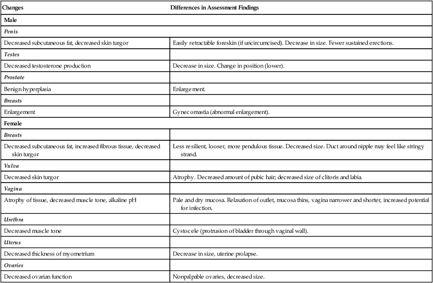
With advancing age, changes occur in the male and female reproductive systems (Table 51-3). In women many of these changes are related to the altered estrogen production that is associated with menopause.6 A reduction in circulating estrogen along with an increase in androgens in postmenopausal women is associated with breast and genital atrophy, reduction in bone mass, and increased rate of atherosclerosis. Vaginal dryness may occur, which can lead to urogenital atrophy and changes in the composition of vaginal secretions.


A gradual testosterone decline in older men occurs. Manifestations of hormonal decline in men can be physical, psychologic, or sexual. Some of the changes include an increase in prostate size and a decrease in testosterone level, sperm production, muscle tone of the scrotum, and size and firmness of the testicles. Erectile dysfunction and sexual dysfunction occur in some men as a result of these changes.7


Gradual changes resulting from advancing age occur in the sexual responses of men and women. The cumulative effects of these changes, as well as the negative social attitude toward sexuality in older adults, can affect the sexual practices of older adults. You have an important role in providing accurate and unbiased information about sexuality and age. Emphasize the normalcy of sexual activity in older adults.


Assessment of Male and Female Reproductive Systems


Subjective Data


Important Health Information.

In addition to general health information, elicit information specifically relating to the reproductive system. Reproduction and sexual issues are often considered extremely personal and private. Develop trust with the patient to elicit such information. A professional demeanor is important when taking a reproductive or sexual history. Be sensitive, use gender-neutral terms when asking about partners, and maintain an awareness of a patient’s culture and beliefs. Begin with the least sensitive information (e.g., menstrual history) before asking questions about more sensitive issues such as sexual practices or STIs.


Past Health History.

The past health history should include information about major illnesses, hospitalizations, immunizations, and surgeries. Inquire about any infections involving the reproductive system, including STIs. Also take a complete obstetric and gynecologic history from the female patient.


Common pediatric illnesses that affect reproductive function are mumps and rubella. The occurrence of mumps in young men has been associated with an increase in sterility. Bilateral testicular atrophy may occur secondary to mumps-related orchitis. In the health history ask male patients if they have had mumps, been immunized with the mumps vaccine, or have any indications of sterility.


Rubella is of primary concern to women of childbearing age. If rubella occurs during the first 3 months of pregnancy, the possibility of congenital anomalies is increased. For this reason, you should encourage immunization for all women of childbearing age who have not been immunized for rubella or have not already had the disease. (Rubella immunity can be determined by antibody titers.) However, women should not be immunized if they are already pregnant.8 Advise women not to conceive for at least 3 months after immunization.


Question the patient regarding current health status and any acute or chronic health problems. Problems in other body systems are often related to problems with the reproductive system. Ask questions relating to possible endocrine disorders, particularly diabetes mellitus (DM), hypothyroidism, and hyperthyroidism, because these disorders directly interfere with women’s menstrual cycles and with sexual performance. Men who have DM may experience erectile dysfunction and retrograde ejaculation. In women with uncontrolled DM, pregnancy may have significant health risks. Many other chronic illnesses such as cardiovascular disease, respiratory disorders, anemia, cancer, and kidney and urinary tract disorders may affect the reproductive system and sexual functioning.


Determine if the patient has a history of a stroke. In men, strokes may cause physiologic or psychologic erectile dysfunction. Men who have suffered a myocardial infarction (MI) may experience erectile dysfunction because of fear that sexual activity could precipitate another MI. Women share this concern, both as partners of someone who has had an MI and as patients recovering from an MI. Although most patients have concerns about sexual activity after an MI, many are not comfortable expressing these fears to the nurse. Be sensitive to this concern. In women, a history of cardiovascular disease (e.g., hypertension, thrombophlebitis, angina) is associated with a higher incidence of morbidity and mortality with pregnancy or the use of oral contraceptives.

Stay updated, free articles. Join our Telegram channel

Nov 17, 2016 | Posted by in NURSING | Comments Off on Nursing Assessment: Reproductive System

Full access? Get Clinical Tree

Get Clinical Tree app for offline access