Chapter 7 Multi-infarct Dementia (MID); Dementia of the Alzheimer Type (DAT) Lou Gehrig’s Disease; Motor Neuron Disease; Progressive Bulbar Palsy; Progressive Muscular Atrophy
Neurological Care Plans
Alzheimer’s Disease/Dementia
 For additional care plans, go to http://evolve.elsevier.com/Gulanick/.
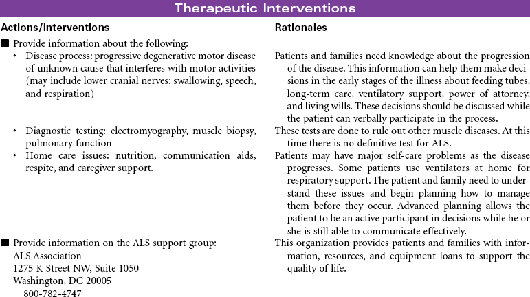
Amyotrophic Lateral Sclerosis
Neurological Care Plans
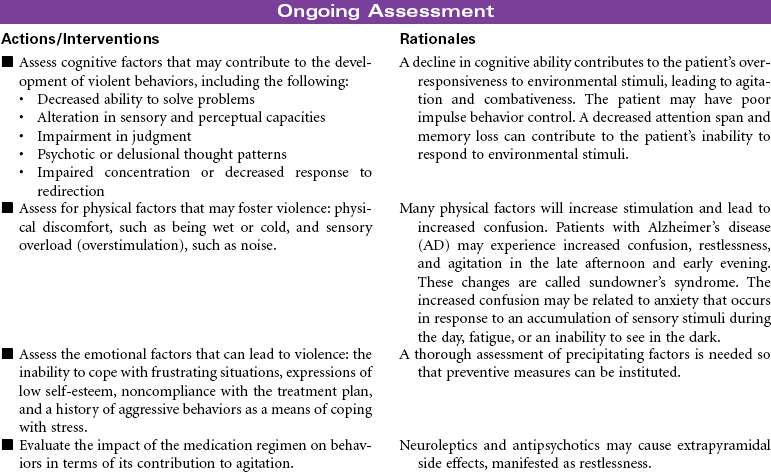
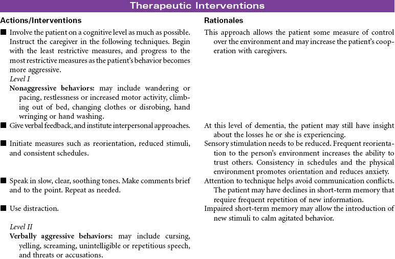
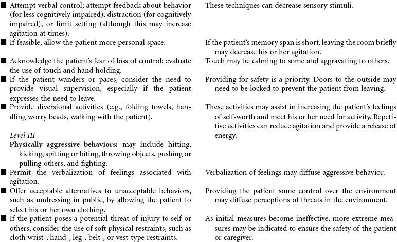
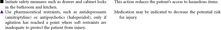
 Risk for Violence: Self-Directed or Other-Directed
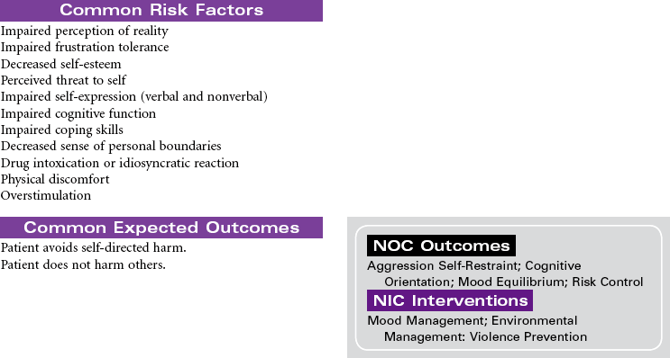
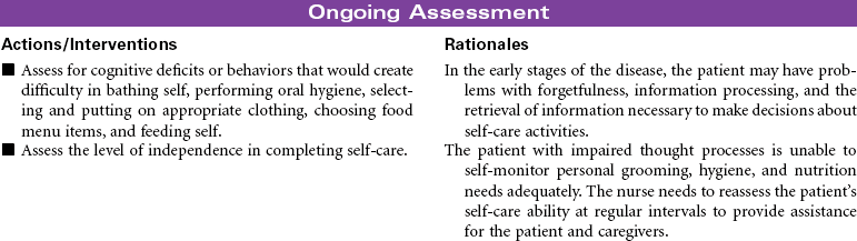
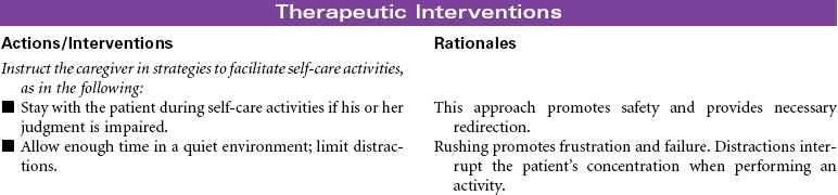
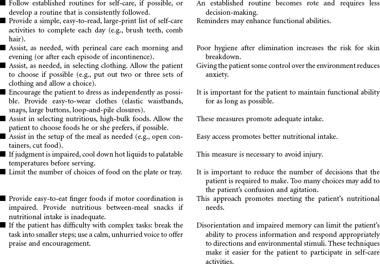
 Self-Care Deficit: Bathing, Dressing, Feeding
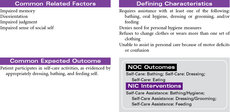
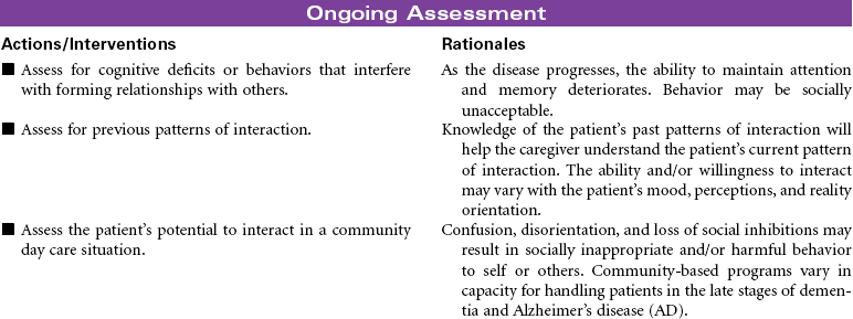
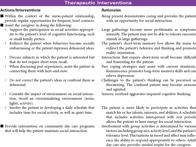
 Impaired Social Interaction
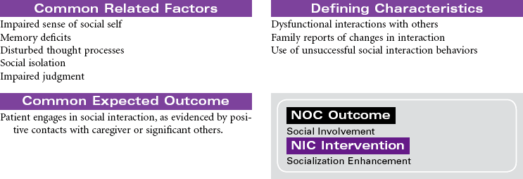
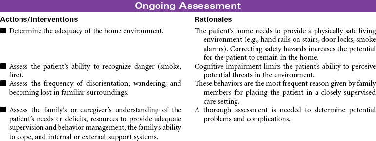
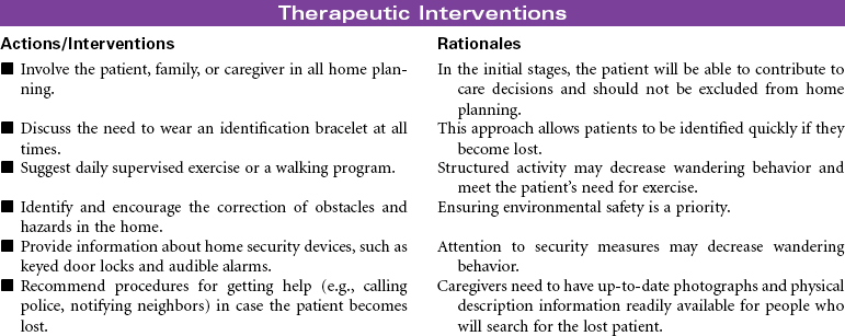
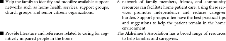
 Impaired Home Maintenance
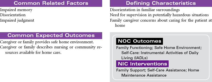
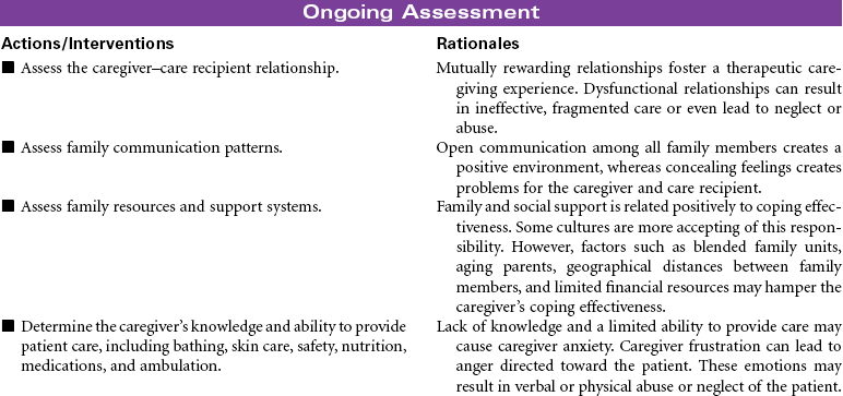
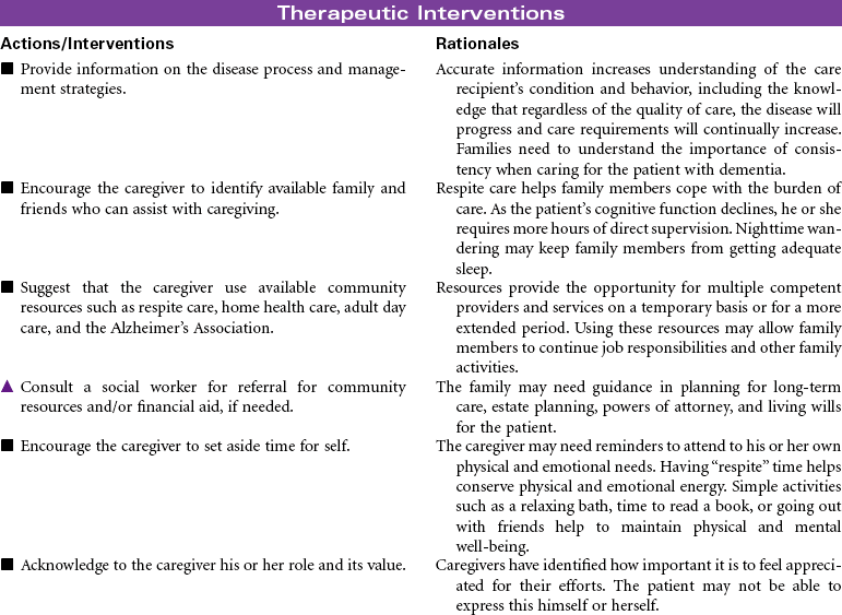
 Caregiver Role Strain
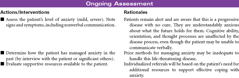
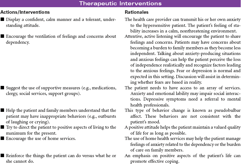
 Anxiety
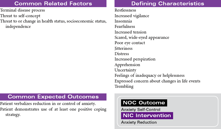
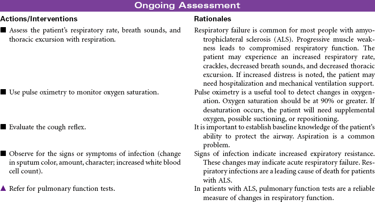
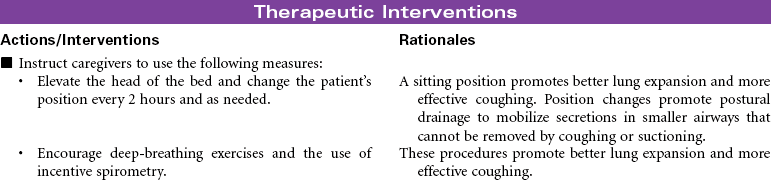
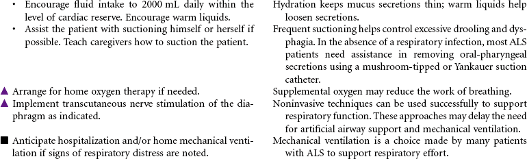
 Ineffective Airway Clearance
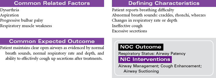
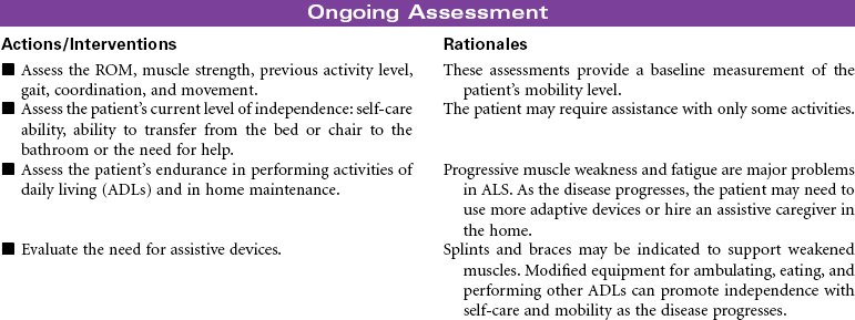
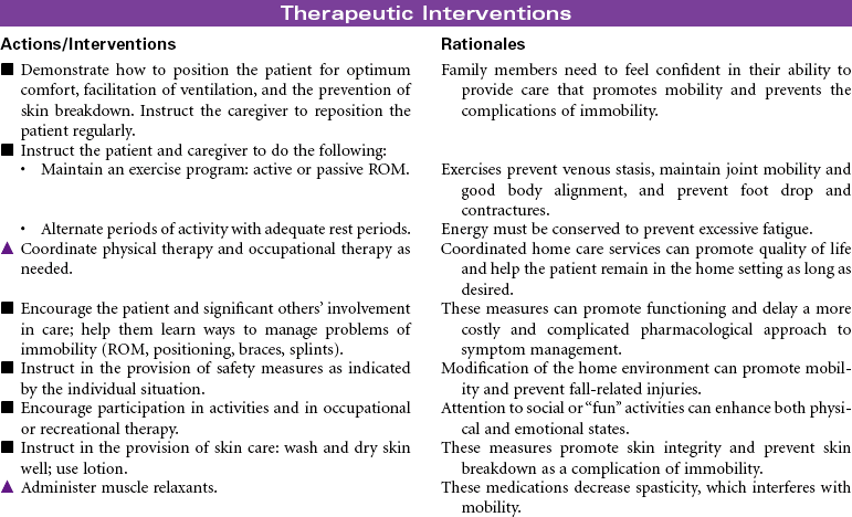
 Impaired Physical Mobility
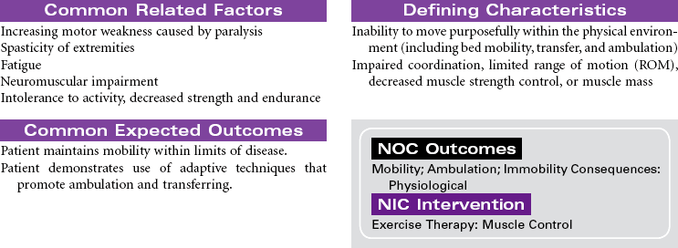
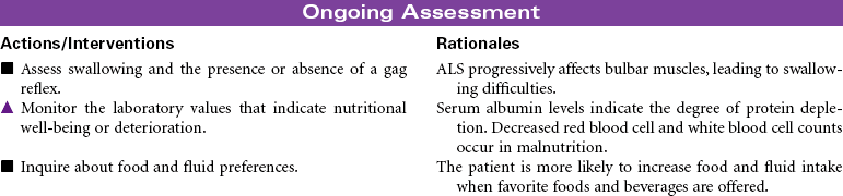
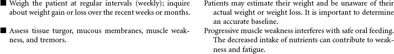
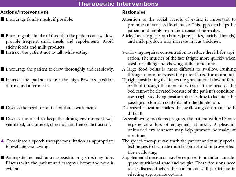
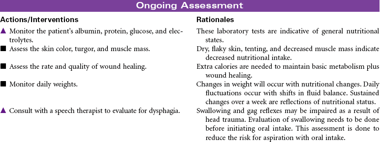
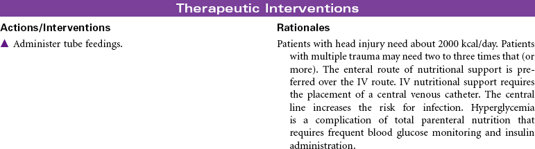
 Imbalanced Nutrition: Less Than Body Requirements
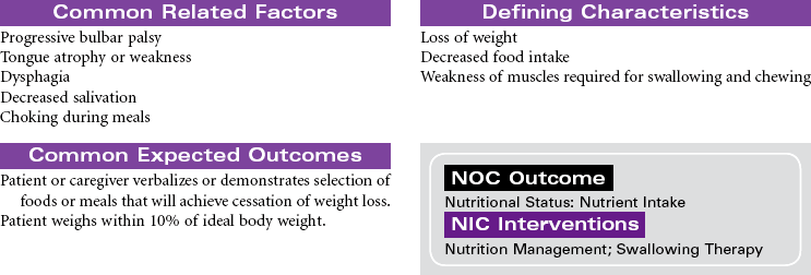
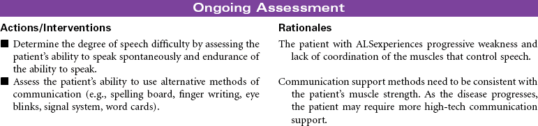
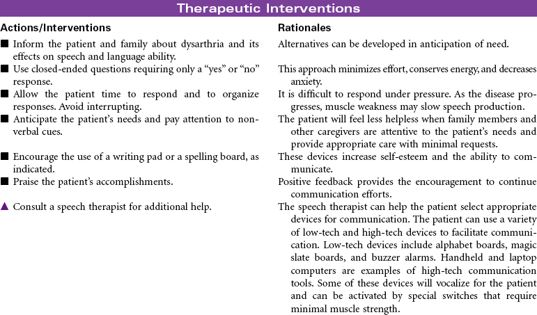
 Impaired Verbal Communication
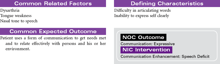
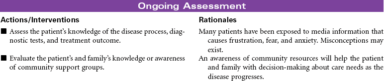
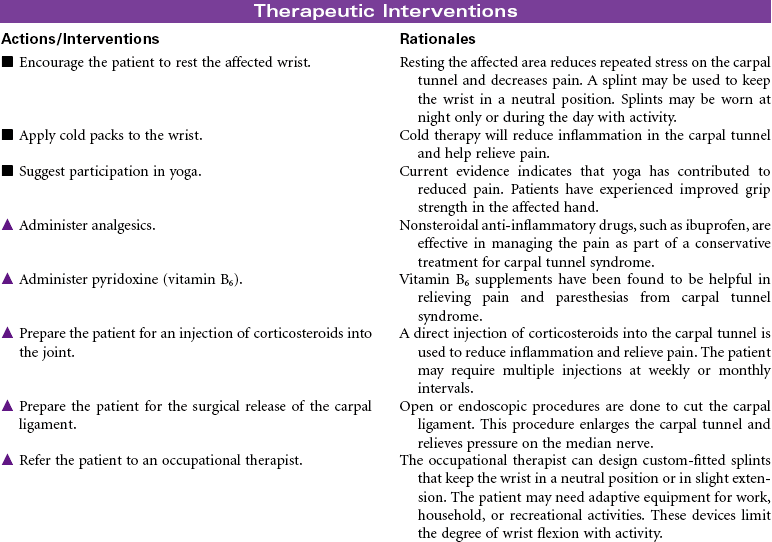
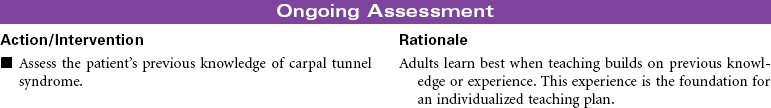
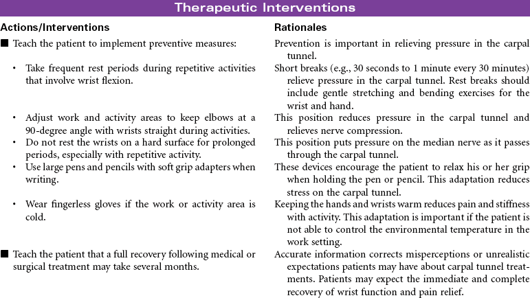
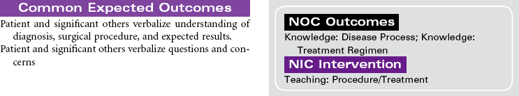
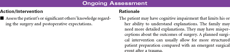
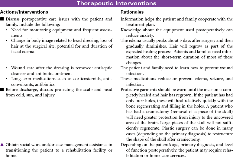
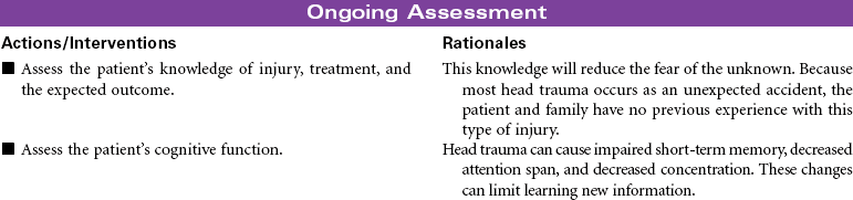
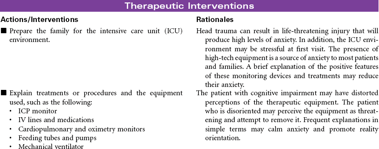
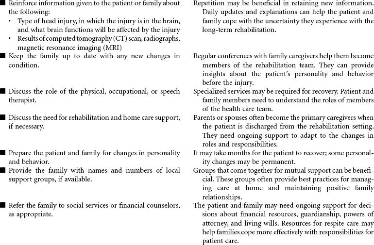
 Deficient Knowledge
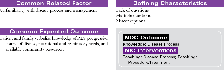
 = Independent
  = Collaborative
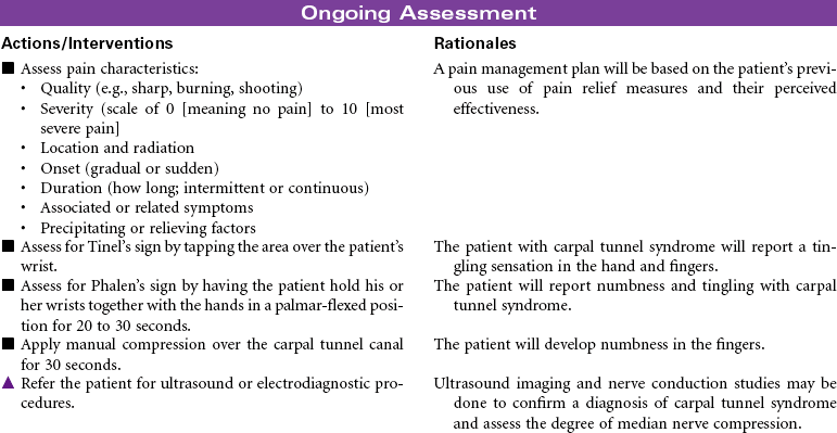
 Acute Pain
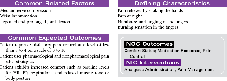
 Deficient Knowledge
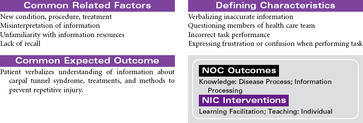
 = Independent
  = Collaborative
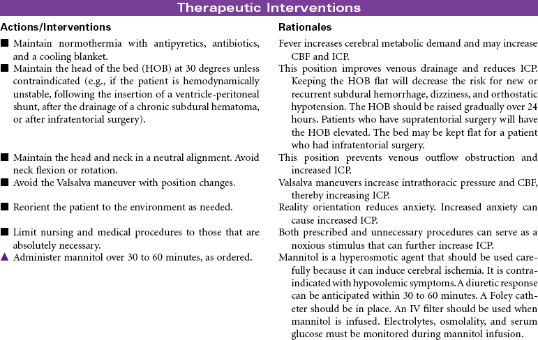
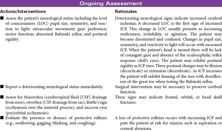
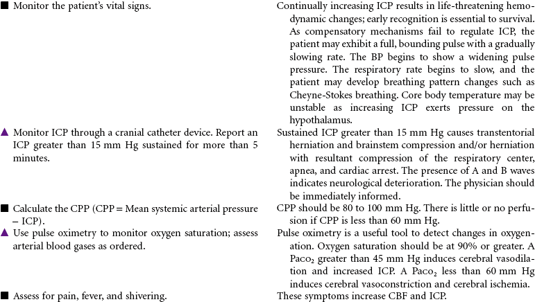
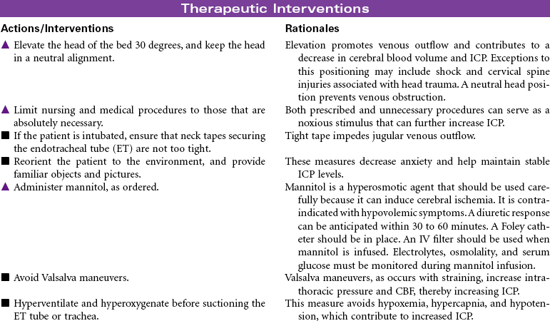
 Decreased Intracranial Adaptive Capacity
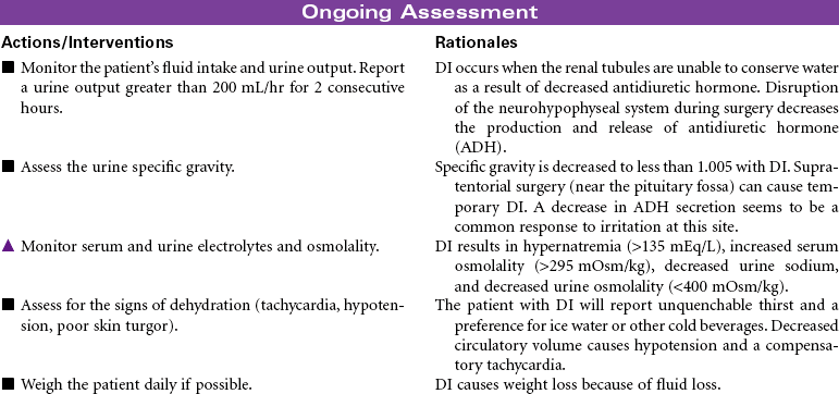
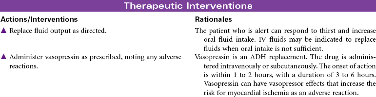
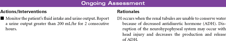
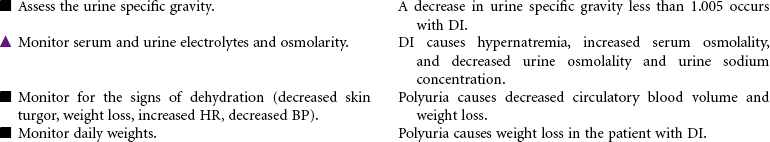
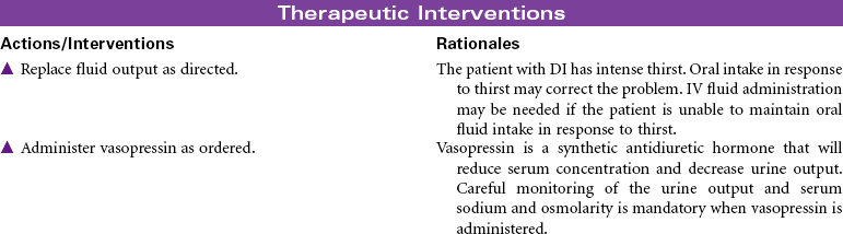
 Risk for Deficient Fluid Volume
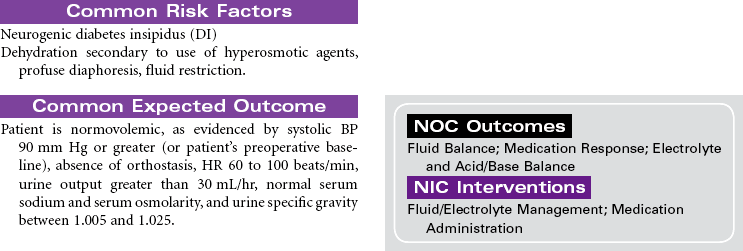
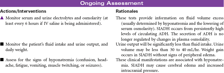
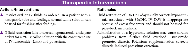
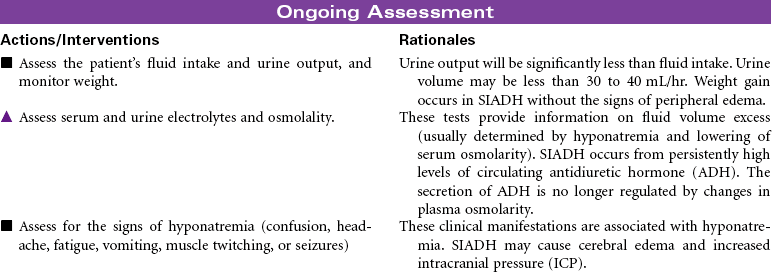
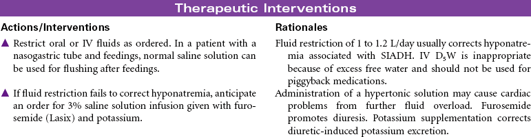
 Risk for Excess Fluid Volume
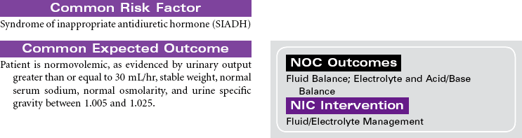
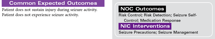
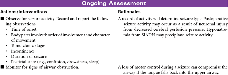
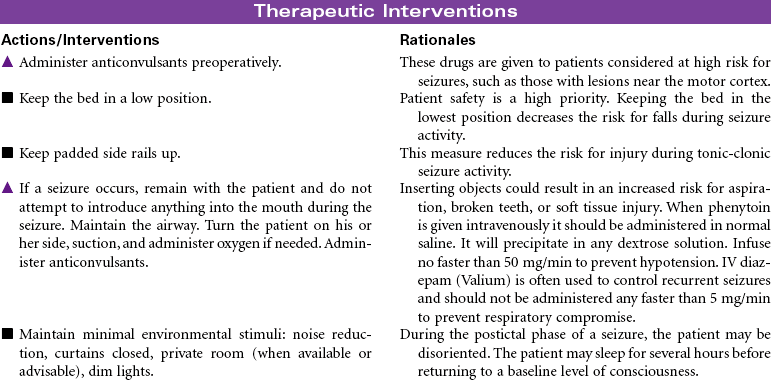
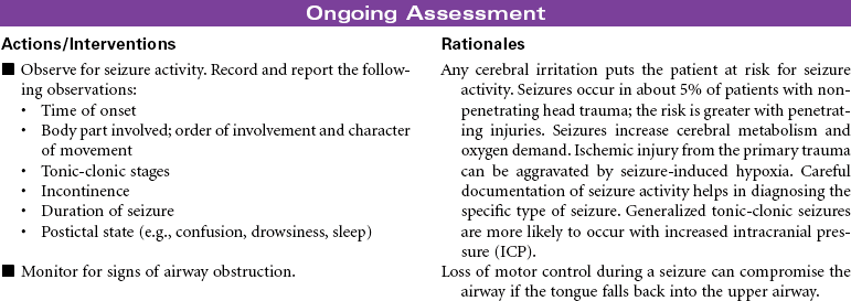
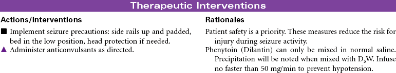
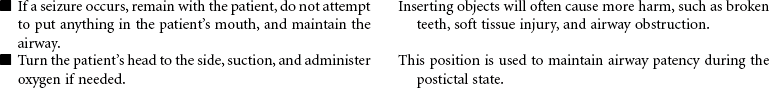
 Risk for Seizures
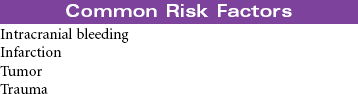
 Deficient Knowledge


 Decreased Intracranial Adaptive Capacity
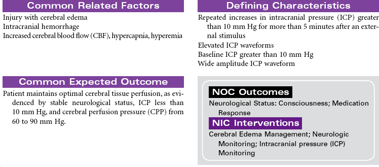
 Risk for Deficient Fluid Volume
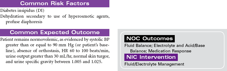
 Risk for Excess Fluid Volume
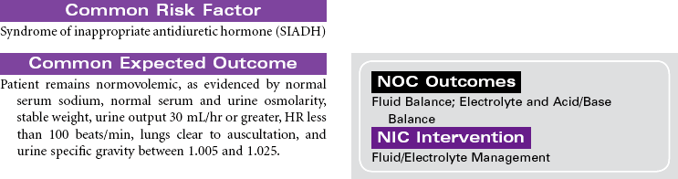
 Risk for Seizures
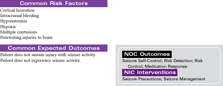
 Risk for Imbalanced Nutrition: Less Than Body Requirements
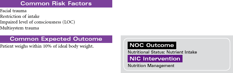
 Deficient Knowledge
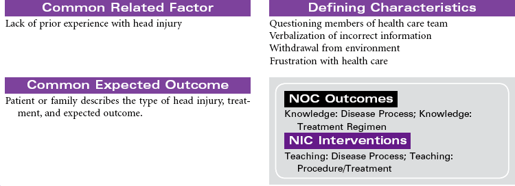
 Deficient Knowledge
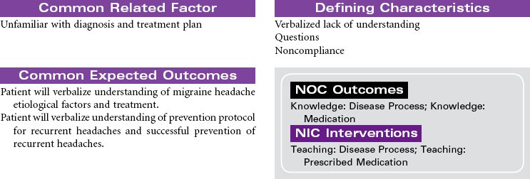


Get Clinical Tree app for offline access
