Chapter 5
Cardiac and Vascular Care Plans
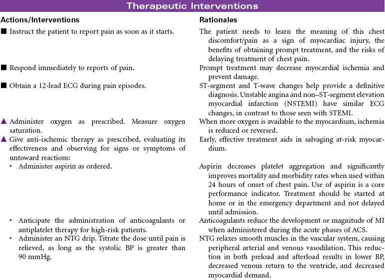
Acute Coronary Syndromes/Myocardial Infarction
 For additional care plans and an Online Care Plan Constructor, go to http://evolve.elsevier.com/Gulanick/.
Nurse Key
Fastest Nurse Insight Engine
 = Independent
  = Collaborative
 Acute Pain
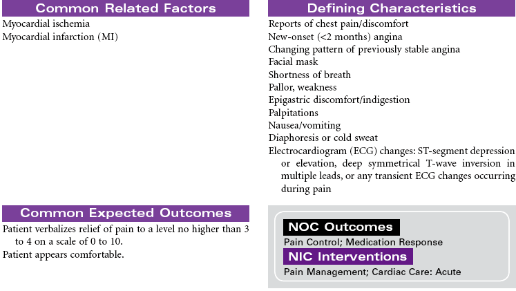
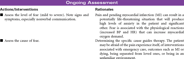
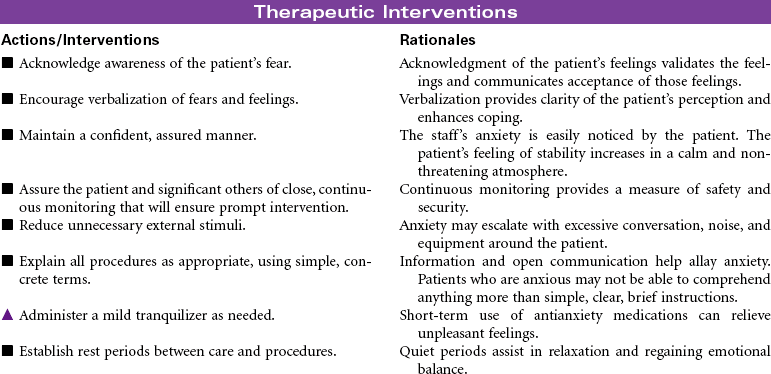
 Fear
Fear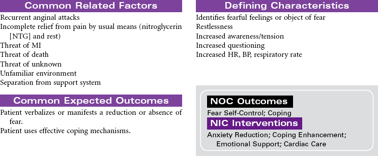
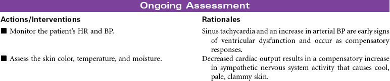
 Risk for Decreased Cardiac Output
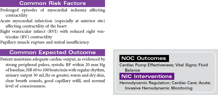
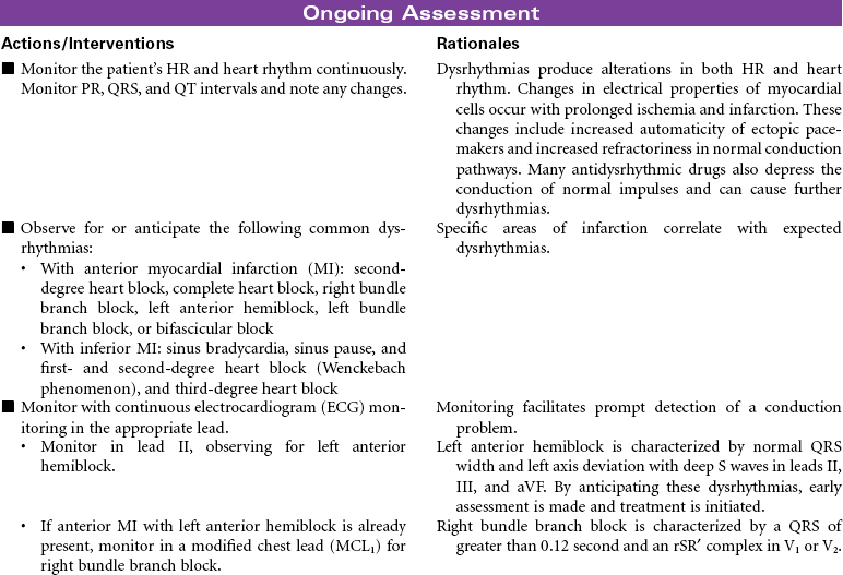
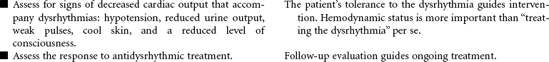
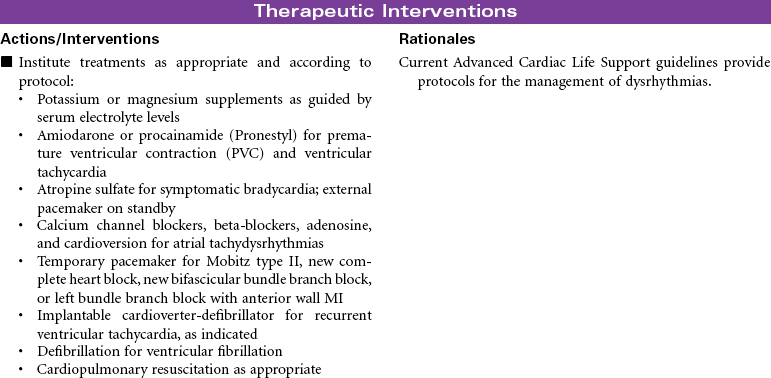
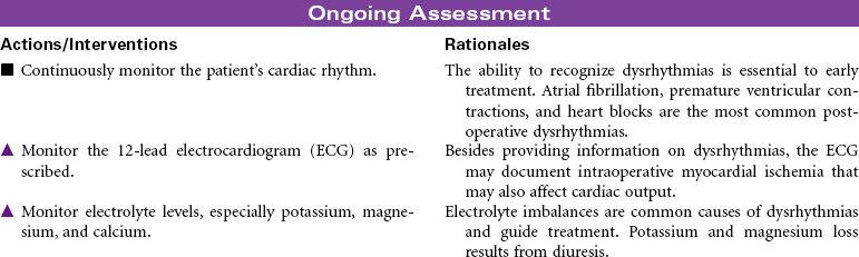
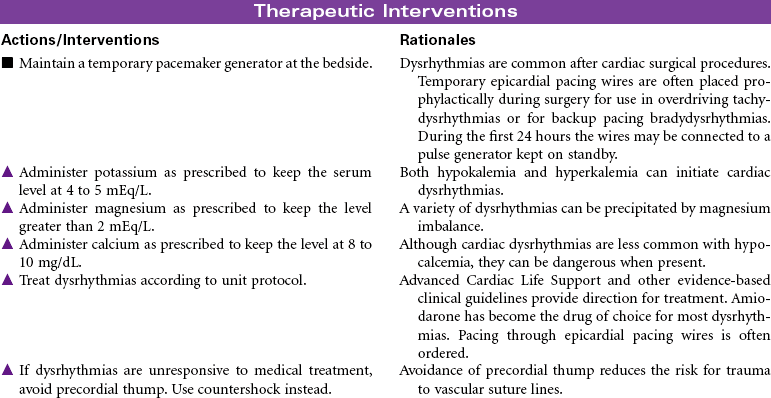
 Risk for Decreased Cardiac Output: Dysrhythmias
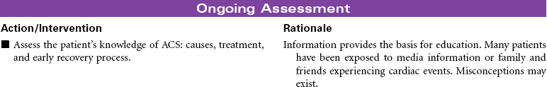
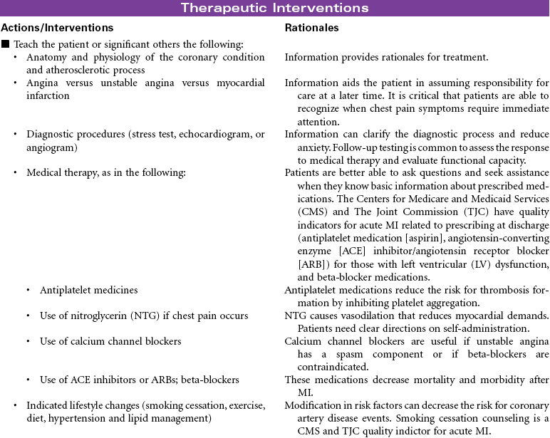
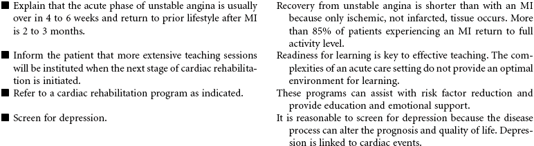
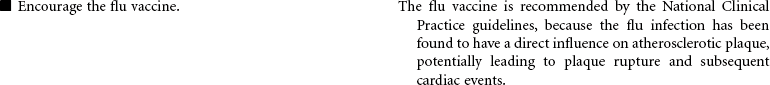
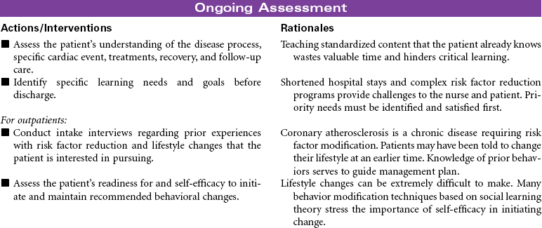
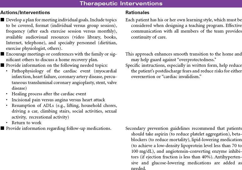
 Deficient Knowledge

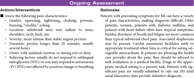
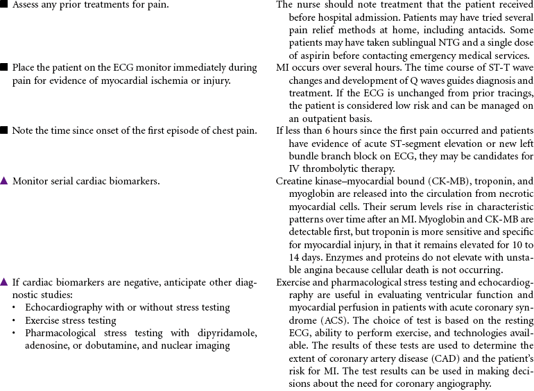
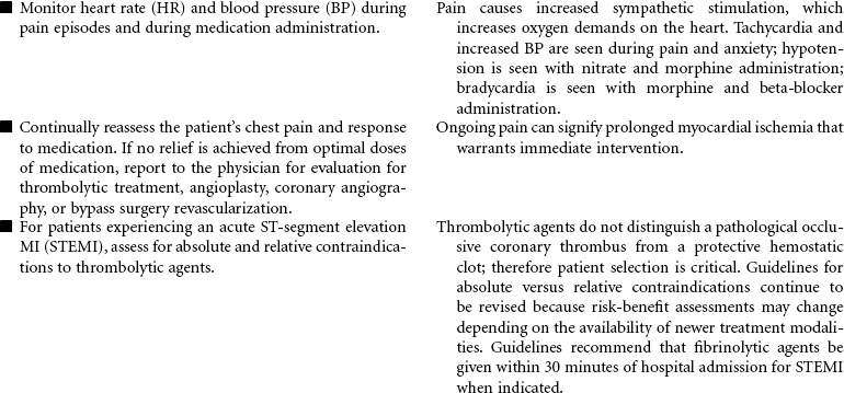
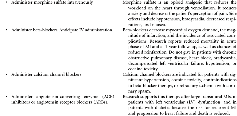
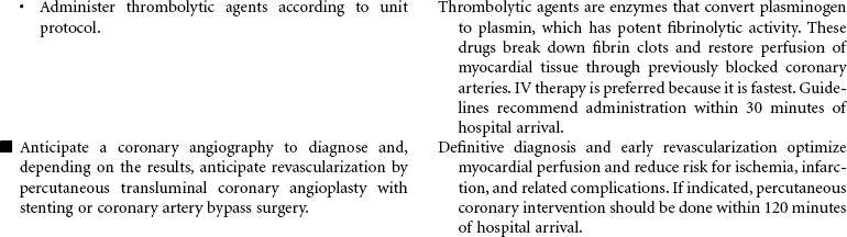
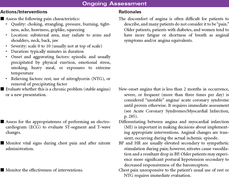
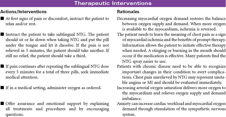
 Acute Pain
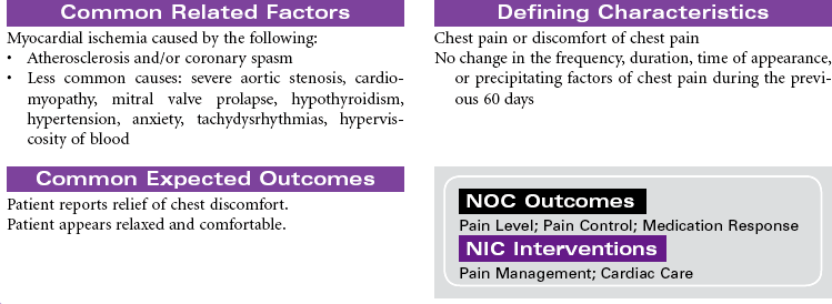
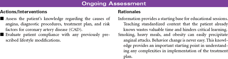
 Deficient Knowledge
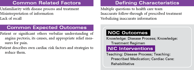

 Activity Intolerance
 Deficient Knowledge
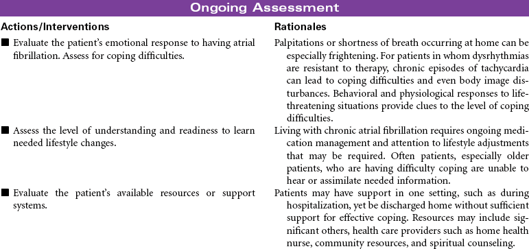
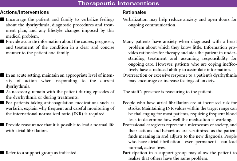
 Risk for Ineffective Coping
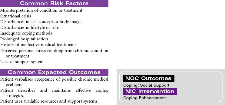

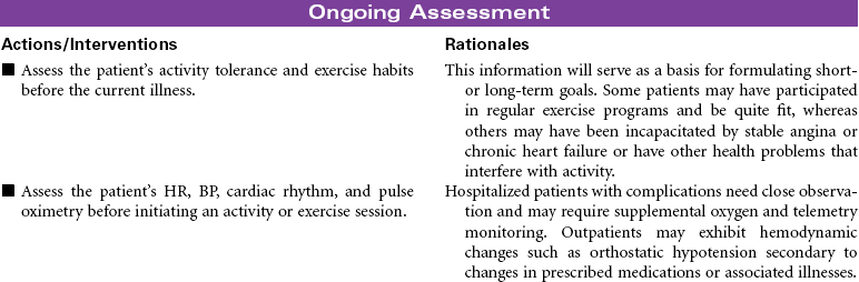
 Activity Intolerance
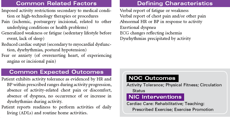
 Deficient Knowledge
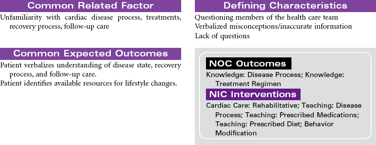
 Risk for Ineffective Coping
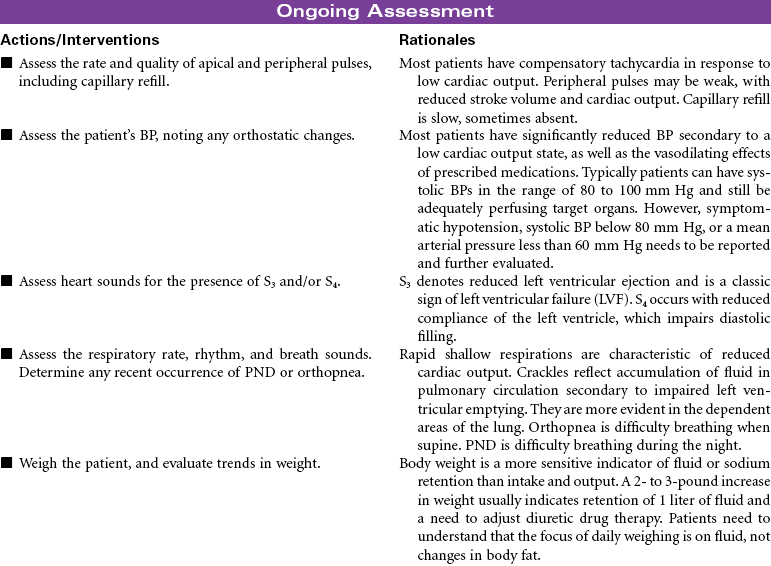
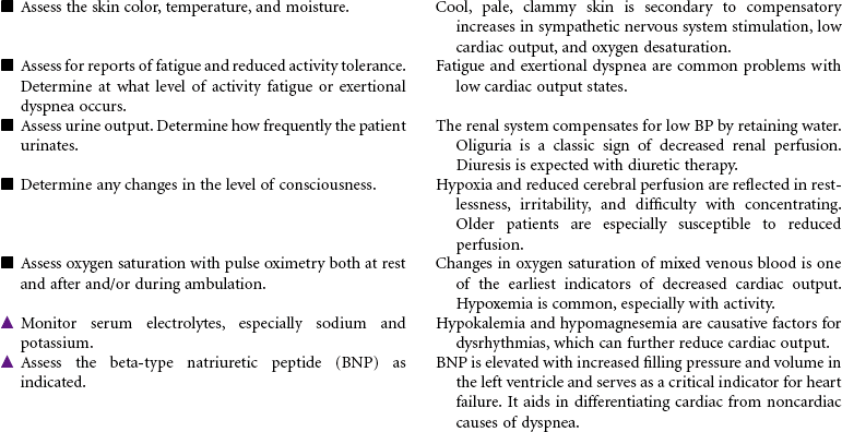
 Decreased Cardiac Output
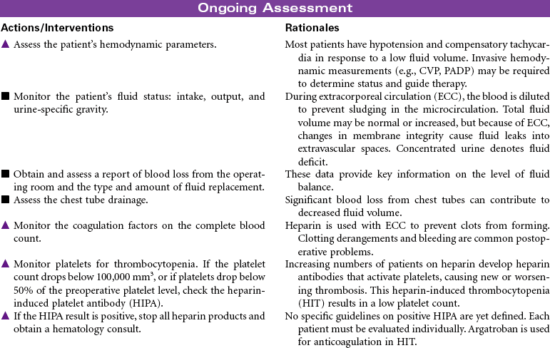
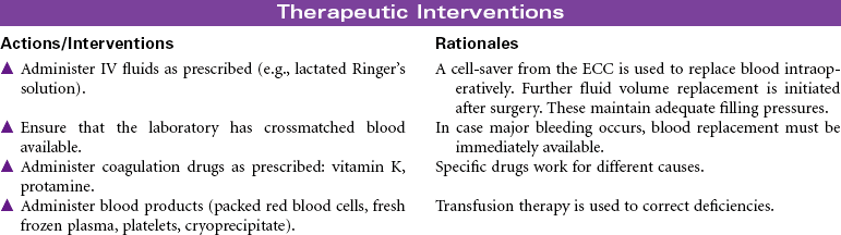
 Deficient Fluid Volume
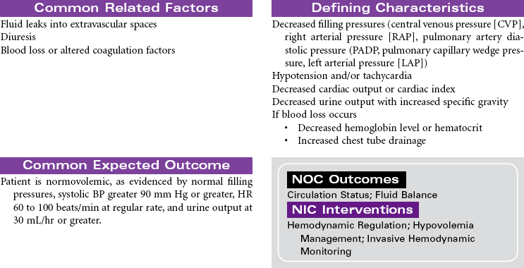
 Risk for Decreased Cardiac Output: Dysrhythmias
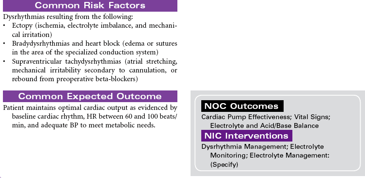
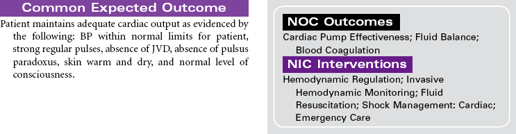
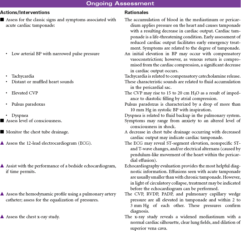
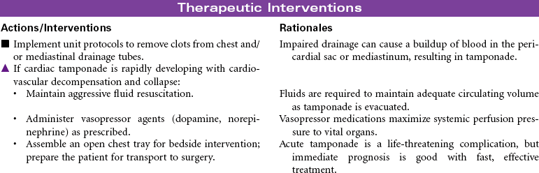
 Decreased Cardiac Output: Cardiac Tamponade
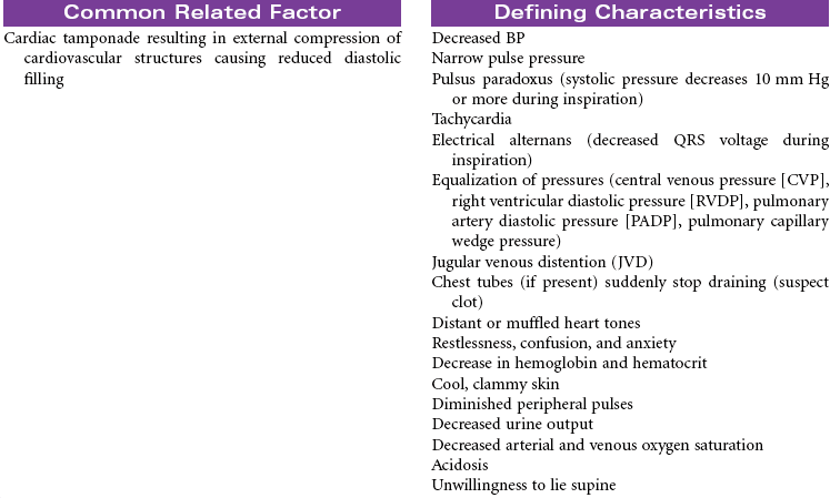
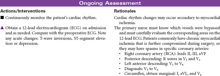
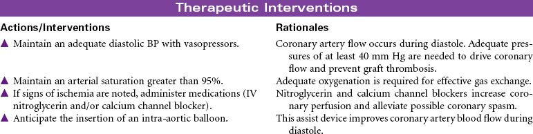
 Risk for Ineffective Myocardial Tissue Perfusion
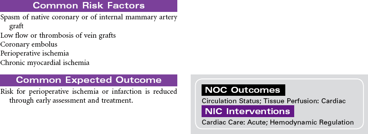
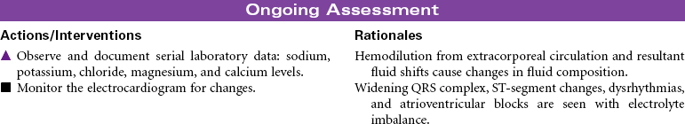
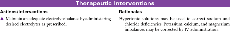
 Risk for Electrolyte Imbalance
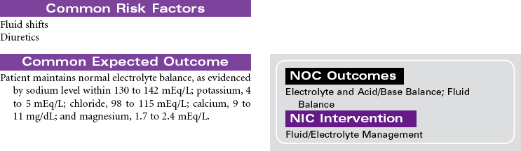
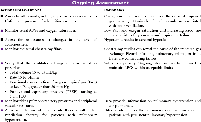
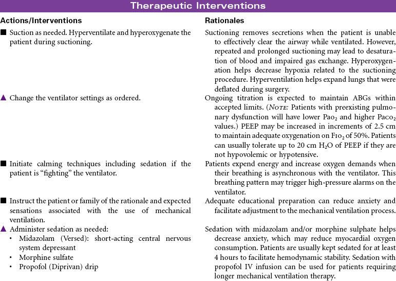
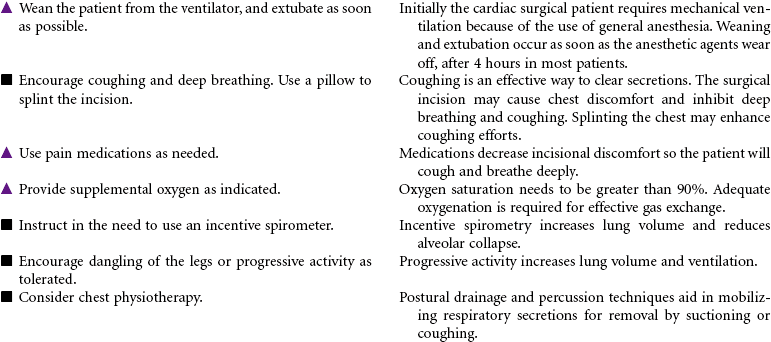
 Risk for Impaired Gas Exchange
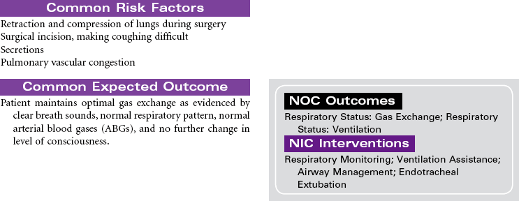
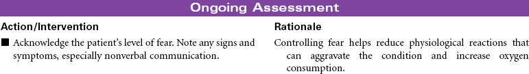
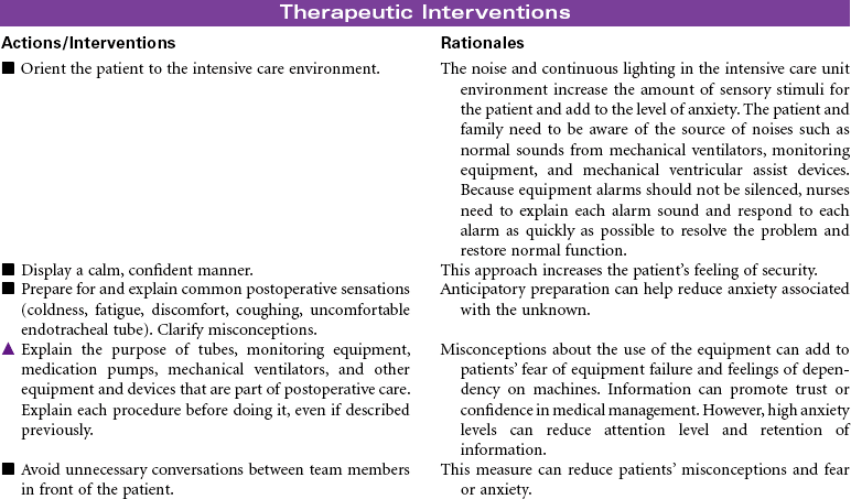
 Fear
Fear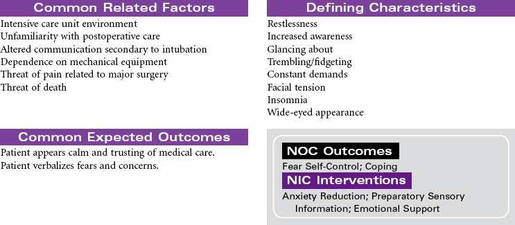

 Decreased Cardiac Output
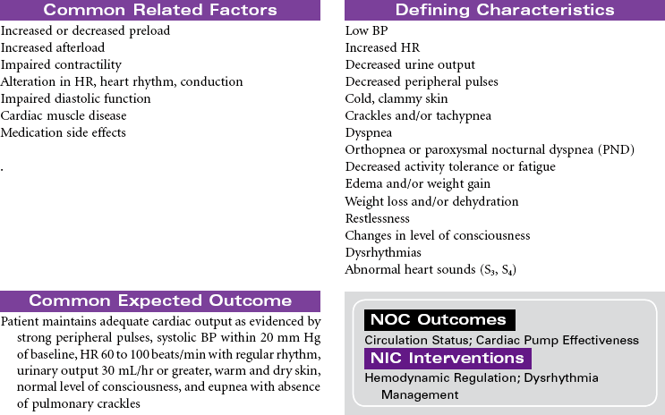
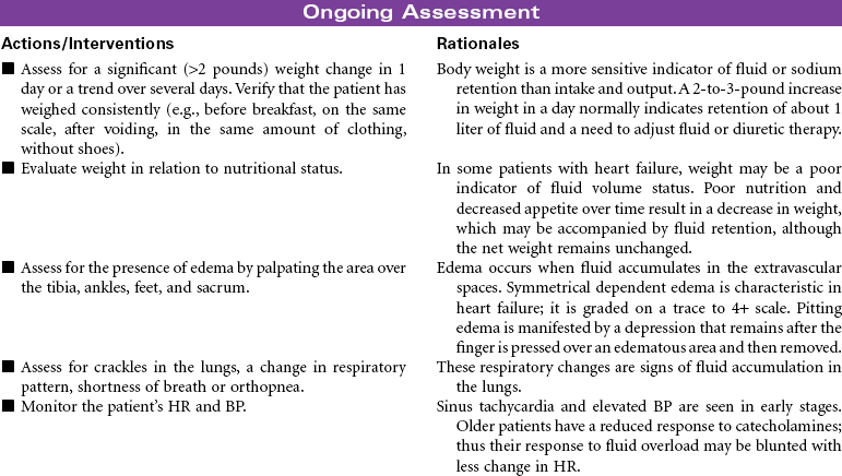
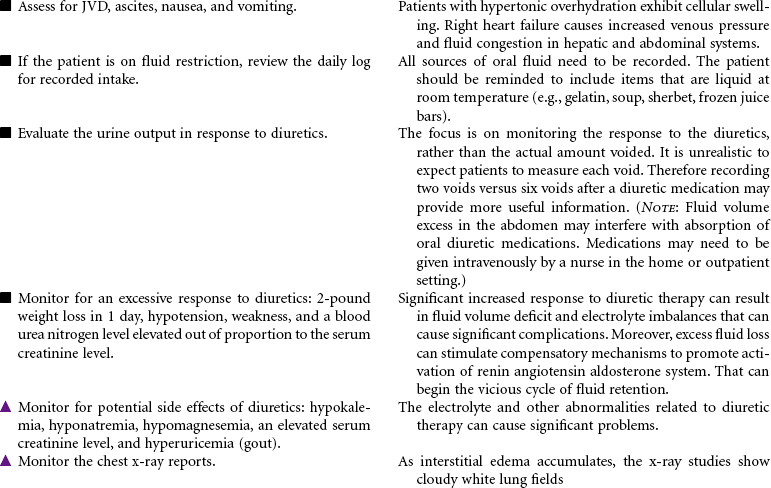
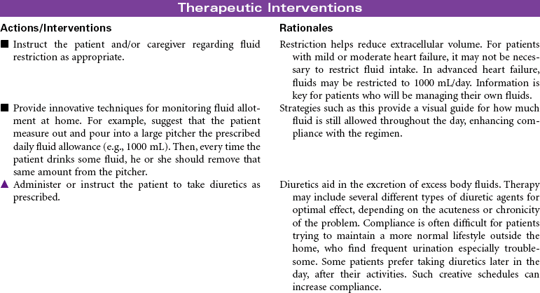
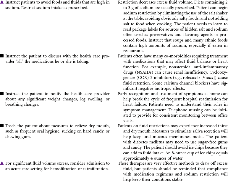
 Excess Fluid Volume
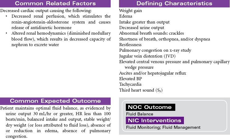
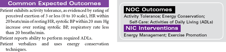
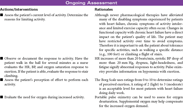
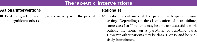
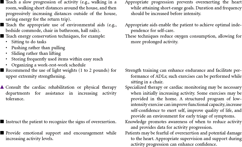
 Activity Intolerance
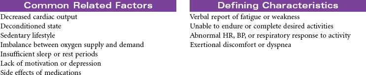
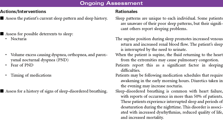
 Insomnia
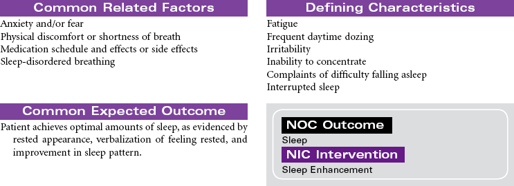
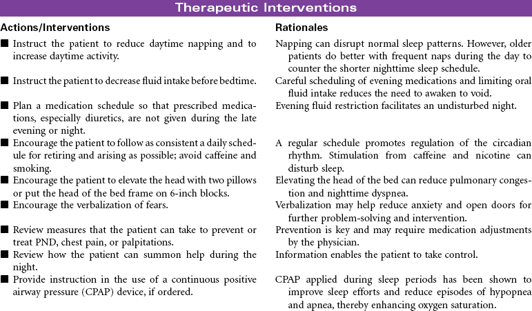
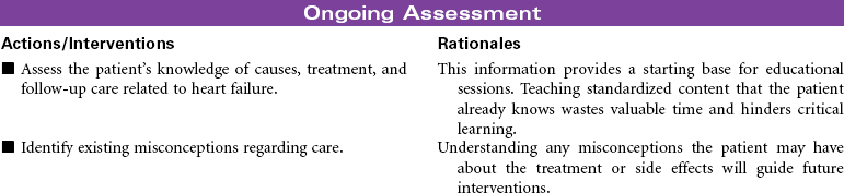
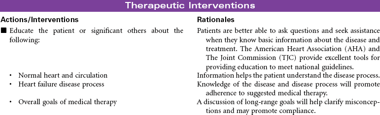
 Deficient Knowledge
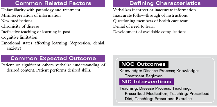


Get Clinical Tree app for offline access
