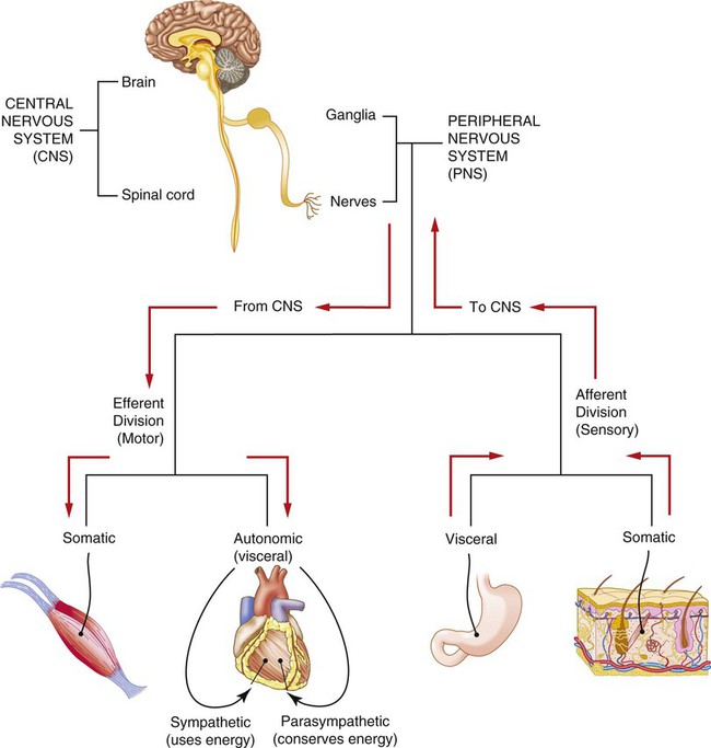
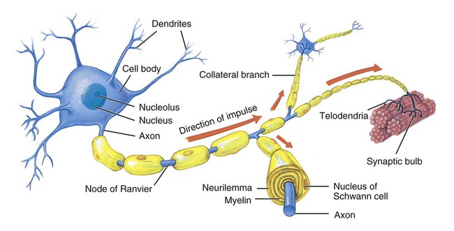
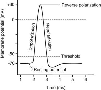
1. Describe the organization and functions of the nervous system. 2. Describe the structure and functions of neurons and neuroglia. 3. Explain how an impulse is conducted along the length of a neuron. 4. List and describe the three layers of meninges around the central nervous system. 5. Describe the location, components, and functional areas of the cerebrum, diencephalon, brain stem, and cerebellum. 6. Compare the composition of gray matter and white matter. 7. Explain the function of cerebrospinal fluid. 8. Describe the structure and functions of the spinal cord. 9. Explain the difference in composition of sensory, motor, and mixed nerves. 10. List the 12 cranial nerves, and state the function of each. 11. Identify the region of the body that is innervated by each of the following spinal nerve plexuses: cervical, brachial, lumbosacral. 12. Explain the difference between the sympathetic and parasympathetic divisions of the autonomic nervous system. 13. Describe ways in which the aging of an individual affects the nervous system. There is really only one nervous system in the body, although terminology seems to indicate otherwise. Although each subdivision of the system is also called a “nervous system,” all of these smaller systems belong to the single, highly integrated nervous system. Each subdivision has structural and functional characteristics that distinguish it from the others. The nervous system as a whole is divided into two subdivisions: the central nervous system (CNS) and the peripheral nervous system (PNS) (Figure 9-1). Each neuron has three basic parts: Figure 9-2 illustrates a typical neuron. The main part of the neuron is the cell body or soma. In many ways the cell body is similar to other types of cells. It has a nucleus with at least one nucleolus and contains many of the typical cytoplasmic organelles. It lacks centrioles, however. Because centrioles function in cell division, the fact that neurons lack these organelles is consistent with the amitotic nature of the cell. An axon may have infrequent branches called axon collaterals. Axons and axon collaterals terminate in many short branches or telodendria (tell-oh-DEN-dree-ah). The distal ends of the telodendria are slightly enlarged to form synaptic bulbs. Many axons are surrounded by a segmented, white, fatty substance called myelin (MY-eh-lin) or the myelin sheath. Myelinated fibers make up the white matter in the CNS, whereas cell bodies and unmyelinated fibers make up the gray matter. The unmyelinated regions between the myelin segments are called the nodes of Ranvier (nodes of ron-vee-AY). In the PNS the myelin is produced by Schwann cells. The cytoplasm, nucleus, and outer cell membrane of the Schwann cell form a tight covering around the myelin and around the axon itself at the nodes of Ranvier. This covering is the neurilemma (noo-rih-LEM-mah), which plays an important role in the regeneration of nerve fibers. In the CNS, oligodendrocytes (ah-lee-go-DEN-droh-sites) produce myelin, but there is no neurilemma, which is why fibers within the CNS do not regenerate. The structure of an axon and its coverings is illustrated in Figure 9-2. Functionally, neurons are classified as afferent, efferent, or interneurons (association neurons) according to the direction in which they transmit impulses relative to the CNS (Table 9-1). Afferent, or sensory, neurons carry impulses from peripheral sense receptors to the CNS. They usually have long dendrites and relatively short axons. Efferent, or motor, neurons transmit impulses from the CNS to effector organs, such as muscles and glands. Efferent neurons usually have short dendrites and long axons. Interneurons, or association neurons, are located entirely within the CNS, where they form the connecting link between the afferent and efferent neurons. They have short dendrites and may have either a short or a long axon. Table 9-1 Types of Neurons Classified According to Function CNS, Central nervous system; PNS, peripheral nervous system. From Applegate E: The anatomy and physiology learning system, ed 4, St Louis, 2011, Saunders. This response to a stimulus—namely, depolarization, reverse polarization, and repolarization—is called the action potential. Electrical measurements show the action potential to peak at approximately +30 mV (Figure 9-3). At the conclusion of the action potential, the sodium-potassium pump actively transports sodium ions out of the cell and potassium ions into the cell to completely restore resting conditions.
Nervous System
Introduction to the Nervous System
Organization of the Nervous System

Nerve Tissue
Neurons

Type of Neuron
Structure
Function
Afferent (sensory)
Long dendrites and short axon; cell body located in ganglia in PNS; dendrites in PNS; axon extends into CNS
Transmits impulses from peripheral sense receptors to CNS
Efferent (motor)
Short dendrites and long axon; dendrites and cell body located within CNS; axons extend to PNS
Transmits impulses from CNS to effectors, such as muscles and glands in periphery
Association (interneurons)
Short dendrites; axon may be short or long; located entirely within CNS
Transmits impulses from afferent neurons to efferent neurons
Nerve Impulses
Stimulation of a Neuron
![]()
Stay updated, free articles. Join our Telegram channel

Full access? Get Clinical Tree


Nervous System
Get Clinical Tree app for offline access


