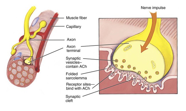
1. State the characteristics and functions of muscle tissue. 2. Describe the structure of a skeletal muscle. 3. List and describe the sequence of events involved in the contraction of a skeletal muscle fiber. 4. Explain how energy is provided for a muscle contraction. 6. Describe and illustrate the movements accomplished by the contraction of skeletal muscle. 7. Identify and describe the major muscles making up the axial skeleton. 8. Identify and describe the major muscles making up the appendicular skeleton. 9. Describe ways in which the aging of an individual affects the muscular system. As described in Chapter 5, there are three types of muscle tissue: skeletal, visceral, and cardiac. These are reviewed in Table 8-1. This chapter takes a closer look at skeletal muscle, which makes up about 40% of an individual’s body weight. It forms more than 600 muscles that are attached to the bones of the skeleton. Skeletal muscles are under conscious control, and when they contract they move the bones. Skeletal muscles also allow us to smile, frown, pout, show surprise, and exhibit other forms of facial expression. Table 8-1 From Applegate E: The anatomy and physiology learning system, ed 4, St Louis, 2011, Saunders. Skeletal muscle has four primary characteristics that relate to its functions: Excitability: Excitability (eks-eye-tah-BILL-ih-tee) is the ability to receive and respond to a stimulus. To function properly, muscles have to respond to a stimulus from the nervous system. Contractility: Contractility (kon-track-TILL-ih-tee) is the ability to shorten or contract. When a muscle responds to a stimulus, it shortens to produce movement. Extensibility: Extensibility (eks-ten-sih-BILL-ih-tee) means that a muscle can be stretched or extended. Skeletal muscles are often arranged in opposing pairs. When one muscle contracts, the other muscle is relaxed and stretched. Elasticity: Elasticity (ee-lass-TISS-ih-tee) is the capacity to recoil or return to the original shape and length after contraction or extension. Muscle contraction fulfills four important functions in the body: Highlight on the Muscular System Rigor mortis: The term rigor mortis means the “stiffness of death.” Within a short time of death, the adenosine triphosphate in muscles breaks down. This causes the myofilaments to remain locked in a contracted position and the body becomes rigid. A day or so later, muscle proteins begin to deteriorate and the rigor mortis disappears. Tetanus: The word tetanus is often confusing because it means different things to different people. In reference to muscle contraction, the term denotes a steady contraction of a muscle fiber, without a relaxation phase. The word also refers to a disease, commonly called “lockjaw,” that is caused by the bacterium Clostridium tetani. The toxin from the bacteria causes nerves to be highly excitable, which, in turn, causes uncontrollable muscle contractions, or spasms. A third use of the word is to denote a condition caused by a deficiency of calcium ions in the extracellular fluid. The lack of calcium increases nerve excitability with resulting muscle spasms, particularly of the extremities. The word tetany is also sometimes used to mean tetanus. Cramps: Cramps are painful, spastic contractions of muscles. They are usually caused by an irritation within the muscles that results in reflex contractions. Local inflammation from the accumulation of lactic acid is one source of irritation. Wryneck: Injury to one of the sternocleidomastoid muscles may result in torticollis, or wryneck. This is characterized by a twisting of the neck and an unnatural position of the head. Diaphragm: Voluntary forceful contractions of the diaphragm increase intraabdominal pressure to assist in urination, defecation, and childbirth. Electrical shock: The muscles that flex the fingers and hand are stronger than the extensor muscles. In a normal relaxed position the fingers are slightly flexed because the normal muscle tone is greater in the flexors. Persons who receive a high-voltage electrical shock through the arms flex their hands tightly and “can’t let go.” All of the flexors and extensors receive the electrical stimulus, but because the flexor muscles are stronger, they contract more forcefully. Intramuscular injections: The gluteus medius is a common site for intramuscular injections. Generally, the injection is given in the center of the upper outer quadrant of the buttock, or gluteal, area. The gluteus medius, rather than the gluteus maximus, is used to avoid damaging the sciatic nerve. Horseback riding: The adductor muscles in the medial compartment are the horse rider’s muscles. These muscles adduct, or press, the thighs together to keep a person on a horse. Quads: The quadriceps femoris group is a powerful knee extensor that is used in climbing, running, and rising from a chair. The region in which an axon terminal meets a muscle fiber is called a neuromuscular junction or myoneural junction, which is illustrated in Figure 8-1. The axon terminal does not actually touch the sarcolemma of the muscle cell but fits into a shallow depression in the cell membrane. The fluid-filled space between the axon terminal and sarcolemma is called a synaptic cleft (gap). Acetylcholine (ACh) (ah-see-till-KOH-leen), a neurotransmitter, is contained within synaptic vesicles in the axon terminal. Receptor sites for the ACh are located on the sarcolemma.
Muscular System
Introduction to the Muscular System
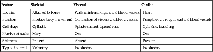
Feature
Skeletal
Visceral
Cardiac
Location
Attached to bones
Walls of internal organs and blood vessels
Heart
Function
Produce body movement
Contraction of viscera and blood vessels
Pump blood through heart and blood vessels
Cell shape
Cylindric
Spindle-shaped; tapered ends
Cylindric, branching
Number of nuclei
Many
One
One
Striations
Present
Absent
Present
Type of control
Voluntary
Involuntary
Involuntary
Characteristics and Functions of the Muscular System
Structure of Skeletal Muscle
Whole Skeletal Muscle

Contraction of Skeletal Muscle
Stimulus for Contraction
![]()
Stay updated, free articles. Join our Telegram channel

Full access? Get Clinical Tree


Muscular System
Get Clinical Tree app for offline access
