Management of Adults with Respiratory Disorders


Management of Adults with Respiratory Disorders


CASE STUDY

Mrs. A is a 57-year-old woman who calls her physician’s office because she “can not breathe.” She has a history of asthma and hypertension. Yesterday, she helped her daughter clean the basement. Mrs. A found that her inhaler did not improve her breathing the previous night. The triage nurse, listening on the phone, hears Mrs. A stopping between words to catch her breath. The triage nurse recommends that Mrs. A have her husband drive her to the office immediately.

When she arrives, her vital signs are: temperature—100.5°F orally, respiration—32 breaths per minute, heart rate—96 beats per minute, and blood pressure 146/92 mmHg. Her oxygen saturation (Sao2) is 90%. On examination, scattered wheezes are noted over both lung fields during inspiration and expiration with diminished breath sounds at the bases. Pulmonary function tests show a 30% decrease in her normal peak expiratory flow rate (PEFR). She is visibly anxious and breathing in rapid, shallow breaths. The physician sees her and recommends a nebulizer treatment with albuterol.


Important Questions to Ask


  • ♦ What physiologic changes cause wheezing?


  • ♦ Why is her PEFR decreased during an asthma attack?


  • ♦ What could have caused this asthma attack?

When oxygenation is disturbed, all tissues are affected. Oxygen is basic to the metabolism of every cell in the body. The process of supplying oxygen to the cells begins with ventilation, the moving of air into and out of the respiratory tract. It continues with the diffusion of gases across the alveolar membrane and into the circulation. The oxygen arrives at the cell by perfusion, the circulation of blood throughout the body. In this chapter, respiratory disorders in the upper and lower respiratory tracts, as well as disorders of diffusion and perfusion, are reviewed. Interventions to maximize oxygenation will be discussed in Chapter 4.


RESPIRATORY TRACT DEFENSES

The respiratory tract has many methods of defense against the neverending assault by the environment. The airways provide filtration of
inspired air and protection of the respiratory tract, as well as humidification and temperature regulation. The first line of defense is air filtration in the nose. The nasal hairs and mucus trap many foreign particles. Obviously, mouth breathing circumvents this system and reduces its effectiveness. The sneezing reflex is initiated in the nose when irritation or particles stimulate the trigeminal nerve. The cough reflex also assists in expelling particles or mucus that is occluding or irritating the airways.


More sophisticated methods of defense in the respiratory tract involve the mucociliary blanket. Cilia are small hairs that line the respiratory tract and constantly beat to move particles and mucus up the respiratory passages to where they can be expelled by coughing, sneezing, or swallowing. Dehydration, smoking, and certain drugs (e.g., atropine) can thicken sputum, rendering this defense mechanism less effective.

The mucus in the airways contains secretory immunoglobulins (IgA) that protect the respiratory tract against bacteria and viruses. Both B and T lymphocytes also protect the respiratory passages. Macrophages in the alveoli engulf and destroy foreign particles. Some particles, such as asbestos, may not be completely removed and can cause tissue changes over time, such as asbestosis or malignancy. Smokers are particularly susceptible to lung damage because much of the debris from habitual smoking remains in the lung tissue.

The large airways of the lower respiratory tract such as the bronchi also protect the lungs. Overreaction to irritants occurs in diseases such as asthma where narrowed airways are caused by bronchoconstriction and excessive mucous production. The smooth muscle surrounding the bronchi may constrict when irritants, such as dust and fumes, are inhaled. When the bronchi are irritated, mucus production increases to encase foreign particles. These protective mechanisms may actually impede normal ventilation.


Despite the elaborate protective mechanisms that exist in the respiratory tract, disorders may develop as a result of multiple causes. Assaults on the respiratory tract by the environment include physical trauma, viral or bacterial infection, and altered cellular processes, such as emphysema
or malignancy. The nurse’s role in treating patients with respiratory disorders is to maintain the patient’s oxygenation by optimizing ventilation, diffusion, and perfusion. Nursing care of patients with respiratory disorders requires the following:


  • ♦ Understanding normal respiratory anatomy and physiology


  • ♦ Distinguishing between different disorders of the upper and lower airways


  • ♦ Assessment of the patient based on the knowledge of different signs and symptoms of respiratory disorders and their impact on the patient’s functioning


  • ♦ Developing nursing and collaborative care plans with appropriate long- and short-term goals with the patient


  • ♦ Implementing the principles of oxygenation into the care plans


  • ♦ Evaluating the effectiveness of the interventions


  • ♦ Revising the treatment plan as necessary

This chapter will review disorders of the upper and lower airways, their clinical presentations, possible diagnostic studies, and collaborative treatment strategies to optimize ventilation and oxygenation.


DISORDERS OF THE UPPER AIRWAYS

The nose, sinuses, and pharynx are the first passages through which air enters the respiratory tract. They provide many defenses against the environment, including air filtration, humidification, and warming. Unfortunately, the filtering abilities of the mucus membranes can lead to irritation and infection of these membranes by the captured intruders.


Acute Rhinitis

The most frequent infection in human beings is acute rhinitis. It is usually caused by the common cold virus or rhinovirus (see Table 3-1). There are more than 100 different rhinoviruses that can cause the annoying symptoms of runny and stuffy nose (rhinorrhea), malaise, sore throat, coughing, and sneezing. Colds are most frequent between
November and March, with most adults having two to three colds per year. The usual incubation period for acute rhinitis is 1 to 4 days after exposure to droplets containing the virus.


Table 3-1 Symptoms of the Common Cold


1.


Red, swollen nasal membranes


2.


Mucoid to thin nasal discharge


3.


Sneezing and coughing


4.


Malaise, headache, and possibly low-grade fever


5.


Decreased sense of taste and smell


Assessment

During the assessment, note the patient’s breathing pattern, especially during speaking, noting any shortness of breath or change in breathing pattern. Clinical findings may include reddened nasal membranes and inferior turbinates, nasal discharge, and dry lips and mouth from mouth breathing. If the patient complains of a sore throat, this should lead to an examination of the posterior pharynx and possible throat culture to rule out beta-hemolytic streptococcal (“strep”) infection (see Table 3-2). The patient’s temperature and white blood cell count usually remain normal with a cold but may be elevated with a bacterial infection.


Elderly patients and those with chronic respiratory disease are particularly prone to complications from the common cold, including sinusitis, bronchitis, and pneumonia (an infection of the lung tissues). These conditions may lead to dyspnea, productive cough, fever, hypoxemia, and disorientation.


Note the color and consistency of nasal discharge in the assessment. Acute rhinitis from a cold virus initially produces clear, runny drainage followed by thicker, milky secretions. Yellow or greenish nasal discharge may indicate a secondary infection of the sinuses. Allergic reactions usually produce thin, clear, or mucoid nasal secretions. Postnasal drip is drainage of nasal secretions down the posterior nasal pharynx causing repeated swallowing and irritation. Other causes of nasal discharge
include sinusitis, drugs such as birth control pills and antihypertensives, as well as smoke and seasonal allergies.


Table 3-2 How to Obtain a Throat Culture


1.


Assemble equipment: tongue depressor, light source, sterile swab, and culture medium.


2.


Explain procedure to patient and warn of gagging sensation when the swab is applied to back of throat.


3.


In a seated position, have the patient tilt head back slightly and open mouth.


4.


Depress tongue with a moistened tongue depressor. Avoid touching the walls of the mouth or throat.


5.


Using a light source, visualize the posterior oropharynx, noting any areas of redness or exudate.


6.


Put down the light source while maintaining the tongue depressor on the tongue and pick up the sterile swab. While the patient says “Ahhh” to elevate the soft palate, put the swab in the mouth without touching the tongue or walls of the mouth and brush the swab over the wall of the posterior pharynx and the tonsils, trying to reach areas of redness or exudate. Discard tongue depressor.


7.


Put swab in appropriate culture medium per individual facility guidelines. Label specimen and send to appropriate laboratory. Wash hands. Document that a culture was obtained.


8.


Tell patient that results will not be ready for 24 to 48 hours, depending on the lab and antibiotics that may be needed for treatment if the culture is positive. Some rapid strep tests may be ready in 10 minutes but are less accurate than a culture.


Treatment

Treatment of acute rhinitis is directed at maintaining ventilation and minimizing symptoms (see Table 3-3). Acute rhinitis is a self-limiting condition that usually resolves in 7 to 10 days. Because the nasal discharge may occlude the airways, the following interventions are directed toward liquefying secretions:


  • ♦ Ensure adequate fluid intake (at least 2500 mL/day or one half the body weight in pounds equals the total ounces per day).


  • ♦ Avoid dairy products that may thicken secretions.


  • ♦ Humidify the air with a vaporizer, and use saline nasal drops to maintain moist nasal mucus membranes.


  • ♦ Allow adequate rest.


  • ♦ Use over-the-counter medications such as antihistamines (chlorpheniramine or pseudoephedrine) to decrease nasal discharge.


Table 3-3 Cold Prevention


1.


Decrease droplet exposure by frequent hand washing, especially after blowing the nose or sneezing.


2.


Use a tissue for blowing the nose, sneezing, and coughing; discard after each use.


3.


Avoid using the drinking glasses and eating utensils of others with a cold.


4.


Take vitamin C supplementation for possible antiviral effects.


Unfortunately, antihistamines may also dry out the mucous membranes, predisposing the patient to mucus stasis and a secondary bacterial infection. Over-the-counter sympathomimetics such as pseudoephedrine may be used to decrease mucus production by constricting the vasculature of the mucous membranes. Nasal drops, such as phenylephrine, produce vasoconstriction but should be limited to less than 72 hours of use because of the risk of rebound congestion (rhinitis medicamentosa). Echinacea (coneflower), an herbal remedy, is thought to be effective as an antimicrobial, but this treatment is still being investigated. Zinc lozenges are advertised as useful in decreasing the length of cold symptoms, but studies concerning the effectiveness of zinc have provided inconsistent results.


Nursing management of acute rhinitis usually occurs in the outpatient setting. Along with an assessment and diagnostic testing, such as a throat culture, the nurse teaches the patient about symptom management. She instructs the patient to seek further health care if the following occurs:


  • ♦ Symptoms last more than 7 days.


  • ♦ Temperature exceeds 100.5°F.


  • ♦ Nasal discharge becomes yellow to green or is accompanied by face pain or headache.


  • ♦ The patient has frequently recurring colds.


Allergic Rhinitis

A frequent cause of nasal discharge is allergic rhinitis. Unlike acute rhinitis in which a virus is responsible for the symptoms, allergic rhinitis is the result of environmental allergens. In allergic rhinitis, inhaled substances (pollen, animal dander, or dust) cause a type I hypersensitivity reaction. This leads to the release of potent vasoactive and inflammatory substances by the mast cells in the nasal mucosa. These substances produce vasodilation and increased capillary permeability in the mucus membranes, causing sneezing, watery eyes, and hypersecretion of thin mucus. It affects 10-20% of adults, most of whom have a family history of allergies, atopic dermatitis, asthma, or eczema.


Assessment

In cases of allergic rhinitis, the assessment focuses on the patient’s symptoms, their relation to possible causative agents, their clinical course, and seasonal variations. When assessing a patient with allergic rhinitis, ask the patient about occupation, smoke exposure, life stresses, alcoholic beverage intake, and drug exposure. The most common symptoms of allergic rhinitis are nasal congestion with thin, clear discharge, frequent sneezing, itchy eyes with increased tearing, and pruritis. The physical examination may show pale, swollen nasal mucosa with swollen turbinates, watery nasal discharge, watery eyes with puffy eyelids, reddened conjunctiva, dark circles under the eyes (allergic shiners), and pharyngitis.


A diagnosis of allergic rhinitis may require further diagnostic tests, such as skin testing, serum immunologic studies, and nasal smear to identify the allergen and look at the number of eosinophils. A large number of eosinophils may indicate an allergic response; serum immunologic studies may also indicate an allergic response when the IgE, a serum immunoglobulin, is elevated. Skin testing involves the application of a dilute allergen into needle scratches or pricks in the upper inner aspect of the arm or back. Normal saline is used as a control on the opposite side. Fifteen minutes later the sites are assessed for a response. A flare (reddened area) or a wheal (a raised area) over a site more than 5 mm larger than the
control would indicate an allergic response. Identification of the allergen provides a direction for treatment.


Treatment

Allergic rhinitis often occurs only during specific times of the year and is usually related to pollen exposure (“hay fever”). The most common offenders are grasses, ragweed, and wind-pollinated trees. Despite the name, hay does not contribute. Skin testing is useful in identifying pollen allergies. Total avoidance of pollen would be difficult at best. Treatment focuses on teaching the patient strategies to avoid pollen exposure (see Table 3-4).

Reduction of exposure to specific allergens may be difficult. Careful questioning of patients with allergic rhinitis may identify specific substances such as foods, food dyes, molds in cheese, wine, dried fruit, and some drugs that stimulate allergic responses. Once identified, the patient should avoid the offending foods and drugs. Decreasing exposure to other allergens such as animal dander may require removing the pet(s) from the household. Sensitivity to house dust and mites requires more involved steps to minimize exposure (see Table 3-5).


If the offending substance(s) cannot be completely avoided or symptoms continue despite measures to avoid allergens, then medications may be useful in minimizing symptoms. Antihistamines are the most frequently used treatment for allergic rhinitis. They help decrease the nasal congestion, mucous production, and excessive tearing and pruritus. Tolerance to these drugs can occur, and patients should be taught to report changes in drug effectiveness so that another class of antihistamine may be initiated. Antihistamines may be used in conjunction with decongestants to alleviate symptoms without the drowsiness that may occur with antihistamines alone. Both cromolyn and glucocorticoid nasal sprays are useful in relieving nasal congestion by stabilizing mast cells in the nasal mucosa. They have a slow onset of action and may take 3 to 5 days to become effective. Prophylactic use of medications may be useful for seasonal allergies to minimize responses to known allergens such as pollen.


Table 3-4 Reducing Exposure to Pollen


1.


Stay indoors during times of high pollen count, especially in centrally airconditioned homes where almost all pollen is filtered out by the system. Go outside only during or immediately after rainfall when the air has been cleared of pollen.


2.


Avoid eating honey, which may contain pollen.


3.


Drive with the windows closed.


4.


Install an air filter in the house to capture pollen.


Table 3-5 Reducing Exposure to Allergens


1.


Remove wall-to-wall carpeting and use hard floors, with washable throw rugs if desired.


2.


Encase mattresses and pillows in airtight covers and change them yearly. Wash bedding three times a week.


3.


Remove all feather-containing pillows and comforters. Use blankets and pillows made out of synthetic materials as they are less likely to harbor mites than feathers and wool.


4.


Avoid sweeping. Dust gently with a damp cloth.


5.


If possible, have someone else vacuum. Change the vacuum cleaner bag and filter regularly.


6.


Remove dust-collecting furniture and draperies.


7.


Reduce high humidity to decrease mite breeding. Do not use humidifiers or vaporizers.


8.


Install a high-efficiency air filter.


9.


Do not have pets. If that is not possible then do not allow pets in the bedroom and bathe them weekly.


10.


Avoid cigarette smoke.


If medication and exposure reduction are not effective in decreasing symptoms, then immunotherapy may provide some relief. Immunotherapy is a desensitization process involving subcutaneous injections of the known allergen in gradually increasing doses to increase the patient’s tolerance to the substance. Because anaphylactic reactions can occur, desensitization treatments are done in a physician’s office where emergency supplies are immediately available. Although the treatment may be effective in many patients, it is lengthy and expensive, requiring 2 to 5 years of treatment. Adherence to the lengthy process may be improved by teaching
patients about the treatment plan and possible side effects, and accommodating the patient’s schedule.


Nursing management of patients with allergic rhinitis usually occurs in the out-patient setting. Important components of the nurse’s role include assessing symptoms and treatment effectiveness, teaching about avoidance to allergens, observing for secondary complications, and encouraging adherence with the treatment plan. These interventions facilitate normal air passage through the nose, allowing the nasal passages to perform their important functions, minimize symptoms, and improve quality of life.


Sinusitis

An infection of the sinuses (sinusitis) may be a bacterial infection or secondary to a viral exposure. Bacteria, viruses, fungi, and allergic reactions may all cause sinusitis. Acute sinusitis usually develops after a primary viral infection or the common cold. The normal drainage paths from the sinuses to the nasopharynx are blocked by swollen mucous membranes and exudate, and thick sputum and bacteria begin to grow. Exudate and white blood cells fill the sinus, causing pressure and pain.


Assessment

The most common presenting symptom of sinusitis is facial pain and headache. The area over the sinus may be swollen and tender to palpation. There may also be toothaches if the maxillary sinuses are involved and a headache if the frontal or ethmoid sinuses are involved. The physical assessment of the nasopharynx will reveal reddened mucosa in the nasal passages with yellow to green exudate.


Treatment

Most cases of sinusitis present in the outpatient setting. Symptoms such as fever and facial swelling may indicate more diffuse infection requiring intravenous antibiotics. The treatment for acute sinusitis includes humidification with a vaporizer to help drain the sinuses, oral antibiotics for
10 to 14 days, and topical vasoconstrictors such as phenylephrine for, at most, 7 days. It is important to teach the patient about finishing the entire course of antibiotics even if he or she is feeling better. This will prevent reinfection with resistant organisms.


Other Disorders of the Nose

Normal ventilation through the nose may be interrupted by nasal fractures, septal deformities, nosebleeds (epistaxis), tumors, and polyps. Nasal fractures are the most common fracture of the facial bones. (Refer to the section on facial trauma later in this chapter.) Septal deformities, such as a deviated septum, may impede airflow. They may result from trauma or developmental deformities and are usually asymptomatic. Restricted airflow through one or both nasal passages or chronic sinusitis caused by the deviated septum blocks the sinus opening and may indicate the need for surgical correction.


Nosebleed or epistaxis occurs commonly and may result from trauma (including nasal fracture and nose picking), infections such as sinusitis and rhinitis, drying of the mucous membranes, bleeding disorders, malignancies of the nose or paranasal sinuses, hypertension, and some systemic infections such as scarlet fever. The diagnosis is obvious from the clinical exam. Treatment involves applying gentle pressure to the nose for 5 to 10 minutes while the patient remains seated with his head tilted forward to prevent aspiration of blood or clots. If this method does not stop the bleeding, topical medications like tetracaine should be applied to vasoconstrict the capillaries in the nasal mucosa. If the bleeding continues, the bleeding site needs to be located and cauterized chemically (silver nitrate) or electrically, or the nasal passage needs
to be packed to apply direct pressure to the bleeding site. Chronic or recurrent nosebleeds may indicate a bleeding tendency and a need for further evaluation.

Nasal polyps are grape-like masses of swollen nasal mucosa. They may block the nasal passages, impede drainage of the sinuses, and promote sinusitis. Polyps are benign growths that may result from chronic sinusitis and allergic rhinitis. The treatment involves topical treatment with corticosteroids using a nasal inhaler. Nursing management involves teaching the patient about the medications using a nasal inhaler and evaluating the effectiveness of the treatment. Surgical treatment may be required to remove the polyps as they tend to recur.

Other growths in the nose may be benign or malignant. The most common benign growth in the nasal passage is a papilloma. The most common malignancy is a squamous cell tumor. Patients with intranasal growths show symptoms of a unilateral airway obstruction, bloody discharge, numbness, or swelling. Surgical excision may be required to determine the pathology of the growth and plan the treatment course.


Pharyngitis

Acute inflammation of the pharynx, pharyngitis, may be caused by viral or bacterial infections. Pharyngitis, or a sore throat, may cause pain, especially when swallowing, and tender lymph glands in the neck. Patients may also have a fever and malaise.


Clinical findings of pharyngitis include mild to severe redness of the pharynx with or without swollen tonsils. Exudate may or may not be present on the tonsils or posterior oropharynx. In viral infections of the pharynx, there is usually no exudate but the pharynx is reddened with a “cobblestone” appearance. In bacterial infections, the pharynx and tonsils are reddened, and a white to yellow exudate may be present over these areas. Cervical lymphadenopathy may also be present.


Assessment

Assessment includes asking the patient about the onset and duration of symptoms, checking the vital signs for fever, and obtaining a throat culture to rule out beta-hemolytic streptococcal infection (see Table 3-2). Strep infections may have serious consequences such as acute glomerulonephritis and rheumatic fever.


Treatment

Treatment of strep infections includes antibiotic therapy, warm saline gargles, a soft diet, rest, analgesics for pain relief, throat lozenges, and plenty of fluids (2 to 4 L per day). Teach patients to finish the course of antibiotics, even though they may be feeling better, to prevent reinfection with antibiotic-resistant organisms. Patients should call their health care provider if they are still having symptoms or fever after 3 days of antibiotic treatment.


Oropharyngeal Cancer

Malignancies of the mouth, tongue, or pharynx often involve complex medical and nursing care. Screening for oral cancers should be done in the following groups of people who are at high risk:


  • ♦ Chronic use of cigarettes


  • ♦ Age over 40 years old


  • ♦ Use of chewing tobacco


  • ♦ Regular use of alcohol

The most common presentations of oropharyngeal cancers are a painless red or white lesion in the oropharynx and cervical lymphadenopathy. The most common malignancy of the oropharynx is squamous cell carcinoma.


Assessment

Patients receiving treatment for head and neck cancers require specialized assessments before, during, and after treatment. All patients who will be receiving radiation therapy or chemotherapy to the head and neck should have a thorough dental examination prior to beginning their treatment. It will be difficult or even impossible to do certain dental procedures, such as extractions, after radiation treatment
because it can destroy the blood vessels in the jawbone. Salivary glands may also be destroyed by radiation therapy, predisposing the patient to a dry mouth (xerostomia), altered taste perceptions, accelerated tooth decay, difficulty chewing, swallowing, and speaking.


Treatment

Medical treatment for oropharyngeal cancers is often complex, involving surgery, radiation therapy, and chemotherapy. Surgery involves excision of the lesion, surrounding tissue, and, perhaps, lymph nodes. Nursing care for patients with oropharyngeal cancers is also complex and may include the following:


  • ♦ Maintaining a patent airway


  • ♦ Ensuring adequate nutrition


  • ♦ Teaching patient about mouth care


  • ♦ Controlling treatment side effects


  • ♦ Providing alternative methods of communication


  • ♦ Recognizing changes in body image and self-esteem that may occur after treatment

One common side effect of treatment for head and neck cancers is mucositis, the breakdown, ulceration, and infection of the oral mucosa. It may be quite painful requiring a break during treatment. Mucositis develops because the cells that line the oral cavity have a rapid turnover rate. The cancer treatment affects rapidly dividing cells and can kill normal cells in the mucosa, as well as malignant cells. The damaged lining can ulcerate and become susceptible to infection.


A common infection in patients who are being treated for malignancies or are immunocompromised is oral candidiasis, or thrush. Candidiasis is the infection of mucous membranes with Candida albicans and creates distinctive whitish patches on the oral mucosa that may be quite painful. Antifungal agents such as fluconozole are used to treat these infections.


Treatment for mucositis involves preventative dental care, thorough mouth care, frequent assessment of the oral cavity, and topical and/or systemic analgesia. Current treatments include antibiotic therapy for bacterial infections, antifungal agents, antiulcer medications (misoprostol), and amino acid (glutamine) mouthwashes. Teaching patients about oral care prior to treatment will minimize side effects and promote early detection of problems (see Table 3-6). (Nursing care of the oncology patient receiving chemotherapy and radiation therapy is reviewed later in this chapter).


Laryngitis

Laryngitis is an acute or chronic inflammation of the mucus membranes lining the larynx and sometimes the vocal cords themselves. It occurs most frequently as a result of viral infection but may also be related to other respiratory infections such as bronchitis and influenza. Hoarseness and unnatural diminution of the voice may occur due to chronic overuse of the voice, exposure to inhaled irritants such as cigarette smoke or volatile gases, allergic reactions, or endotracheal intubation.


Table 3-6 Care of the Mouth During Cancer Treatment


1.


Brush teeth gently after every meal and before bed with a soft toothbrush rinsed in warm water to further soften the bristles, or use a sponge toothbrush.


2.


Do not use mouthwashes that contain alcohol as they can dry out the mucosa.


3.


Eat soft foods that are warm, not too hot or too cold, and avoid spicy foods.


4.


Avoid alcohol and all tobacco products.


5.


Use a saliva substitute to keep the mouth moist if dryness is a problem.


Assessment

Assessment of the patient with acute laryngitis focuses on the symptoms of hoarseness, difficulty swallowing (dysphagia), and any other symptoms of respiratory infections. Laryngeal examination is performed indirectly with a laryngeal mirror or directly with a fiber-optic laryngoscope. When any disorder other than acute laryngitis is suspected or hoarseness lasts more than 2 weeks, patients should be referred to an otolaryngologist.


Treatment

The primary treatment of laryngitis is voice rest. Nursing management of the patient with laryngitis involves teaching the patient about voice rest, increased fluid intake (2 to 4 L per day), topical lozenges, and steam inhalation (such as sitting in a steamy shower or use of a humidifier) to relieve some of the symptoms. Voice rest involves not only refraining from talking but also from whispering and heavy lifting, which strain the larynx. Antibiotics may be ordered if other respiratory infections like bronchitis are suspected. Avoidance of inhaled irritants, such as cigarettes or noxious fumes, is important in treating chronic laryngitis. Recurrent bouts of laryngitis that do not respond to conventional treatment require further medical evaluation.


Vocal Cord Nodules and Polyps

Vocal cord nodules are frequently seen in people who use their voice frequently and loudly, such as singers and actors. Vocal cord polyps develop as a result of chronic voice abuse or inhalation of irritants such as cigarette smoke (see Figure 3-1). Symptoms in both cases include hoarseness and a breathy voice quality.


Assessment

Assessment of patients with symptoms of chronic hoarseness should be performed by an otolaryngologist. Direct or indirect laryngoscopy and possibly biopsy may be necessary to identify the source of the symptoms.


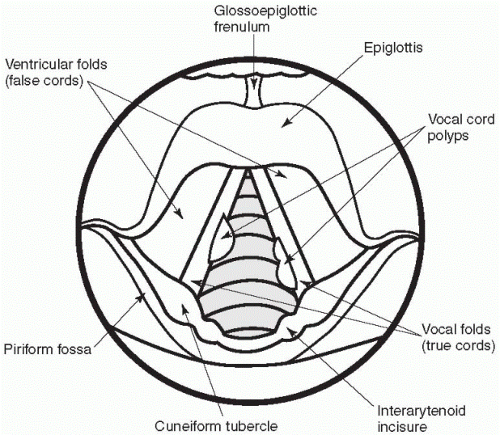
Figure 3-1 Vocal cord polyps.


Treatment

Treatment for vocal cord nodules and polyps involves surgical removal and voice therapy. Malignancy needs to be ruled out in both cases, especially in cases of chronic cigarette abuse. The nurse focuses on teaching the patient how to prevent voice abuse and methods of smoking cessation. If surgical removal of the polyp or nodule is done, then complete voice rest will require the patient to use alternative methods of communication such as picture boards, slate, and alphabet boards for 2 weeks. Preoperative education of the patient and family is important because most of these procedures are done on an ambulatory basis, putting the burden of the communication challenges on the family.


Vocal Cord Paralysis

Vocal cord paralysis may result from neck or chest tumors, central nervous system tumors, trauma, or viral illness. Other causes include prolonged intubation, total thyroidectomy, lung tumors, aortic aneurysms, and an enlarged right atrium. The patient with vocal cord paralysis presents
with a diminished or hoarse voice and possibly difficulty breathing and swallowing. Identification of the cause is vitally important to protect the airway.


Assessment

To accurately assess the patient with vocal cord paralysis, direct laryngoscopy is necessary to visualize the vocal cords. Nursing care focuses on airway protection. Putting the patient in a high Fowler’s position and suctioning as necessary to remove secretions facilitates the maintenance of a patent airway. Stridor, coarse loud sounds when breathing, may indicate difficulty in moving air through the paralyzed cords and emergency intubation or tracheotomy may be necessary.


Treatment

Treatment focuses on maintaining the airway even at the expense of the voice. Patients are taught supraglottic swallowing (i.e., taking a deep breath and holding it prior to swallowing). This maneuver allows the larynx to elevate and the epiglottis to close, preventing food and fluids from entering the lower respiratory tract during swallowing. Because patients with vocal cord paralysis are at high risk for aspiration, they must be carefully assessed for aspiration pneumonia.


Laryngeal Trauma

Laryngeal trauma can occur from blunt force, fracture, or from prolonged endotracheal intubation. Symptoms may be an obvious laceration or hemoptysis; swelling and laceration; or symptoms of hoarseness, dyspnea, aphonia, and subcutaneous emphysema. Nursing care focuses on maintaining a patent airway, frequent evaluation of vital signs and pulse oximetry, and observation for symptoms of increased respiratory difficulty such as stridor, tachypnea, dyspnea, and restlessness. Respiratory distress resulting from laryngeal trauma may require emergency intubation. Prolonged intubation may result in further laryngeal damage, and may necessitate the creation of a tracheostomy to bypass the larynx.


Facial Trauma

One of the most common injuries seen in emergency rooms is facial trauma. Whether from physical fights, automobile accidents, falls, or sports injuries, the bones in the face are susceptible to fracture. Nasal fractures are the most common facial injury. Patients complain of pain, tenderness, and swelling over the nose and may have epistaxis and rhinorrhea. The nurse should quickly assess the adequacy of the airway and any other more serious trauma. A history of the event that caused the accident is important, including details about the patient’s response to the injuries, especially any loss of consciousness. Blows to the head that are forceful enough to break facial bones may also cause head trauma. The nurse should be alert to signs of head trauma and increasing intracranial pressure.


Assessment

Physical assessment may reveal ecchymosis (bluish discoloration of the skin caused by extravasation of blood into the subcutaneous tissues), swelling, asymmetry, and bony fragments if a compound fracture occurred. Palpation of the area may reveal unusual mobility of the nasal bones or displacement, and crepitus.


Treatment

Treatment of nasal fractures involves reducing the fracture if displacement occurred, ice packs, and analgesics. Care for the nasal fracture includes ice packs for 20 minutes of each hour for the first 24 hours. This reduces swelling and hematoma formation. Closed reduction of a displaced nasal fracture usually returns the bones to their normal position,
and packing may be inserted to stabilize the area. If there was not any displacement of the nasal bones, placement of a cast over the dorsum of the nose may protect it from further trauma.

Nursing management includes evaluating the patient for serious injuries such as airway obstruction and/or head trauma that may have occurred with the nasal fracture. Patients are instructed not to blow their nose or pinch the nostrils together, as this could force air into the subcutaneous tissues. The swelling and hematoma should resolve in 2 to 3 weeks. Patients should avoid situations where reinjury could occur, such as contact sports.


Maxillofacial Fractures

Maxillofacial fractures are more serious than nasal fractures. Rapid assessment of the patient is required. Hemorrhage and airway obstruction are the two most common life-threatening complications. The nurse presumes that cervical injuries are present with maxillofacial fractures until X-rays are negative because the force required to cause maxillofacial trauma is sufficient to cause a cervical spine injury. Bone chips may perforate the dura, causing cerebrospinal fluid (CSF) leaks. The patient may perceive CSF leaks as a salty postnasal drip or rhinorrhea. The fluid in the nose should be checked for glucose by dipstick, which is present in CSF but not in nasal mucus.


Treatment

Treatment of maxillofacial fractures involves maintaining a patent airway, controlling bleeding, protecting the cervical spine, and treating the head injuries. Nursing management involves trauma care: maintaining a patent airway with suctioning if needed, elevating the head of the bed (if injuries allow) to decrease bleeding, observing for head injuries and CSF leaks, assessing for orbital injuries, and evaluating changes in vision and eye movement. Patients with jaw fractures who require intermaxillary fixation need ongoing care after discharge to assess oral hygiene and nutritional status. Intermaxillary fixation is the
wiring together of the upper and lower jawbones by a series of stainless steel wires and elastics.


Cancer of the Head and Neck

Caring for patients with head and neck cancer may be one of the greatest challenges for nurses. The disease and treatment may leave the patient with difficulty eating, swallowing, breathing, and speaking. Surgical excision may change the patient’s body, altering his appearance and body image. Treatment depends on a multidisciplinary team to identify the tumor, decide the appropriate treatment(s), support the patient during treatment, and return the patient to his optimum functioning.

Squamous cell carcinomas account for 90% of head and neck tumors. The average patient is a male and over 50 years. Over 85% of all patients with head and neck cancer have a history of tobacco or alcohol use. Patients with these risk factors should have a careful oral exam annually. Head and neck tumors usually remain confined to the region and then spread in an orderly fashion along the associated lymphatic chains. Early detection of tumors while they are still relatively small and confined can greatly improve the prognosis. Even when the cancer has spread to the lymph nodes, a cure is possible.


Head and neck tumors may present in a variety of ways. Oral cancers may be detected by a dentist, a healthcare provider during routine assessment, or by the patient (see Table 3-7). Oral cancers usually begin with red patches (erythroplasia) or whitish patches (leukoplakia) on the mucous membrane. Nasal tumors may cause facial swelling, numbness, facial pain, and epistaxis. Patients with oropharyngeal cancer may complain of hoarseness, a lesion in the mouth, pain when swallowing, unilateral ear pain (from destruction of the glossopharyngeal nerve), and fullness in the face. Because of the variety of ways that head and neck cancers present, it is important for the nurse to understand the warning signs and assess patients who are at risk for developing any of these cancers.


Table 3-7 Warning Signs of Head and Neck Cancer


1.


Oral lesion or sore that does not heal in 2 weeks


2.


Persistent or unexplained oral bleeding


3.


Color changes (red, white, black, or brown) on the mouth or tongue


4.


Difficulty swallowing


5.


Persistent hoarseness or changes in voice quality


6.


Persistent or recurrent sore throat that does not respond to treatment


7.


A lump in the mouth, throat, or neck


8.


Pain in the mouth, lips, throat, neck, or under the dentures


Assessment

The first contact that a nurse may have with a patient with head and neck cancer is during the assessment. Sensitive questioning about tobacco (all forms) and alcohol use is important. Calculate the number of packs per year of cigarette use with the formula in Chapter 2. Ask about other risk factors to potential carcinogens, such as occupational exposure (woodworkers, asbestos exposure, and petroleum workers). Review the course of the presenting symptoms and note their onset and duration.


Treatment

The treatment course for head and neck cancers varies depending on the location and stage of the disease, the physician’s recommendation, and the patient’s desires. The treatment course for head and neck cancers is determined by the stage of the disease. Tumors are biopsied to determine the histology, and follow-up CAT scans or MRls are used to assess tumor size and local spread. Head and neck tumors are staged according to the TNM system. T indicates the size and site of the primary tumor, N denotes the number and size of local lymphatic spread, and M is used to indicate the presence of distant metastases.


Many tumors of the head and neck require some surgery to biopsy and remove the tumor. More extensive surgery or radiation therapy is required to treat the tumor bed and the associated lymph nodes that may have disease. Chemotherapy has an increasing role in advanced cancers. Because there are so many different tumors and treatments, this section will focus on the patient with laryngeal cancer.

Laryngeal cancer is the most common head and neck cancer, 95% of which is squamous cell carcinomas. The cancer may occur in the true vocal cords, the epiglottis, the pyriform sinus, or the postcricoid area. Patients usually present symptoms of hoarseness, pain, a lump in the neck, or difficulty swallowing.


Early stages of cancer in the vocal cord and epiglottis are treated with radiation therapy. Daily radiation treatments for 6 to 7 weeks can be tiring for the patient. The treatments themselves are not painful, but the side effects can cause many difficulties for the patient as the treatment progresses. Because radiation affects all anatomic structures within the treatment field, the pharynx, larynx, and esophagus may all be affected. Side effects of radiation therapy appear as the treatment progresses and the total dose of radiation to the area increases. Side effects include skin reactions, esophagitis (inflammation of the lining of the esophagus with pain on swallowing), laryngitis, and pharyngitis. If the oropharynx is in the treatment field, then the patient may experience xerostomia (dry mouth), decreased taste, and possibly infections of the oropharynx. Mucositis may be very painful and may make it difficult for the patient to eat and swallow. The challenge for nurses is to maintain optimum nutrition, control pain, and watch for treatment reactions.

Surgical treatment of laryngeal cancer depends on the stage of the tumor. More advanced glottic cancers with cartilage invasion may require complete laryngectomy with a radical neck dissection to remove the associated lymph nodes. A complete laryngectomy is performed when previous treatment with radiation or chemotherapy and radiation therapy have failed (see Figure 3-2). After the laryngectomy, the patient can no longer speak in the normal way. Later, a tracheoesophageal fistula may be surgically created into which a one-way valve can be inserted for speech. Other methods of creating speech are by eructation or esophageal speech and the use of an electrolarynx. When a tracheostomy is created, an opening
is made from the trachea to the anterior neck to maintain the airway. The pharynx is sutured to the esophagus to permit swallowing.


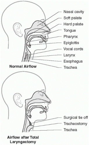
Figure 3-2 Altered airflow after total laryngectomy.


DISORDERS OF THE LOWER RESPIRATORY TRACT

Disorders of the lower respiratory tract may be acute or chronic processes. Acute processes include brief infections such as pneumonia and chronic processes include emphysema and asthma. Chronic conditions often have a disease course marked with remissions and exacerbations. The nursing care for acute and chronic respiratory disorders is a collaborative process with the nurse, the physician, and the respiratory
therapist all contributing to the development of the best care plan for the patient. In this section, acute infections (influenza, acute bronchitis, pneumonia, and tuberculosis) will be reviewed first followed by diseases of chronic airflow limitation (asthma, chronic bronchitis, and emphysema), acute respiratory failure, adult respiratory distress syndrome (ARDS), pulmonary vascular disorders (pulmonary embolus and pulmonary hypertension), pleural effusion, lung cancer, and chest trauma including pneumothorax. (Specific interventions to maintain oxygenation such as oxygen therapy, tracheostomy, mechanical ventilation, thoracentesis, and chest tube maintenance and endotracheal suctioning are reviewed in Chapter 5.)


Influenza

Influenza is a viral infection of the respiratory tract. It usually occurs in epidemics during the fall and winter. Influenza may be a serious infection, particularly in those over 65 years of age, immunocompromised patients, and those with chronic lung and heart disease. There are three common types of the influenza virus: A, B, and C, as well as numerous subtypes. The incubation period is 1 to 4 days after exposure to droplet nuclei spread by coughing or sneezing. The presenting symptoms fall into three syndromes: a rhinotracheitis, a viral respiratory infection, and a viral pneumonia. The presentation depends on the type of droplet exposure. Fine droplet exposure inhaled into the nasal passages may produce a rhinotracheitis. A larger exposure of viral-laden droplets directly into the lower airways may produce a viral respiratory infection or pneumonia. Basic hygiene (hand washing, covered sneezes, and coughs) may decrease the spread of influenza.


Assessment

Assessment of the patient requires an understanding of the normal course of influenza versus other respiratory tract disorders. The first symptoms of influenza are often abrupt onset of fever, chills, and malaise followed by a profusely runny nose (rhinotracheitis), muscle aches, and headache. The symptoms of rhinotracheitis peak in 3 to 5 days and
resolve spontaneously in 7 days. Secondary complications may develop after the acute infection and may be related to bacterial infections that may include sinusitis, otitis media (infection of the middle ear), bacterial pneumonia, and bronchitis. These secondary bacterial infections occur just as the patient is starting to feel better or when symptoms are prolonged after the normal influenza course.

The symptoms of influenza pneumonia are more severe and can rapidly progress to hypoxemia and even death. The symptoms of cough, fever, chills, and malaise come on abruptly and can be quite severe.


Treatment

Treatment for influenza is usually symptomatic including antipyretics, decongestants, fluids, and rest. Amantadine, an antiviral drug, may be used to shorten the course of an influenza infection or for prophylaxis. Prolonged courses of influenza indicate additional assessment, especially in the elderly or otherwise compromised patients.


Treatment of patients with influenza includes rest, extra fluids, and acetaminophen for high temperatures (greater than 101°F). Acetaminophen is preferred over aspirin as an analgesic and antipyretic because of the risk of Reye’s syndrome, a rare complication of influenza that causes liver failure and encephalitis. It is seen more often when aspirin is used during an influenzal illness, particularly in children.


Many influenza infections can be prevented by use of a vaccine. Atrisk individuals should be vaccinated annually in the fall. The vaccine is reformulated each year to include the most common strains of the influenza virus from the previous year’s data. Populations at high risk for complications from influenza include people over 65 years of age; residents of nursing homes and chronic care facilities; patients with chronic heart, lung, metabolic, or immunologic problems; and children or adults receiving chronic aspirin therapy (because of the risk of Reye’s syndrome). The
influenza vaccine should not be given to individuals with an allergy to egg whites. Side effects to the vaccine are infrequent and include redness and tenderness at the vaccination site and, rarely, malaise and fever.


Acute Bronchitis

Acute bronchitis is the inflammation of the large airways in the lower respiratory tract. It may be caused by bacteria, viruses, or exposure to inhaled irritants. Bronchitis can be classified as acute or chronic. Chronic bronchitis will be reviewed later in the chapter.


Acute bronchitis may result from a previous infection with a virus (i.e., influenza) predisposing a patient to a secondary bacterial infection. The most common infective agents in acute bronchitis are Staphylococcus aureus, Pneumococcus, and Haemophilus influenza. The bacteria are usually passed from the nasopharynx to the bronchi by small amounts of aspirant. The organisms cause an inflammatory response in the bronchi with swelling and excessive mucus production.


Assessment

Symptoms of acute bronchitis include a productive cough, fever, malaise, substernal pain especially when coughing, and auscultatory crackles and wheezes. Wheezes may indicate some degree of bronchoconstriction. A dry cough often progresses to a productive cough with purulent and/or blood-streaked sputum. Acute bronchitis can progress to a severe illness with high fever, dyspnea, and cyanosis requiring hospitalization.


Treatment

Treatment is based on the clinical findings and includes antibiotics, extra fluids, humidity, rest, acetaminophen for fever and pain, and sometimes oxygen therapy. Cough suppressants should be used cautiously because
excessive secretions need to be cleared from the lungs. Expectorants (such as guaifenesin) may be useful in relieving chest congestion. Cigarette smokers are encouraged to stop smoking, as this further irritates the lining of the bronchi.


Pneumonia

Pneumonia is an inflammatory process of the parenchymal structures of the lung, such as the bronchioles and alveoli, and may impair gas exchange. Bacteria, viruses, fumes, and even gastric contents can cause pneumonia. Pneumonia is classified by location, infectious agent, and other factors.

Normally, respiratory defense mechanisms such as the cough reflex and the mucociliary blanket protect the lower airways. Some factors that can impair the effectiveness of these defense mechanisms are immunodeficiency, smoking, viral diseases, and loss of the cough reflex due to neuromuscular disease or anesthesia. Although antibiotics have decreased the mortality associated with pneumonia, it is still a leading cause of death in adults in the United States. In 2003, a new deadly form of pneumonia emerged, severe acute respiratory syndrome (SARS).


Assessment

Pneumonia may present with a variety of symptoms depending on the type and location. Signs and symptoms include cough, fever, pleuritic chest pain, and adventitious lung sounds. Older adults with pneumonia may present with confusion and lethargy rather than fever and cough. The cough may be dry or productive. On auscultation, there may be decreased breath sounds and adventitious sounds such as wheezes or crackles over the affected area.


Chest X-ray reveals pulmonary infiltrates in cases of pneumonia. Other diagnostic tests include sputum for culture and sensitivity, white blood cell count (and differential), pulse oximetry, arterial blood gases (ABG), and possibly bronchoscopy.

Pneumonia is classified according to the anatomic distribution and the causative agent. Acute bacterial pneumonia has one of two anatomic
patterns: either lobar or bronchial pneumonia (see Figure 3-3). Lobar pneumonia is so named because a chest X-ray reveals inflammation of a lobe of the lung. Approximately 90% of all forms of lobar pneumonia are caused by Streptococcus pneumoniae. The symptoms of lobar pneumonia are rapid onset of malaise, chills, high fever, and leukocytosis (e.g., increased white blood cell count). Initially, the cough may produce watery sputum, and the breath sounds may be diminished due to congestion in the alveolar walls. Later, the sputum becomes rusty colored or purulent. Pleuritic pain, especially on deep respiratory movements, may be present.


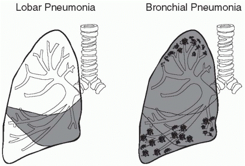
Figure 3-3 Lobar and bronchial pneumonia.


Bronchial pneumonia differs from lobar pneumonia in both the presentation and the course of the illness. On chest radiography, a bronchial pneumonia appears as patchy consolidation in several lobules. It is usually the extension of a preexisting bronchitis. Bronchial pneumonia tends to be a disease of the very young, the very old, and the immunocompromised. It presents insidiously with a low-grade fever, cough, crackles, and leukocytosis. Many different organisms can cause bronchial pneumonia, including the previously mentioned Streptococcus pneumoniae,
as well as Staphylococcus aureus, Haemophilus influenzae, and Pseudomonas aeruginosa. Nosocomial pneumonia is pneumonia that develops within 48 hours of admission to a healthcare facility.

Only gold members can continue reading. Log In or Register to continue

Stay updated, free articles. Join our Telegram channel

Oct 17, 2016 | Posted by in NURSING | Comments Off on Management of Adults with Respiratory Disorders

Full access? Get Clinical Tree

Get Clinical Tree app for offline access