Assessment of the Cardiovascular System


Assessment of the Cardiovascular System


CASE STUDY

Mr. H has a long-standing history of chronic obstructive lung disease and high blood pressure, and is 60 pounds overweight. He has continued to smoke despite urgings from his physician to quit. He called the office this morning because his chest felt tight and he could not catch his breath. When the nurse asked him what he did to help the pain, he said that he laid down an hour ago but the pain did not go away. Suspecting that his pain could be cardiac in origin, the nurse asked if his son was available to take him to the hospital or if he could call an ambulance. This scares Mr. H, but the nurse reassured him that this is the best place for him because they can get to the root of his pain and make him feel better. The nurse reminded him not to smoke any more cigarettes. Vital signs, an EKG, blood work, and pulse oximetry were done on Mr. H’s arrival in the emergency department. His vital signs were as follows: HR—108 bpm, RR—32, and BP—176/94 mmHg. His Sao2 was 89%. The nurse started oxygen via nasal cannula at 2 L/min and gave him aspirin per the protocol. His EKG showed ST segment elevations and his chest pain was not fully relieved by sublingual nitroglycerine, so a cardiac catheterization was ordered.


Important Questions to Ask


  • ♦ How would you explain the purpose of the cardiac catheterization to Mr. H?


  • ♦ What allergies would you ask Mr. H about prior to the procedure?


  • ♦ What feelings should Mr. H report to the nurses during the procedure?


  • ♦ Why is bed rest important after the catheterization?

Assessment of the cardiovascular system involves evaluating the adequacy of the heart’s ability to pump blood and perfuse tissues. The perfusion process is similar to a plumbing system: the heart acts as the pump to deliver oxygenated blood to the cells, and the vascular network functions as the pipes. Perfusion through the cardiovascular network is a closed system that responds to changes in pressure. Unlike plumbing, the cardiovascular system is also responsive to changes in the
tissue demands and can alter flow to meet the demands of the body. This chapter will review the assessment of the cardiovascular system as it pertains to tissue perfusion and cell oxygenation.


GENERAL ASSESSMENT

A general overview of the patient’s health provides clues to the overall functioning and the efficiency of the cardiovascular system. Examine the patient and estimate his apparent age. A comparison of his apparent and stated age may reveal that the patient looks older because of smoking, sun exposure, or poor health. Notice any cyanosis around the mouth and nose or in the extremities as seen in clubbing of the nails. Central cyanosis could indicate poor oxygenation or circulation. Assess the patient’s facial expression, posture, and body language. These may allude to problems with breathing, pain, or anxiety. Pallor may indicate anemia or low cardiac output. Diaphoresis may suggest hypotension or myocardial infarction. Note the patient’s respiratory patterns during activity. Increased breathlessness during a mild activity like disrobing may indicate problems with the respiratory or cardiovascular systems. While these are only general clues, they can direct a more detailed assessment of the heart and circulatory system.


ASSESSMENT OF THE CARDIOVASCULAR SYSTEM

An assessment of the cardiovascular system begins with questions about the patient’s family health history. Any history of heart disease in the patient’s family is important in understanding the patient’s health risks. Family history would include congenital heart disease, angina, myocardial infarction, elevated cholesterol levels (dyslipidemia), high blood pressure, and strokes. Other diseases associated with heart problems include a family history of diabetes, hypertension, and thyroid disease.

Ask about the patient’s health history, including the following:


  • ♦ Heart disease—Acute, chronic, or congenital heart problems, hypertension, diabetes, dyslipidemia, heart murmurs, rheumatic fever, or varicose veins


  • ♦ Lifestyle—Occupation, hobbies, sleep habits, stressors, exercise, smoking, and alcohol intake


  • ♦ Medications—Cardiac medications, antihypertensives, over-thecounter medications, oral contraceptives, herbal remedies, and nutritional supplements


  • ♦ Nutritional habits—Caloric intake, fat, salt, and caffeine consumption

The patient’s health history should include questions about the presenting problem plus nonspecific symptoms. Ask the patient about what brought him to seek a healthcare provider and use follow-up questions to determine the timing of the symptom(s), alleviating factors, and how they affect the patient’s functioning. Nonspecific symptoms might include fatigue, cough (refer to Chapter 2), dizziness, palpitations, or problems with sleep including snoring and sleep apnea. Some common signs and symptoms of patients with cardiovascular disorders include the following:


  • ♦ Chest pain—Including arm, shoulder, and neck pain, and epigastric discomfort, as well as timing of the pain to activities


  • ♦ Pain in the extremities on exertion—Intermittent claudication is relieved by rest, and heaviness in the extremities caused by varicose veins is relieved by rest and elevation.


  • Dyspnea on exertion or with ordinary activities—Note the timing, the precipitating activity, and the alleviating factors.


  • Orthopnea or difficulty breathing when recumbent—Note the timing of the orthopnea, including paroxysmal nocturnal dyspnea (PND). Ask about the number of pillows the patient uses when sleeping.


  • ♦ Palpitations (awareness of heart beating)—Note any precipitating factors like caffeine, nicotine, alcohol, sugar, or stress. Also ask about awareness of skipped beats.


  • ♦ Edema of the extremities—Note location of unilateral or bilateral swelling, any discoloration of the extremities, presence of ulcerations, and time during the day when the edema is noticeable.


  • ♦ Episodes of dizziness or fainting—These may indicate cardiac arrhythmias, postural hypotension, or a vasovagal response.


To begin an examination of the cardiovascular system, have the patient disrobe. The examining room should be warm, well-lit,
and quiet. Start by assessing the skin and mucus membranes: color, lesions, ulcers, pigmentation changes, temperature, and hair distribution on the extremities. Patients with venous insufficiency may have brownish pigmentation or ulcers on the lower extremities. Those with arterial insufficiency may present with reddened, cool, or swollen extremities that are hairless. Check the nail beds for clubbing, a symptom of central cyanosis.


Pulses

Assess the adequacy of arterial perfusion by palpating the peripheral pulses (see Figure 6-1). The pulses that are palpable include the temporal, carotid, brachial, radial, femoral, popliteal, dorsalis pedis, and posterior tibial pulses. Using the pads of the index and middle fingers, gently palpate over the pulse and grade it on a scale of 0 to 4 (see the section on grading pulses). Too much pressure can obliterate the pulse during assessment. Assessing and grading the pulses allows the nurse to evaluate perfusion by documenting the patient’s baseline status, monitoring changes in circulation, and comparing the corresponding pulses on the opposite side of the body. Compare findings with the medical record and document the present findings.


Sometimes the pulses are difficult to palpate, especially when there is arterial insufficiency of the lower extremities. A Doppler ultrasound (DUS) may be used to listen for the presence of a pulse. The DUS apparatus has a special stethoscope with a transducer and an audio unit. The transducer is applied to the skin, and ultrasound waves detect the movement of red blood cells in the blood vessels. The pressure wave of increased blood flow after the heart contracts is detectable by the transducer as a pulse. A water-soluble gel is used on the skin to enhance the transmission of the sounds. The pulse sites may be marked with a waterproof marker so that they are easier to locate during subsequent assessments.

With arterial insufficiency, the pulses may be weak or absent in the extremities. Capillary filling may be slow (return to normal color greater
than 1 second after tissue compression), and the affected extremity starts to blanch when elevated above the heart for 1 to 2 minutes. When patients have venous insufficiency of the extremities, the pulses are present but may be difficult to palpate due to peripheral edema.


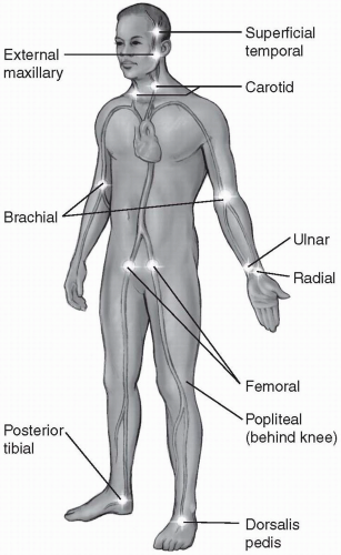
Figure 6-1 Sites for palpating arterial pulses.


Pulse Rate

Using the patient’s radial pulse on the wrist, count the pulsation for a full minute to get a sense of the heart rate and regularity. Use the finger pads of the middle fingers pressed lightly over the pulse. If the pulse is regular, count the pulsation for 30 seconds and multiply by 2 to get the 1-minute pulse rate. If the pulse is irregular, count the pulsation for a full minute.


The most accurate method for obtaining the heart rate, especially when the pulse is irregular, is by listening to the apical pulse with a stethoscope for a full minute (see Cardiac Assessment section later in this chapter.) An irregular heart rate can represent premature beats that might not be palpable peripherally at the radial pulse because left ventricular filling was incomplete prior to systole. When a pulse is irregular, note the type of irregularity: regularly irregular (e.g., beat, beat, pause, beat, beat, pause), irregular with respiration (e.g., an increasing rate with inspiration and decreasing rate with expiration), or totally irregular. Alternately, take a radial and apical pulse rate and note any discrepancies as they may vary with cardiac output.


Blood Pressure

The blood pressure is an important determinant of cardiovascular functioning. As the heart relaxes and contracts, it sends a pressure wave of blood through the circulatory system. The pressure wave produces the impulse detected as an arterial pulse and the Korotkoff sounds that are heard when auscultating a blood pressure with a stethoscope. Normally, the blood pressure is finely controlled by neural and hormonal influences that maintain adequate tissue perfusion. Measuring the blood pressure can assess the adequacy of the circulation and the regulatory mechanisms.

Blood pressure can be measured either directly or indirectly. Direct measurement requires the insertion of a catheter into an artery (see Figure 6-2). Indirect blood pressure readings are done with a blood pressure cuff and auscultation of Korotkoff sounds or Doppler reading of the pressure wave. Using a sphygmomanometer (a blood pressure cuff attached to a manometer) and a stethoscope, the blood pressure is usually taken on the upper arm. A mercury sphygmomanometer aneroid instrument may be used to take a blood pressure (see Table 6-1).


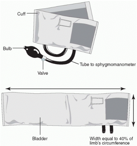
Figure 6-2 Blood pressure cuff-appropriate size for accurate mesurement.

The blood pressure measurement should be taken on both arms. When hypertension is suspected, the blood pressure should be repeated on three separate occasions. The patient should refrain from smoking cigarettes or drinking caffeine for 30 minutes before the blood pressure measurements. In patients with symptoms of dizziness and syncope, the blood pressure should be taken in the reclined, sitting, and standing positions to assess for orthostatic hypotension. Changing from the reclined to the standing position does not normally change the systolic pressure and may only slightly increase the diastolic pressure. But a decrease in the systolic blood pressure of more than 20 mmHg when changing from the sitting to standing positions may indicate orthostatic hypotension. Dehydration, hypovolemia, and certain antihypertensive medications are common causes of postural hypotension.


Table 6-1 Measuring Blood Pressure


1.


Choose a cuff of the appropriate size; the width should be 40% of the upper arm circumference (about 12 to 14 cm) (Figure 6-2). The bladder should be 80% of the circumference. A cuff that is too small may give an abnormally high reading.


2.


Have the patient uncross his or her legs. Palpate the brachial pulse and position the arm at the level of the patient’s heart with the palm facing up.


3.


Wrap the cuff around the upper arm and brachial artery so that the bottom of the cuff is about 2.5 cm above the brachial pulse. It is best to have no clothing on the arm when taking a blood pressure.


4.


To know how high to inflate the cuff, first take a palpable systolic reading. Palpate the radial pulse and rapidly inflate the cuff until the radial pulse disappears. Then, while deflating the cuff, palpate the return of the radial pulse. This is the palpable systolic pressure. Add 30 to this number, and use this as the highest pressure to which the cuff is to be inflated.


5.


Place the stethoscope over the brachial pulse. The bell of the stethoscope picks up the low-pitched sounds (Korotkoff sounds) better than the diaphragm.


6.


Inflate the cuff to the predetermined number (30 above the palpable systolic pressure), and slowly deflate the cuff (2 to 3 mmHg per second), listening for two consecutive sounds. Note this number as the systolic pressure. Continue deflating the cuff until the sounds become muffled and disappear. This is the diastolic pressure. The point of disappearance gives the best measure of diastolic pressure.


Paradoxical blood pressure is a decrease of greater than 10 mmHg in the systolic pressure during inspiration. This change is sometimes called pulsus paradoxus. It can be assessed by taking the blood pressure twice, once during inspiration and again at rest. First, palpate the systolic pressure while the patient stops breathing for a moment. Then, auscultate the blood pressure during inspiration and note when the sounds are first heard. Subtract the second number from the first and note any discrepancy between the two systolic pressures. Causes of pulsus paradoxus include pericardial tamponade, pulmonary hypertension, and restrictive pericarditis.


Blood pressure ranges for normal and high blood pressure have been labeled by the Joint National Committee on Detection, Evaluation, and Treatment of High Blood Pressure in the United States. Values for normal and abnormal blood pressures are seen in Table 6-2.

High blood pressure can be categorized in different ways. If both the systolic and diastolic pressures are elevated, the highest pressure determines the category of hypertension. When only the systolic or diastolic pressure is elevated, it is called isolated systolic or isolated diastolic hypertension.


Examination of the Chest

The examination of the chest allows assessment of the structure and functioning of the cardiovascular system as well as the respiratory system. To review, the landmarks for both cardiac and respiratory assessments are established by the anatomy of the thorax: the midsternal line, the midclavicular line, the sternal notch, the midscapular line, the midaxillary line, and the intercostal spaces between the ribs (see Figure 6-3). Several key landmarks are used in cardiac assessment as they approximate the location of cardiac structures within the bony thorax (see Figure 6-4). These cardiac landmarks are used during palpation of the chest wall and auscultation of the heart.


Table 6-2 Normal and Abnormal Blood Pressures


Category


Systolic


Diastolic


Normal


< 130


< 85


High normal


130-139


85-89


Mild hypertension


140-159


90-99


Moderate hypertension


160-179


100-109


Severe hypertension


180-209


110-119


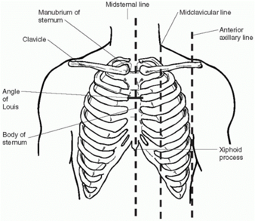
Figure 6-3 Thoracic landmarks.


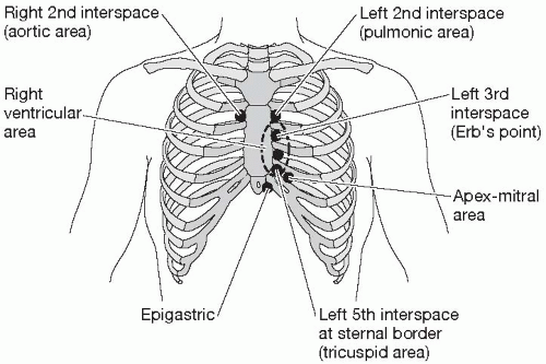
Figure 6-4 Cardiac landmarks.


Inspection

Start examining the patient by looking at the anterior chest while the patient is sitting. Take care to drape female patients as much as possible while allowing for adequate visualization of the thorax. Notice any pulsation, retractions, or movements of the chest wall. Inspect the large vessels of the neck: the carotid arteries and jugular veins. Note their location and how high up the neck the pulsations are visible. The jugular veins may also be used to estimate the venous pressure and the pressure in the right atrium of the heart. A noninvasive method of estimating venous pressure is to observe the jugular vein in the neck.


Assess the jugular venous distention using the following steps:

Oct 17, 2016 | Posted by in NURSING | Comments Off on Assessment of the Cardiovascular System

Full access? Get Clinical Tree

Get Clinical Tree app for offline access