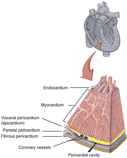
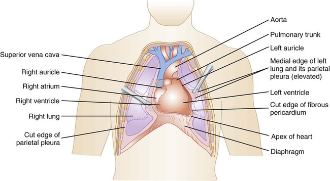
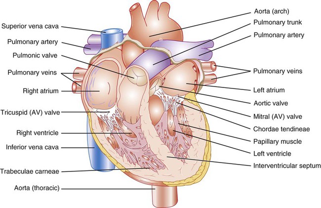
1. Describe the size and location of the heart. 2. Identify the layers of the heart wall, and state the type of tissue in each layer. 3. Label a diagram of the heart, including the chambers, valves, and associated vessels. 4. Trace the pathway of blood flow through the heart. 5. Describe the components and function of the conduction system of the heart. 6. Summarize the events of a cardiac cycle, and correlate the heart sounds heard with these events. 7. Describe the physical characteristics and functions of blood. 8. Identify the composition of blood plasma. 9. Identify the formed elements of the blood. 10. State the function of each formed element in blood. 11. Describe the life cycle of an erythrocyte. 12. List and describe the five types of leukocytes. 13. Explain the blood clotting mechanism of the body. 14. Explain the basis of blood types. 15. Describe the structure and function of arteries. 16. Describe the structure and function of capillaries. 17. Describe the structure and function of veins. 18. Describe ways in which the aging of an individual affects the circulatory system. Knowledge of the heart’s position in the thoracic cavity is important in hearing heart sounds, obtaining electrocardiograms (ECGs), and performing cardiopulmonary resuscitation (CPR). The heart, illustrated in Figure 12-1, is located in the thoracic cavity between the two lungs. It is posterior to the sternum and anterior to the vertebral column, and it rests on the diaphragm. About two thirds of the heart mass is to the left of the body’s midline, and one third is to the right. The apex, or pointed end of the heart, extends downward to the level of the fifth intercostal space. The opposite end, the base, is larger and less pointed than the apex and has several large vessels attached to it. Its most superior portion is at the level of the second rib. The size of the heart varies with the size of the individual. On average, it is about 9 cm wide and 12 cm long, which is about the size of a closed fist. The thick middle layer is the myocardium (my-oh-KAR-dee-um). It forms the bulk of the heart wall and is composed of cardiac muscle tissue. Refer to Chapter 5 for a review of the different types of muscle tissue. Contractions of the myocardium provide the force that ejects blood from the heart and moves it through the vessels. The smooth inner lining of the heart wall is the endocardium (en-doh-KAR-dee-um). Its smooth surface permits blood to move easily through the heart. The endocardium also forms the valves of the heart and is continuous with the lining of the blood vessels. Figure 12-2 illustrates the layers of the heart wall.
Circulatory System
Introduction to the Circulatory System
Heart
Overview of the Heart
Form, Size, and Location of the Heart
Structure of the Heart
Layers of the Heart Wall


Circulatory System
Get Clinical Tree app for offline access


