Introduction to anatomy and physiology
Barbara Lauritsen Christensen and M. Christine Neff
Objectives
1. Define the difference between anatomy and physiology.
2. Define the term anatomical position.
4. Use each word in a given list of anatomical terms in a sentence.
5. List the nine abdominopelvic regions and the abdominopelvic quadrants.
6. List and discuss in order of increasing complexity the levels of organization of the body.
7. Differentiate among tissues, organs, and systems.
8. Identify and define three major components of the cell.
9. Discuss the stages of mitosis and explain the importance of cellular reproduction.
11. Describe the four types of body tissues.
12. Discuss the two types of epithelial membranes.
13. List the 11 major organ systems of the body and briefly describe the major functions of each.
Key terms
cytoplasm (C -t
-t -pl
-pl zm, p. 6)
 -F
-F -zh
-zh n,
n, homeostasis (h -m
-m –
– -ST
-ST -s
-s s, p. 5)
 -T
-T -s
-s s,
s,  z-M
z-M -s
-s s,
s, phagocytosis (f g-
g- -s
-s -T
-T -s
-s s, p. 8)
physiology (f z-
z- –
– L-
L- -j
-j , p. 1)
pinocytosis (p-n -s
-s -T
-T -s
-s s, p. 8)
Caring for a person with a disease process requires an understanding of the normal functioning of the human body, so the nurse must know basic human anatomy and physiology. Anatomy is the study, classification, and description of structures and organs of the body. Physiology explains the processes and functions of the various structures and how they interrelate. The normal, healthy human body is like a finely tuned machine, with each part performing a special function to accomplish a goal. As with the machine, when the body malfunctions, the repairer must understand how it works. Without the necessary repairs to return the body to homeostasis, illness, disease, or death may result.
Anatomical terminology
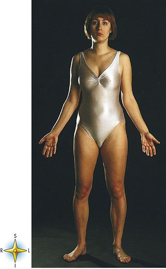
Study of the human body first requires one to master certain terms that aid in locating specific structures. To understand the following terms, consider the body in a normal anatomical position, that is, standing erect with the face and palms facing forward (Figure 1-1):
Anterior (or ventral): To face forward; the front of the body. The chest is located anterior to the spine (Figure 1-2).
Posterior (or dorsal): Toward the back. The kidneys are posterior to the peritoneum.
Cranial: Toward the head. The brain is located in the cranial portion of the body.
Caudal: Toward the “tail”; the distal portion of the spine. A caudal anesthetic may be given.
Superior: Toward the head or above. The neck is superior to the shoulders.
Inferior: Lower, toward the feet, or below another. The foot is inferior to the ankle.
Medial: Toward the midline. The sternum (breastbone) is located in the medial portion of the chest.
Superficial: Nearer the surface. The skin of the arm is superficial to the muscles below it.
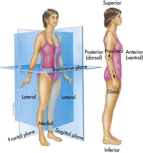
Body planes
To make it easier to study individual organs or the body as a whole, divide the body into three imaginary planes: the sagittal, the coronal (frontal), and the transverse (see Figure 1-2):
Body cavities
From the outside, the body appears to be a solid structure, but it is not. It is made up of open spaces, or cavities, that contain compact, well-ordered arrangements of internal organs. The body has two major cavities that are, in turn, subdivided and contain compact, well-ordered arrangements of internal organs. The two major cavities are the ventral and the dorsal body cavities (Figure 1-3 and Table 1-1).
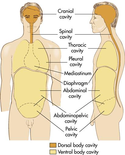
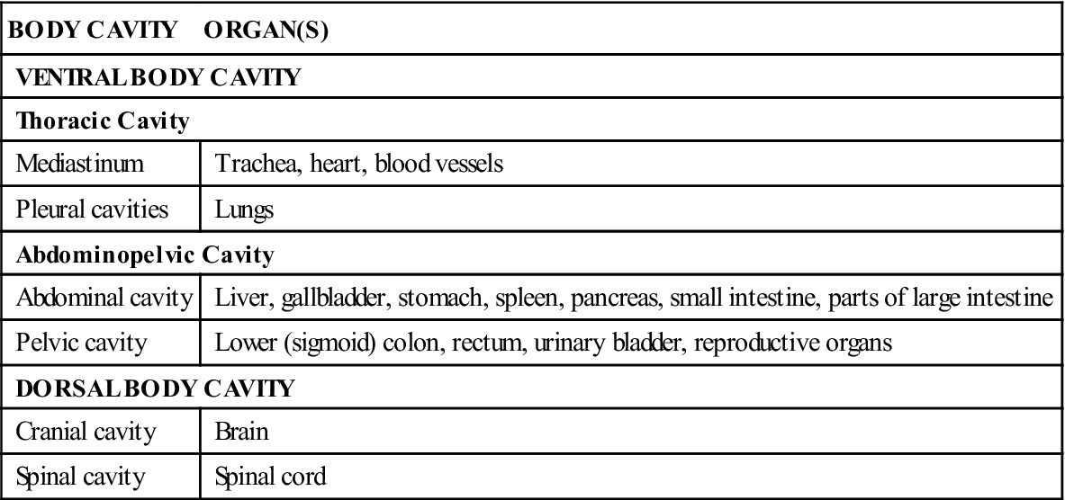
Table 1-1
| BODY CAVITY | ORGAN(S) |
| VENTRAL BODY CAVITY | |
| Thoracic Cavity | |
| Mediastinum | Trachea, heart, blood vessels |
| Pleural cavities | Lungs |
| Abdominopelvic Cavity | |
| Abdominal cavity | Liver, gallbladder, stomach, spleen, pancreas, small intestine, parts of large intestine |
| Pelvic cavity | Lower (sigmoid) colon, rectum, urinary bladder, reproductive organs |
| DORSAL BODY CAVITY | |
| Cranial cavity | Brain |
| Spinal cavity | Spinal cord |
Ventral cavity
The ventral cavity consists of the thoracic (or chest) cavity and the abdominopelvic cavity (see Figure 1-3), which are separated by the diaphragm (a muscle directly beneath the lungs).
The thoracic cavity contains the heart and the lungs. Its midportion is a subdivision of the thoracic cavity, the mediastinum, which contains the trachea, the heart, and the blood vessels. Its other subdivisions are the right and left pleural cavities, which contain the lungs.
The abdominal cavity contains the stomach, the liver, the gallbladder, the spleen, the pancreas, the small intestine, and parts of the large intestine. A subdivision called the pelvic cavity contains the lower portion of the large intestine (lower sigmoid colon, rectum), the urinary bladder, and the internal structures of the reproductive system. The abdominal and pelvic cavities are not separated by any structure and therefore are referred to as the abdominopelvic cavity (see Table 1-1).
Dorsal cavity
The dorsal cavity is composed of the cranial and spinal body cavities. The cranial body cavity houses the brain, whereas the spinal cavity contains the spinal cord. The dorsal body cavity is smaller than the ventral cavity (see Table 1-1).
Abdominal regions
For convenience in locating abdominal organs, anatomists divide the abdomen into nine imaginary regions. The nine regions (Figure 1-4), identified from right to left and from top to bottom, are the following:
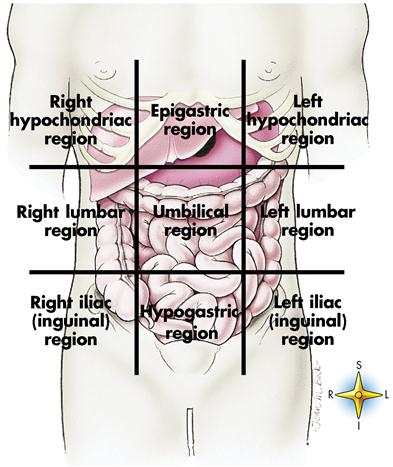
The most superficial organs located in each of the nine abdominal regions are shown in Figure 1-4. The visible organs in each region are as follows: (1) right hypochondriac region, the right lobe of the liver and the gallbladder; (2) epigastric region, parts of the right and left lobes of the liver and a large portion of the stomach; (3) left hypochondriac region, a small portion of the stomach and large intestine; (4) right lumbar region, parts of the large and small intestine; (5) umbilical region, a portion of the transverse colon and loops of the small intestine; (6) left lumbar region, additional loops of the small intestine and a part of the colon; (7) right iliac region, the cecum and parts of the small intestine; (8) hypogastric region, loops of the small intestine, the urinary bladder, and the appendix; and (9) left iliac region, portions of the colon and the small intestine.
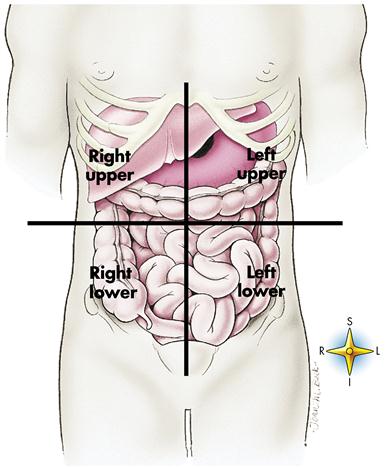
Abdominopelvic quadrants
Health professionals frequently divide the abdomen into four quadrants to describe the site of abdominopelvic pain or locate an internal pathologic condition such as a tumor or abscess (Figure 1-5). Horizontal and vertical lines passing through the umbilicus (navel) divide the abdomen into right and left upper quadrants and right and left lower quadrants.
Structural levels of organization
Before studying the structure and function of the human body and its many parts, think about how those parts are organized and how they might fit together into a functioning whole. Figure 1-6 illustrates the different levels of organization that influence body structure and function. The levels of organization progress from the least complex (chemical level) to the most complex (body as a whole). The structural levels of organization in the body are cells, tissues, organs, and systems.
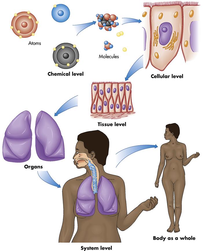
Although the body is a single structure, it is made up of billions of smaller structures. Atoms and molecules are often referred to as the chemical level of organization (see Figure 1-6). Atoms are small particles that form the building blocks of matter, the smallest complete units of which all matter is made. When two or more atoms unite through their electron structures, they form a molecule. A molecule can be made of atoms that are alike (e.g., the oxygen molecule is made of two identical atoms), but more often a molecule is made of two or more different atoms (e.g., a molecule of water [H2O] contains one atom of oxygen [O] and two atoms of hydrogen [H]) (see Figure 1-6).
The existence of life depends on the proper levels and proportions of many chemical substances in the cytoplasm of cells. Cells are considered the smallest living units of structure and function in our body. Although considered the simplest units of living matter, cells are far from simple; they are extremely complex.
Tissues are even more complex than cells. By definition a tissue is an organization of many similar cells that act together to perform a common function. Cells are held together and surrounded by varying amounts and types of gluelike, nonliving intercellular substances.
Organs are more complex than tissues. An organ is a group of several different kinds of tissues arranged to perform a special function. The lungs shown in Figure 1-6 are an example of organization at the organ level.
Systems are the most complex units that make up the body. A system is an organization of varying numbers and kinds of organs arranged to perform complex functions for the body. The organs of the respiratory system shown in Figure 1-6 permit air to enter the body and travel to the lungs, where oxygen and carbon dioxide are exchanged. Organs of the respiratory system include the nose, the windpipe (or trachea), and the complex series of bronchial tubes that permit passage of air into the lungs.
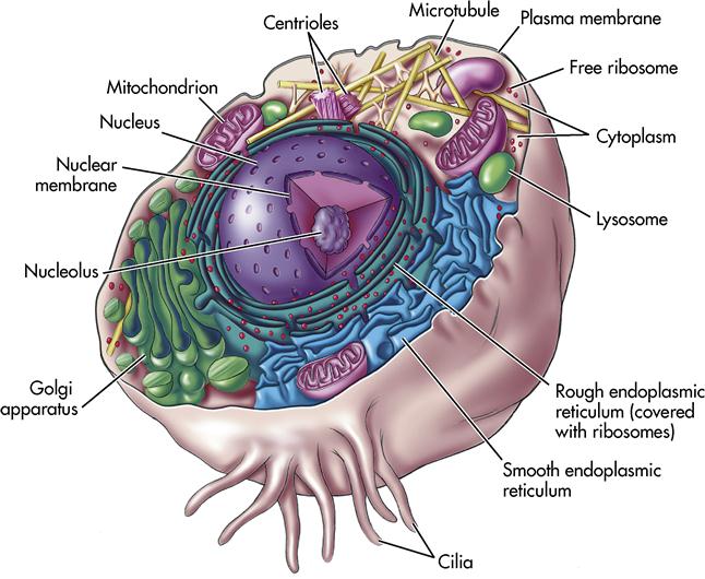
Cells
Almost 350 years ago Robert Hooke discovered the first cell while examining plant fragments under the microscope. The structures reminded him of tiny, individual miniature prison cells, so he coined the term cell (the fundamental unit of all living tissue) (Figure 1-7). Many living things are so simple that they consist of just one cell. The human body, however, is so complex that it has trillions of these tiny powerhouses of life.
All cells are microscopic but differ widely in size and shape. Despite their differences, all cells exhibit five unique characteristics of life: growth, metabolism, responsiveness, reproduction, and homeostasis. Homeostasis is when the body’s internal environment is relatively constant; this state is naturally maintained by adaptive responses that promote healthy survival.
Structural parts of cells
The three main parts of a cell are the plasma membrane, the cytoplasm, and the nucleus (see Figure 1-7).
Plasma membrane
The plasma membrane encloses the cytoplasm and forms the outer boundary of the cell. It is an incredibly delicate structure—only about 7 nm (nanometers), or 3/10,000,000 inch, thick! Yet it has a precise, orderly structure.
Even though it seems fragile, the plasma membrane is strong enough to keep the cell whole and intact. It also performs other life-preserving functions for the cell, serving as a gateway between the fluid inside the cell and the fluid around it. The plasma membrane is selectively permeable. This means the membrane permits certain substances to enter and leave while not allowing other substances to cross. This membrane separates the cell contents from the dilute saltwater solution called interstitial fluid, or simply tissue fluid, which bathes every cell in the body. The plasma membrane also has distinct surface proteins that identify a cell as coming from one particular individual. This fact is the basis of tissue typing, a procedure performed before an organ from one person is transplanted into another. Carbohydrate chains attached to the surface of cells often help identify cell types.
Cytoplasm
Cytoplasm is the internal living material of cells. Cytoplasm (protoplasm) is a sticky, fluidlike substance that lies between the plasma membrane and the nucleus of the cell. Numerous organelles (tiny functioning structures) are located within the cytoplasm. Cells contain cytoplasm, or “living matter,” a substance that exists only in cells. Cytoplasm is composed largely of a gel-like substance that contains water, food, minerals, enzymes, and other specialized materials. The term cyto- is a combining form from the Greek and denotes a relationship to a cell. These organelles were not discovered until the development of the powerful electron microscope.
Cytoplasm is composed of 70% water with traces of proteins, lipids, carbohydrates, minerals, and salts. (Table 1-2 lists major cell structures and their functions.)
Table 1-2
Some Major Cell Structures and Their Functions
| CELL STRUCTURE | FUNCTION(S) |
| Plasma membrane | Serves as the cell’s boundary; protein and carbohydrate molecules on outer surface of plasma membrane perform various functions (e.g., serve as markers that identify cells of each individual or as receptor molecules for certain hormones) |
| Endoplasmic reticulum (ER) | Ribosomes attach to rough ER to synthesize proteins; smooth ER synthesizes lipids and certain carbohydrates |
| Ribosomes | Synthesize proteins; the cell’s “protein factories” |
| Mitochondria | Synthesize adenosine triphosphate (ATP); the cell’s “powerhouses” |
| Lysosomes | Serve as cell’s “digestive system” |
| Golgi apparatus | Synthesizes carbohydrate, combines it with protein, and packages the product as globules of glycoprotein |
| Centrioles | Function in cell reproduction |
| Cilia | Short, hairlike extensions on the free surfaces of some cells capable of movement; often have specialized functions such as propelling mucus upward over cells that line the respiratory tract |
| Flagella | Single projections of cell surfaces, much larger than cilia; an example in humans is the “tail” of a sperm cell; propulsive movement makes it possible for sperm to “swim” or move toward the ovum once they are deposited in the female reproductive tract |
| Nucleus | Dictates protein synthesis, thereby playing an essential role in other cell activities, namely, active transport, metabolism, growth, and heredity |
| Nucleoli | Play an essential role in the formation of ribosomes |
Nucleus
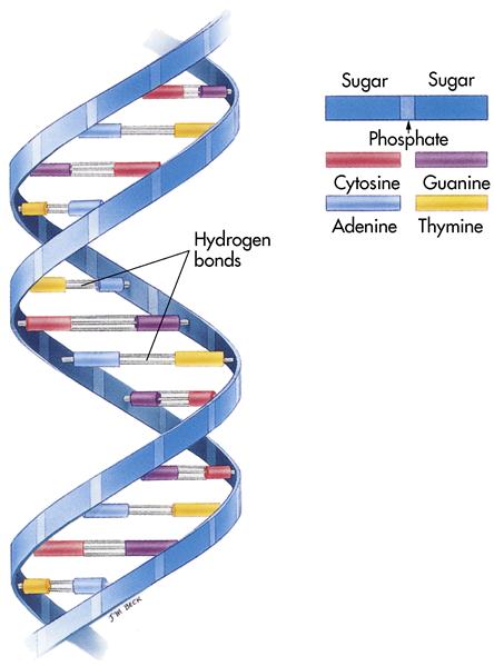
The nucleus is the largest organelle within the cell. It is responsible for cell reproduction and control of the other organelles. The nucleus is surrounded by the nuclear membrane. It contains nucleoplasm, a refined form of cytoplasm. The nucleus contains two specialized structures: the nucleolus and the chromatin granules. The nucleolus is critical in the formation of protein. The chromatin granules are composed of protein and deoxyribonucleic acid (DNA). DNA contains the genetic code, or blueprint, of the body.
Endoplasmic reticulum
Throughout the cytoplasm lies a system of membranes, or canals, called the endoplasmic reticulum (ER). ER functions as a miniature circulating system for the cell by carrying substances from one part of the cell to another. There are two types of ER: (1) smooth, which is found in cells that deal with fatty substances; and (2) rough, which is found in cells that manufacture proteins.
Ribosomes
Ribosomes are tiny structures floating free in the cytoplasm or attached to the rough ER. They are called protein factories because they produce enzymes and other proteins.
Mitochondria
The mitochondria are the powerhouses of the cells. They are bean shaped with a folded interior membrane. They take food and convert it to a complex energy form, adenosine triphosphate (ATP), for use by the cell. ATP is described as the “energy currency” of the cells because it supplies the energy for all activities.
Lysosomes
Lysosomes are small saclike structures containing enzymes that digest food compounds and microbes that have invaded the cell.
Golgi apparatus
The Golgi apparatus is usually located near the nucleus. It is the “packaging plant” of the cell. It packages certain carbohydrate and protein compounds into globules. Then it moves outward through the cell membrane, where it breaks open and releases its contents.
Centrioles
The centrioles are paired, rod-shaped organelles. During cell division (mitosis) they aid in the formation of the spindle, a structure necessary for cell reproduction.
Protein synthesis
Protein is a vital component of every cell in the body. Protein production relies on nucleic acids in the cell’s cytoplasm and nucleus. Two important nucleic acids are (1) DNA (Figure 1-8), which is located in the nucleus; and (2) ribonucleic acid (RNA), which is located in the cytoplasm. The DNA encodes the message for protein synthesis and sends it to the RNA, which transports it to the ribosomes, where the protein is produced. Hence DNA is called the chemical blueprint, and RNA is called the chemical messenger.
Stay updated, free articles. Join our Telegram channel

Full access? Get Clinical Tree


