Organ Donation and Donor Management


Organ Donation and Donor Management


Maureen T. Smith

Joanne V. Hickey


The Uniform Anatomical Gift Act of 1968 (revised 2006) made it clear that a person, or their designee, may donate all or part of their body for science or transplantation.1 Since the act passed, there has been a growing disproportion between the number of people waiting for a life-saving organ transplant and the number of organs available for transplantation in the United States and the world. According to The Organ Procurement and Transplantation Network (OPTN) there are more than 113,000 people on the organ transplant waiting list.2 Conversely, from January to November of 2011 there were a total of 26,247 organs transplanted from a total of 12,960 donors.2

Over the past several years, the number of people on waiting lists has grown while the number of donors has been stagnant. In an effort to improve this disparity, the Department of Health and Human Services in 2003 initiated the Organ Donation Breakthrough Collaborative. The goal of the Collaborative is “saving or enhancing thousands of lives a year by spreading known best practices to the nation’s largest hospitals, to achieve organ donation rates of 75% or higher in these hospitals.”3 Since the original charter was developed, the goals of the Collaborative have expanded to improve: (1) the number of organs made available; (2) the capacity of organ procurement organizations (OPO) and transplant centers to effectively manage more organ donors and perform more organ transplants; and (3) efforts to expand the use of other types of organ donors such as cardiac death donors and expanded criteria donors.3


There are two categories of organ donation from deceased donors based on the criteria identified as the cause of death. First, death based on the criteria of absence of cardiopulmonary function is identified as cardiac death. Second, death based on the criteria of absence of neurological function is identified as brain death. Each cause of death will be discussed.


ORGAN DONATION AFTER CARDIAC OR CIRCULATORY DEATH (DCD)

The definition of organ donation after cardiac or circulatory death (DCD) is defined as the surgical removal of organs after the pronouncement of death based on the absence of cardiopulmonary function. Patients considered for DCD usually have sustained a nonrecoverable neurological injury that does not progress to brain death, but the next of kin elects to withdraw life-sustaining therapies. After the next of kin makes the decision to withdraw life-sustaining therapies, evaluation of the patient occurs to determine whether death will occur within a predetermined period of time after withdrawal of support. The time frame from the withdrawal of support to organ recovery varies according to individual institutional policy, but should consider the maximum acceptable interval for minimizing ischemic organ injury.

To ensure that there is no conflict of interest, the decision to limit, withhold, or withdraw life-sustaining therapy should be independent of any discussion regarding organ and tissue donation. After attaining consent for DCD, physiological support is maintained through the evaluation and organ allocation period. Once organ allocation review is completed and procurement teams are available and a recipient has been notified/prepared for transplant (especially if there is a small organ viability period once procurement has occurred), the patient is transferred to the operating room and life support is withdrawn in the presence of the care team.

When cardiopulmonary function has ceased, the patient is pronounced dead by a provider not associated with procurement teams or the OPO. The transplant team must wait a pre-established time and then begins the surgical recovery of the organ or organs consented for donation. The Institute of Medicine, the American College of Critical Care Medicine, and the Society for Critical Care Medicine recommend waiting 2 to 5 minutes after cessation of cardiopulmonary function before beginning organ procurement.4


ORGAN DONATION AFTER NEUROLOGICAL DEATH

For some unfortunate patients, injury to the nervous system is so severe that the injuries are incompatible with life. Neuroscience nurses are often the first health professionals to recognize a clinical pattern that could lead to brain death. Potentially fatal brain injury (e.g., severe cerebral trauma, major stroke, severe subarachnoid hemorrhage), a Glasgow Coma Score of less than 5, absence of two or more brainstem reflexes, or sustained intracranial pressure of more than 30 mm Hg are ominous red flags of neurological demise. The appropriate OPO should be called as early as possible when organ donation is even a remote possibility. The OPO will assess each case to determine possible donation. It is the goal of the OPO to rule in as many people as possible for organ donation and, therefore, it is critical to contact the OPO so that they can be involved early to evaluate the patient and make necessary arrangements. For more information call 1-800-558-LIFE (5433).

In 1995, the American Academy of Neurology published practice parameters for determining brain death in adults.5 Brain death is a clinical diagnosis made by a physician. Brain death is defined as irreversible cessation of all functions of the entire brain, including the brainstem.6, 7 Diagnosis of brain death is made based on fulfillment of strict, well-defined clinical criteria of irreversible coma, absence of cortical activity, absence of motor response to pain, loss of brainstem reflexes, and apnea. Ancillary diagnostic tests may be used to support the clinical diagnosis, but they are not strictly required to make the diagnosis. It is critical that the cause of the coma be investigated, and that a cause can be identified that is capable of causing irreversible apneic coma when there is no evidence of cerebral or brainstem function.8 The guidelines underscore three key clinical findings necessary to confirm irreversible cessation of all functions of the entire brain. The three key characteristics in brain death are coma or unresponsiveness, absence of brainstem reflexes, and apnea.5 Each finding is discussed briefly below.


Coma or Unresponsiveness

Definite clinical, neuroimaging, or cerebrospinal fluid evidence of an acute central nervous system catastrophic event compatible with death must be found. All reversible causes of coma must be excluded before a diagnosis of brain death can be made. Potentially reversible confounding factors of coma include hypothermia (core temperature below 32°C related to blunted brainstem reflexes which can be addressed with a warming blanket, fluid warmers, gastric lavage, and/or coronary bypass to raise the temperature to more than 36°C); drug intoxication or poisoning; use of neuromuscular blocking agents (check presence of neuromuscular blockade with the train of four twitches with maximal nerve stimulation); severe electrolyte imbalance; severe acid-base abnormalities; and severe metabolic or endocrine imbalance. If barbiturates have been used (e.g., barbiturate-induced coma), the diagnosis of brain death can still be made if the levels of drug are subtherapeutic. Exclusion of the presence and effect of a central nervous system depressant can be made by history, drug screen, calculation of the clearance using five times the drug’s half-life (assuming normal hepatic and renal function) or drug plasma levels.9 In addition, Wijdicks recommends that definite clinical, neuroimaging, or cerebrospinal fluid evidence of an acute central nervous system catastrophic event compatible with brain death must be found.8


Absence of Brainstem Reflexes

Loss of brainstem function is incompatible with life. The brainstem reflexes that are assessed include pupillary reaction to light, corneal reflex, gag reflex, and oculovestibular reflex. All reflexes should be absent in brain death. There should not be a light response to a bright light introduced to the pupil. Most pupils are midposition (4 to 6 mm), although there may be variations from 4 to 9 mm, all of which are compatible with brain death. In addition, pupils may be round, ovoid, or irregularly shaped. The corneal reflex is tested with a wisp of cotton or saline drops, and the reflex should be absent. With the application of deep pain, observe for any grimacing in response to the deep pain. The gag reflex is tested with a tongue blade. Along with an absent gag reflex, the cough reflex should also be absent. Ocular movements, as tested by head turning and caloric testing, are absent in brain death. (See Chapter 7 for a description of caloric testing and Chapter 8 for assessment of ocular movement, including precautions to be taken before testing.) Note that sedatives, aminoglycosides, tricyclic antidepressants, anticholinergics,
anticonvulsants, and chemotherapeutic agents can diminish or completely abolish the caloric response.5, 8 Loss of brainstem function also results in loss of breathing and vasomotor control, which results in apnea and hypotension to loss of blood pressure.


Apnea

In diagnosing brain death, demonstration of apnea to evaluate respiratory drive (brainstem function) is critical. Severe hypotension and cardiac arrhythmias (e.g., premature ventricular contractions, ventricular tachycardia) may occur during apnea testing, either spontaneously due to acidosis or related to inadequate precautions. Therefore, there are prerequisites recommended when testing for apnea: (1) core temperature of 36°C (97°F) or higher; (2) systolic blood pressure 90 mm Hg or more; (3) positive fluid balance in the past 6 hours; (4) arterial PCO2 normalized between 35 and 45 mm Hg; and (5) arterial PO2 200 mm Hg or more.5, 8

Although there may be slight variations in institutional protocol for apnea testing, the following outlines a model apnea testing protocol that the physician may follow5:


  • Preoxygenate with 100% FiO2 for 30 minutes.


  • Disconnect the patient from the ventilator (a PCO2 rise of 3 to 6 mm Hg/min is estimated in the apneic patient).


  • Immediately upon disconnection, place an oxygen cannula at the level of the carina and administer 100% oxygen at 9 to 12 L/min by tracheal cannula. Observe for respiratory movement of the chest/abdomen for 8 minutes (a respiration is defined as abdominal or chest movement that produces adequate tidal volume).


  • After 10 minutes, draw arterial blood gases. If there is no respiratory movement and the PCO2 is 60 mm Hg or higher, the clinical diagnosis of brain death is made. If the PCO2 has not met the target level of 60 mm Hg or higher, apnea testing is repeated after a period of time and confirmatory testing may also be ordered. Note that the target PCO2 may be higher in patients with chronic hypercapnia (e.g., severe chronic obstructive pulmonary disease, bronchiectasis, sleep apnea, morbid obesity) because the patient’s PCO2 baseline is higher. If chronic hypercapnia is suspected, additional noninvasive confirmatory tests are strongly recommended.5, 8


REFLEX MOTOR ACTIVITY

Spontaneous motor responses of spinal origin sometimes called the “Lazarus sign,” observed as limb movements, may be seen in brain death.10 Possible movements seen, especially in younger patients, can include any of the following: rapid flexion of the arms; raising of one or all of the limbs off of the bed; grasping movements; or jerking of one leg. In addition, multifocal vigorous myoclonus may be noted. Although unexpected, these movements are not purposeful and should not be interpreted as such.


CONFIRMATORY TESTING

As mentioned earlier, brain death is a clinical diagnosis made by a physician. In most instances, the clinical evaluation is conducted two times with an interval of hours (e.g., 6 to 8 hours) elapsing between examinations. Many hospitals have developed guidelines that often include a requirement of two independent clinical evaluations by two different physicians. Figure 4-1 provides a sample guideline. Confirmatory testing is not required to make the diagnosis of brain death in the United States. However, a physician may choose to include confirmatory tests for patients in whom specific components of the clinical evaluation cannot be reliably tested. Confirmatory tests that are accepted based on clinical experience and reliability include conventional angiography, blood-flow studies (e.g., technetium 99 m), electroencephalography, and more recently, transcranial Doppler ultrasonography. Absence of blood flow and electrical activity of the brain are confirmatory findings of brain death. Wijdicks, et al.9 provides an excellent discussion of use, validity, and disadvantages of confirmatory tests and brain death.8, 11

After brain death has been confirmed, the patient is pronounced dead and all life-sustaining treatment is stopped.12 The use of ventilators, medications, and warming therapies will continue if organ donation is planned. After brain death has been confirmed, the patient is pronounced dead.

The 2010 evidence-based guideline update for determining brain death in adults expands the 1995 guidelines published by the American Academy of Nursing.9 The objectives of the 2010 publication were (1) to provide an update of the 1995 guidelines regarding determination of brain death in adults and (2) to use evidence-based methods to answer five questions related to variations in brain death determination to promote uniformity in diagnosis. A systematic review of the literature including classification and rating of the strength of evidence was used to answer the five questions (Table 4-1). In addition, the authors note that many of the details of the clinical neurological examination used to establish brain death cannot be established by evidence-based methods, but are based on practical guidance. A fourstep process is provided for the determination of brain death. The first is the prerequisites of the clinical evaluation. It includes establishing an irreversible and proximate cause of coma, achieving normal core temperature, achieving normal systolic blood pressure, and performance of one neurological examination (sufficient to pronounce brain death in most states in the United States). The second component is the neurological assessment which includes establishing coma, absence of brainstem reflexes, and apnea (previously addressed in this chapter). The third component is ancillary testing such as EEG, CTA, TCD, MRI/MRA, and nuclear scan. Finally, documentation of brain death in the record is critical including the details of when ancillary tests were conducted and results, apnea testing and results, time of death signed including date and time by the physician. The nurses’ notes usually provide additional information such as supportive nursing care, family present and response, notifications, and other related and complimentary information.


ROLE OF THE NEUROSCIENCE NURSE RELATED TO BRAIN DEATH

The nurse has a major independent and collaborative role in caring for the patient with impending and pronounced brain death. First, the nurse must understand what brain death is and how it manifests itself. This includes understanding the reflex movements and other aberrations that can occur in brain death that can be confusing and alarming to family members. Second, the nurse provides empathetic family support to an often stunned, immobilized, and grieving family. Listening and hearing what the family is saying provides direction for emotional support, education, and identification and mobilization of resources to support the family. Because the process of declaration of brain death often occurs over hours, the basic needs, comfort, spiritual support, and privacy of a family are often left to the nurse to address.

Third, communications become complex and involve multiple care providers, support services, family members, and others connected to
the patient. The nurse is often central in communications with the interdisciplinary practice team and interface with the family. Referrals may need to be made to the ethics committee, social worker, case manager, and others which include sharing of critical information. The interface with the family requires interpretation, clarification, and reinforcement of information throughout the process. It includes arranging for visitation and helping family in saying “good-bye” to the patient.


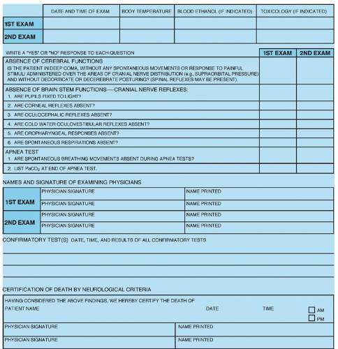
Figure 4-1 ▪ A sample checklist for determination of death by neurological criteria. The cause of coma must be established and sufficient to account for the loss of all brain function. Reversible conditions, such as drug sedation, metabolic disturbance, hypothermia (below 32.2°C), neuromuscular blockage, and shock, must be searched for and appropriately treated. Two separate clinical examinations must be completed, the second no sooner than 6 hours after the first. Each examination must be conducted by two physicians, independent of each other, who shall be licensed to practice medicine in the state. (Note that in this sample form, the institution requires two physicians to examine the patient each time.) The physicians may or may not choose to perform cold calorics.

Fourth, coordination and transition of care fall to the nurse. The transition in care from therapeutic to supportive care occurs as the diagnosis of brain death becomes clear. Care with attention to basic care needs for a presentable, clean, and groomed physical appearance is important to convey respect and a peaceful image to the family. The physical environment must also be addressed to further convey an uncluttered environment where the last moments can be spent with the loved one by the family in privacy and comfort.

Finally nurses must work collaboratively with the organ donation organization to make organ donation an option for those who wish to participate. Hospital policies and procedures should be
designed to support the nurse with necessary clear information to provide efficient and effective care for the patient and family.


TABLE 4-1 FIVE QUESTIONS AND RECOMMENDATIONS ADDRESSED IN 2010 UPDATE RELATED TO BRAIN DEATH


QUESTION ADDRESSED


RECOMMENDATION


Are there patients who fulfill clinical criteria of brain death who recover?


The 1995 criteria for brain death are still valid; no evidence of recovery has been found.


What is an adequate observation period to ensure cessation of neurologic function is permanent?


There is insufficient evidence to determine the minimally acceptable observation period.


Are complex motor movements that falsely suggest retained brain function sometimes observed in brain death?


Yes; false-positive triggering on the ventilator may occur in brain-dead patients.


What is the comparative safety of technique for determining apnea?


There is insufficient evidence to determine comparative safety of technique used for apnea testing.


Are there new ancillary tests that accurately identify patients with brain death?


There is insufficient evidence to change current practices.


From: Wijdicks, E. F., Varelas, P. N., Gronseth, G. S., & Greer, D. M., American Academy of Neurology. (2010). Evidence-based guideline update: Determining brain death in adults: Report of the Quality Standards Subcommittee of the American Academy of Neurology. Neurology, 74(23), 1911-1918, with permission.

Only gold members can continue reading. Log In or Register to continue

Stay updated, free articles. Join our Telegram channel

Jul 14, 2016 | Posted by in NURSING | Comments Off on Organ Donation and Donor Management

Full access? Get Clinical Tree

Get Clinical Tree app for offline access