nucleus, and red nucleus. Symptoms of PD are caused by loss of nerve cells in the pigmented substantia nigra pars compacta, including the locus ceruleus in the midbrain. Cell loss also occurs in the globus pallidus and putamen.3 Depletion of the dopaminergic neurons (pars compacta) of the substantia nigra results in reduction of dopamine, the main biochemical abnormality in PD. Normally, the pars compacta neurons of the substantia nigra provide dopaminergic input to the striatum, a part of the basal ganglia. In PD, loss of pars compacta neurons leads to striatal dopamine depletion and reduced thalamic excitation of the motor cortex.3 Although the central problem in PD is dopaminergic loss in the basal ganglia, other neurotransmitters are also involved and the disease extends to other areas of the brain.5
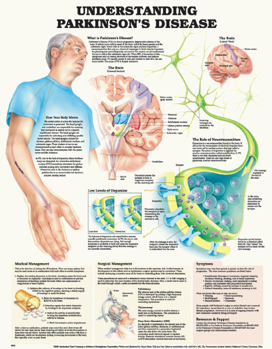 Figure 31-1 ▪ Understanding Parkinson’s disease. Asset provided by Anatomical Chart Co. |
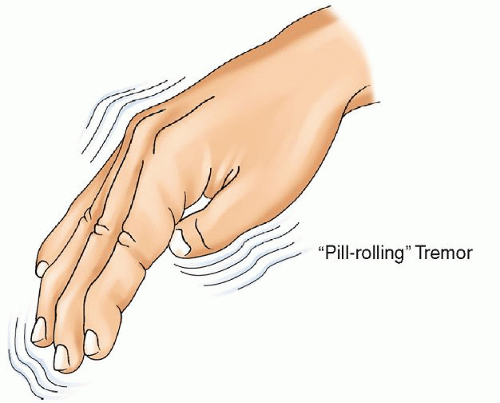 Figure 31-2 ▪ Tremors of Parkinson’s disease. |
 Figure 31-3 ▪ The clinical features of Parkinson’s disease. |
scale described by Hoehn and Yahr is frequently used to characterize the severity of illness (Table 31-2).12, 13 The standard tool for tracking PD progress and response to therapy is the United Parkinson’s Disease Rating Scale (UPDRS). The UPDRS is a standard rating scale used for tracking PD progress and response to therapy in patients with PD. The UPDRS is subdivided into three scales including cognitive and mood aspects, motor aspects, and ADLs. The UPDRS is available at http://www.mdvu.org/library/ratingscales/pd/. Items include mentation, behavior, and mood (e.g., intellectual impairment, thought disorder, depression, motivation/initiative); ADLs (e.g., speech, salivation, swallowing handwriting, etc.); and motor examination (e.g., facial expression, rigidity, hand movements, arising from a chair, etc.). Most items scored on a scale from 0 to 4 (0 = normal to 4 = most severe). There are a few items scored on a scale of 0 to 1. The scores on the subscales are added together to yield a total score (higher scores indicate a higher degree of disability).14 The scale has good inter-rater reliability. In addition, other standardized scales, such as the Beck Depression Inventory for depression are helpful for assessing and monitoring other symptoms common in PD.
TABLE 31-1 SECONDARY MANIFESTATIONS OF PARKINSON’S DISEASE | ||||||||||||||||||||||||||||||||||||||||||||||||||||||||
|---|---|---|---|---|---|---|---|---|---|---|---|---|---|---|---|---|---|---|---|---|---|---|---|---|---|---|---|---|---|---|---|---|---|---|---|---|---|---|---|---|---|---|---|---|---|---|---|---|---|---|---|---|---|---|---|---|
| ||||||||||||||||||||||||||||||||||||||||||||||||||||||||
tomography (SPECT) imaging with DaTSCAN has been recently approved for diagnosis of PD. DaTSCAN is a dopamine transporter ligand that tags presynaptic dopaminergic neurons in the basal ganglia; in patients with PD, there is a decreased signal on SPECT.5
Stay updated, free articles. Join our Telegram channel

Full access? Get Clinical Tree


