Neuroendocrine Disorders in Neuroscience Patients


Neuroendocrine Disorders in Neuroscience Patients


Laura J. Griffin

Joanne V. Hickey


Several interrelated, complex physiologic mechanisms work together to maintain homeostasis. The hypothalamic-pituitary-adrenal (HPA) axis plays an integral role in creating this delicate balance. Any central neurological process can result in injury, inflammation, or compression of the hypothalamus or pituitary gland, resulting in endocrine dysfunction. As many as 15% to 68% of patients with traumatic brain injury (TBI) and 37% to 55% with subarachnoid hemorrhage (SAH) have been found to have some form of endocrine dysfunction.1 Since untreated hypopituitary functioning has been associated with increased mortality and morbidity, the nurse must vigilantly assess patients for signs and symptoms of neuroendocrine dysfunction.1 This chapter will address neuroendocrine dysfunction including corresponding fluid and electrolyte disorders such as diabetes insipidus (DI), syndrome of inappropriate secretion of antidiuretic hormone (SIADH), and cerebral salt wasting (CSW). Neuroendocrine dysfunction related to catecholamine excess including sympathetic storming, neurogenic stress cardiomyopathy (NSC), and neurogenic pulmonary edema (NPE) will also be discussed.


HYPOTHALAMIC-PITUITARY AXIS

The term hypothalamic-pituitary axis is used to describe the intricate relationship between the hypothalamus and the pituitary gland. Through production of five hypophysiotropic hormones (e.g., releasing and inhibiting factors) and the neurotransmitter dopamine, the hypothalamus exerts a direct influence on the secretion of anterior pituitary hormones. The hypothalamus is regulated by the autonomic nervous system (ANS), environmental stimuli, and hormone-mediated signals received from peripheral endocrine feedback. Input is mediated by several neurotransmitters including acetylcholine, norepinephrine, dopamine, serotonin, and gammaaminobutyric acid (GABA). The hypothalamus uses the information received to regulate homeostasis by sending signals via releasing and inhibiting neuropeptides to the pituitary gland down the pituitary stalk, or infundibulum. The pituitary gland responds to the hypothalamic neuropeptides by the regulation of pituitary hormones which directly affect the function of target organs.


Hypothalamus

The hypothalamus located below the thalamus on either side of the third ventricle is responsible for maintaining homeostasis through its ability to coordinate both the endocrine and the ANS. The hypothalamus controls temperature regulation, food and water intake, cyclic body functions, sexual behavior, reproduction, and the release of eight major hormones by the pituitary gland.

The hypothalamus is made up of several major groups of neurons including the preoptic, supraoptic, paraventricular, and the arcuate nuclei. Each region is responsible for the production of certain neuromediators and hormones. The preoptic nuclear region contains fibers that carry several neuromediators including angiotensin II and endorphins. In addition, reproduction and sexual arousal are regulated by this region through the production of gonadotropin releasing hormone (GRH). The supraoptic and paraventricular nuclei, located above the optic chiasm, are responsible for the release of several hormones that affect the pituitary gland including arginine vasopressin, oxytocin, thyrotropin releasing hormone (TRH), and corticotropin releasing hormone (CRH) (Fig. 10-1). Once created, vasopressin and oxytocin are transported through the infundibulum to the posterior pituitary where they are stored until needed for release into the bloodstream. Conversely, CRH is taken up by a hypothalamic-hypophyseal portal vein where it is transported to the anterior lobe of the pituitary gland influencing the production of adrenocorticotropic hormone (ACTH). The arcuate nucleus also referred to as the periventricular nucleus, is responsible for the production of growth hormone-releasing hormone (GHRH) and the growth hormone inhibiting factor, somatostatin. Releasing and inhibiting factors produced by the hypothalamus connect with the pituitary where they directly influence the production of pituitary hormones.

In addition to the above endocrine functions, the hypothalamus is also responsible for the synthesis of various neurotransmitters which affect the ANS including acetylcholine, GABA, glutamate, serotonin, dopamine, norepinephrine and others.


Pituitary Gland

The pituitary gland (hypophysis) lies in the sella turcica, or “saddle” of the sphenoid bone just below the hypothalamus and is divided into two major parts, the anterior (adenohypophysis) and posterior (neurohypophysis).


Adenohypophysis

The adenohypophysis is the largest portion of the pituitary gland accounting for 75% of its total weight. Regulated by feedback loops
involving hypophysiotropic and corresponding target organ hormones, the adenophysis produces hormones that act on a variety of target organs (Fig. 10-2).


Figure 10-1 ▪ Within the hypothalamus are the supraoptic nuclei and paraventricular nuclei, which produce antidiuretic hormone (ADH). This hormone combines with neurophysin and travels down the terminal nerve fibers and terminal nerve endings to be stored in large secretory granules in the nerve endings of the posterior pituitary gland (neurohypophysis). (The lateral hypothalamic area, where the thirst center is located, is not shown.) (From: De Graaff, K. M., & Fox, S. I. (1988). Concepts of human anatomy and physiology (2nd ed.). Dubuque, IA: Wm. C. Brown.)


Adenohypophyseal Hormones

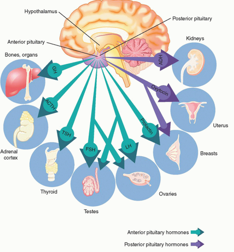
ACTH, regulated by CRH excreted from the hypothalamus, stimulates the production of glucocorticoids from the adrenal cortex. ACTH has a diurnal pattern of excretion and typically peaks just before awakening from sleep. Physical and emotional stress can increase production of ACTH. Alterations in ACTH affect the end production of cortisol. See Adrenal section for further discussion on the effects of alterations of cortisol.

Growth hormone (GH), regulated by the hypothalamus via GHRH and somatostatin, is responsible for linear growth in children, as well as metabolism and tissue integrity in both adults and children. GH levels can be impacted by several nonhypothalamic factors. Sleep deprivation can impact GH levels since 70% of the daily secretion of GH occurs during sleep stages 3 and 4. In addition, GH levels are impacted by dopamine. Dopamine agonists such as levodopa and bromocritpine increase GH levels whereas dopamine antagonists such as metoclopramide can decrease the levels. See Table 10-1 for list of dopaminergic medications. Hypofunction may result from deficiency of GHRH or from a lack of GH receptors in the liver. Hypofunction is most commonly found in children and can result in dwarfism. Since intrauterine growth is independent of GH, children deficient in GH are normal size at birth and may not show signs of deficiency until adolescence. Hyperfunction, or excessive GH, in children where long bones are still growing can result in gigantism. In adults, long bone growth plates are inactive and excess GH instead leads to acromegaly. Acromegaly is most commonly associated with enlarged facial features, but can lead to serious cardiac arrhythmias.

Prolactin, responsible for lactation in the postpartum period is primarily inhibited by the hypothalamus through the neurotransmitter dopamine. Granulomas or pituitary adenomas compressing the pituitary stalk can prevent the free flow of dopamine down the hypophyseal portal veins resulting in hyperprolactinemia. Galactorrhea, or unexpected lactation, may be a presenting symptom in patients with pituitary adenoma, although several medications can result in hyperprolactinemia including those that affect dopamine levels.

Thyroid stimulating hormone (TSH) released from the adenohypophysis binds with the TSH receptors on the thyroid where they stimulate the thyroid gland to increase iodide uptake and release T3 and T4. TSH is regulated by both TRH and somatostatin via a negative feedback loop. Dopamine antagonists may increase TSH levels while dopamine agonists may have the opposite effect. In addition, octreotide, a somatostatin analog, can inhibit TSH as well. Primary hypothyroidism occurs due to a failure in the thyroid itself. Secondary hypothyroidism can occur as a result of hypothalamic or pituitary dysfunction. Patients with symptoms of hypothyroidism and a normal TSH level should be screened for primary hypothyroidism using T3 and T4 levels since the abnormality is occurring at the thyroid level and not the pituitary level.

Gonadotropins, luteinizing hormone (LH) and follicle stimulating hormone (FSH), regulate gonadal function by promoting steroids responsible for sexual function and gametogenesis. LH excretion results in testosterone production in males and estrogen and progesterone in females. In males, FSH aids in maturation of spermatozoa, while in females promotes development of the ovarian follicle. Reproduction, while important to quality of life, is not an immediate concern for the neurological patient in the acute or rehabilitation phase and, therefore, will not be discussed in detail in this text.


Neurohypophysis

The neurohypophysis is the distal axon terminal of the hypothalamic supraoptic and paraventricular nuclei and not a true gland. Unlike the adenohyophysis, the neurohypophysis is not controlled by the use of releasing or inhibiting hormones. Acting as a storage container for vasopressin and oxytocin, the neurohypophysial release of these two hormones is controlled by nerve impulses from the hypothalamus. Since the pituitary gland does not control their excretion, pathophysiological disorders resulting from over- or underproduction of vasopressin and oxytocin are a result of hypothalamic injury and not the posterior pituitary. Working as a unit, the hypothalamic-neurohypophyseal system controls serum osmolality through both oral intake and vasopressin secretion affecting renal excretion of water.


Neurohypophyseal Hormones

Arginine vasopressin, also called 
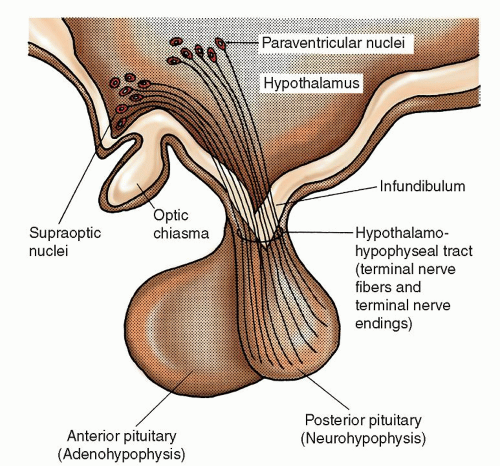
antidiuretic hormone (ADH), affects serum osmolality and total body water (TBW) by increasing water reabsorption in the collecting tubules of the kidney. The perikarya of the magnocellular neurons in the supraoptic and paraventricular nuclei synthesize most ADH. ADH then bonds loosely with a carrier protein called neurophysin and is transported down the pituitary stalk or infundibulum through terminal nerve fibers and terminal nerve endings. Here ADH is stored in large secretory granules in the nerve endings of the posterior pituitary gland. Once the serum osmolality threshold of greater than 280 to 290 mOsmol/kg is reached, electrical impulses generated by the supraoptic and paraventricular nuclei control release of ADH from the nerve
endings. Because of the loose bonding of neurophysin to ADH, the neurophysin immediately separates from ADH, and adjacent capillaries absorb ADH releasing it into the bloodstream.


Figure 10-2 ▪ Hormones of the pituitary gland and corresponding target organs. (From: Scanlon, V. C. (2011). Essentials of Anatomy and Physiology (6th ed.). Philadelphia, PA: F. A. Davis Company. Used with permission.)

ADH is very potent, and even minute amounts (as little as 2 mcg) have an appreciable effect on water balance. The half-life of ADH is 15 to 20 minutes, with metabolic degradation occurring in the liver and kidney. Even minute changes in volume, concentration, and composition of body fluids respond quickly to ADH. The immediate release and rapid breakdown of ADH account for this quick response.

Three receptor sites exist for vasopressin, V1, V2, and V3. The V1 receptor activation causes vasoconstriction, while the V2 receptor is responsible for water reabsorption. The V3 receptor is the least understood but appears to play a role in the pituitary release of ACTH. Vasoconstriction through the activation of the V1 receptor is the primary use of the continuous infusion of intravenous (IV) vasopressin in patients with hypotension. Activation of the V2 receptor regulates ADH secretion maintaining serum osmolality in a narrow range by two major processes. As serum osmolality rises, ADH stimulates sodium chloride (NaCl) reabsorption in the thick ascending limb of the Loop of Henle while also causing water channels called aquaporins to open in the collecting ducts allowing water to be reabsorbed. Reabsorption of water normalizes serum osmolality, completing the feedback loop, and leading to a decreased secretion of ADH.2 Conversely, without ADH present, the renal collecting ducts and tubules are almost totally impermeable to water so that fluid is not reabsorbed and is excreted in the urine. Desmopressin, a vasopressin analog, to be discussed in the section regarding DI may be administered to increase water reabsorption in the kidney. Desmopressin also increases factor VII and von Willebrand factor and is sometimes used to treat bleeding resulting from platelet dysfunction.


TABLE 10-1 DOPAMINERGIC MEDICATIONS


AGONISTS


ANTAGONISTS


Dopamine infusion


Haloperidol


Levadopa


Metoclopramide


Bromocriptine


Thorazine


Perphenazine


Certain conditions stimulate secretion of ADH, thereby conserving water in the body. They include the upright position, hyperthermia, hypotension, hypovolemia (especially caused by severe blood loss), pain, severe stress, anxiety, nausea, emesis, hypoxia, and trauma. Drugs that increase ADH release include acetaminophen (Tylenol), amitriptyline, anesthetic agents, angiotensin II agents, barbiturates, β-adrenergic agents, bromocriptine, chlorpromazine (Thorazine), chlorothiazide, cholinergic drugs, clofibrate (Atromid-S),
cyclophosphamide, haloperidol, histamines, monoamine oxidase inhibitors, meperidine hydrochloride, metoclopramide, morphine, nicotine, phenothiazines, prostaglandin E2, and vincristine sulfate.2, 3 Other conditions inhibit release of ADH. They include the recumbent position, hypothermia, hypertension, hypoosmolality, increased blood volume, and sleep. Drugs that decrease ADH secretion include ethanol, α-adrenergic agents, anticholinergic agents, demeclocycline, glucocorticosteroids (e.g., dexamethasone), lithium carbonate, narcotic antagonists, phenytoin, tolazamide, and vinblastine.2, 3 Finally, chlorpropamide, carbamazepine, nonsteroidal anti-inflammatory drugs (NSAIDs), and tolbutamide enhance the effect of ADH.

Like vasopressin, oxytocin is synthesized in the magnocellular neurons of the supraoptic and paraventricular nuclei traveling down the infundibulum to be stored in the posterior pituitary. Oxytocin has two major targets, the mammary alveoli and the smooth muscle cells of the uterus. Responsible for species survival, oxytocin stimulates contractions of the uterus during the birthing process, stimulates milk ejection from the mammary glands in lactating women, and is thought to play a role in the bonding of mother to child.


Adrenal Gland

Sitting atop bilateral kidneys, the adrenals are responsible for a variety of endocrine functions. The chief function of the adrenal gland is to provide a response to stress through the production of corticosteroids and catecholamines. Secondary functions include the regulation of serum osmolality, electrolytes, androgens, and blood pressure. The adrenals are regulated by three feedback systems including the HPA axis responsible for the maintenance of cortisol and androgen levels, the renin-angiotensin-aldosterone (RAA) system responsible for the regulation of aldosterone, and the ANS.

The adrenal gland comprises two distinct regions, the outer cortex and the inner medulla. The cortex consists of three functional zones, each responsible for the synthesis of one type of steroids. The zona fasciculata is mainly responsible for the synthesis of glucocorticoids, while the production of mineralocorticoids occurs in the adjacent zona glomerulosa. The most inner zone of the cortex, the zona reticularis, is responsible for androgen production. At the core of the adrenal gland, the medulla, made up of masses of neurons, regulates the fight or flight response through the secretion of epinephrine and norepinephrine.


Adrenal Steroids

The main glucocorticoid, cortisol, effects metabolism through the stimulation of gluconeogenesis in the liver while mobilizing amino acids and adipose tissue to be used for energy production. In addition to metabolic control, cortisol has a direct effect on the immune and inflammatory processes and is most often given exogenously for this effect. Hypofunction of the zona fasciculata, often caused by infection or autoimmune attack, can lead to Addison’s disease and adrenal insufficiency. Patients with Addison’s disease present with low blood pressure, and hyperpigmentation in the skin creases. Addisonian or adrenal crisis is a life-threatening condition presenting with severe hypotension resistant to vasopressors. Adrenal crisis can be caused by local adrenal injury or catastrophic HPA axis failure. Exogenous administration of glucocorticoids is the mainstay of treatment. Hyperfunction, referred to as Cushing’s syndrome, can be caused by excessive production of ACTH by the anterior pituitary or other sources such as tumor, or through exogenous administration of glucocorticoids. Symptoms associated with Cushing’s syndrome include central obesity, moon facies, fat pads on the base of the neck, and striae on the trunk. Treatment focuses on removal of the inciting factor with either steroid medication or tumor resection.

Regulated by the RAA system, the primary mineralocorticoid, aldosterone, maintains adequate blood pressure by increasing the number of sodium pumps in the tubular epithelial cells of the distal tubule.

Increased reabsorption of sodium indirectly causes reabsorption of water leading to increased extracellular volume and blood pressure. In an effort to maintain electroneutrality, a balanced number of positive and negative ions, aldosterone must exchange sodium for potassium and hydrogen ions which may result in hypokalemia and metabolic acidosis. Hypofunction can be caused by destruction of the adrenal gland or induced by medication such as ACE inhibitors, nonsteroidal anti-inflammatory medications and cyclosporine. Patients present with hyperkalemia, metabolic alkalosis, edema, and hypertension. Treatment is focused at removing the inciting factor. Loop diuretics can be used as an adjunct to control hyperkalemia and edema.

Hyperfunction referred to as aldosteronism is frequently caused by an adrenal adenoma, known as Conn’s syndrome, or renal artery stenosis. The mechanism leading to alteration in aldosterone levels in renal artery stenosis is caused by reduced blood supply through the stenosed artery causing decreased intra-arterial pressure in the juxtaglomerulus apparatus (JGA), which in turn, stimulates renin and aldosterone. Patients with aldosteronism present with hypertension, hypokalemia, and metabolic alkalosis. Treatment is focused on the cause including reduction of pituitary ACTH production, bilateral adrenalectomy or medications to suppress adrenal function such as metyrapone, mitotane, aminoglutethimide, ketoconazole.4

Androgens are not only steroids primarily responsible for the development of male sex characteristics but also regulate the menstrual cycle in females. In addition to sexual development, androgens affect memory, fat deposition, muscle mass development, and regulation of aggression and libido. Androgens, produced by the adrenals, include dehydroepiandrosterone, androstenedione, androstenediol, androsterone, and dihydrotestosterone.


HYPOTHALMIC-PITUITARY-ADRENAL AXIS

The HPA is an integrated system that allows the body to respond to internal and external stressors. The hypothalamus directs the neuroendocrine response to stress through both the HPA axis and the sympathetic nervous system (SNS). During emotional or physical stress, sensory input interpreted by the hypothalamus causes a chain of events including release of norepinephrine from the locus coeruleus (LC) in the brainstem, release of CRH from the paraventricular nucleus of the hypothalamus, and activation of the SNS. Increased levels of CRH stimulate the pituitary gland to release ACTH which ultimately leads to the release of cortisol from the adrenal cortex. Circulating norepinephrine further activates the SNS in the preganglionic neurons in the thoracic and lumbar regions causing the classic response known as fight or flight (Fig. 10-3). Nerve impulses continue to travel from the cell bodies in the thoracic spine down the greater splanchnic nerve to the adrenal medulla where the synapse ends at the chromaffin cells. The chromaffin cells convert the amino acid tyrosine into norepinephrine and epinephrine to be released into the bloodstream to further aid in the fight or flight response to stress.


FLUID AND SODIUM REGULATION

The human body requires fluid and electrolyte concentrations within narrow limits in order to function properly. The nervous system in conjunction with the kidneys, cardiovascular and endocrine systems
work together to maintain fluid and electrolyte homeostasis. In the patient with neurological alterations, sodium and fluid balance are especially important for regulating electrical impulses, managing intracranial pressure (ICP), and maintaining adequate blood pressure for efficient oxygen delivery to prevent secondary injuries.


Figure 10-3 ▪ Results of activation of sympathetic nervous system. (From: LifeART Collection, Lippincott, Williams & Wilkins. Used with permission.)


Distribution of Water

The human body comprises approximately 60% water though age, gender, and percentage of body fat influence the exact percentage. Two thirds of TBW is intracellular, while only one third is extracellular (Fig. 10-4). The body maintains its fluid volume through regulation of water intake and output. The majority of water gain comes from oral intake regulated by the hypothalamus through a thirst mechanism. Renal control of sodium and water secretion is the largest contributor to fluid volume management. Most water loss occurs primarily in the form of urine, though some insensible loss also occurs through respiration and perspiration.


Figure 10-4 ▪ Body water compartments.


Basic Concepts

Osmolarity is a measure of the osmoles of solute per liter of solution whereas osmolality is a measure of the osmoles of solute per kilogram of solvent or water. Serum osmolality is a sensitive measure of hydration status. The normal range of plasma osmolality is 282 to 295 mOsm/kg, although it can vary slightly from laboratory to laboratory. The body maintains a near-constant body fluid osmolality primarily by regulating water balance within a narrow range rather than by regulating solute balance.3 Sodium, BUN, glucose, and urea comprise most of the solutes found in the extracellular
fluid (ECF). Plasma osmolality can be directly measured or estimated using the following formula.


(Na is sodium and BUN is blood urea nitrogen; 18 and 2.8 are derived from the conversion of mg/dL to mOsm/L) Sodium is ×2 in order to calculate for the accompanying anions.

Comparable results can usually be derived by simply doubling the serum sodium.5

A discrepancy between measured and calculated osmolarity (known as an osmolar gap) may help identify exogenous solutes such as mannitol, ethanol, ethylene glycol, and methanol.

Tonicity is often confused with osmolality. Osmolality is simply a count of the number of dissolved particles in a solution; whereas, tonicity describes the osmotic pressure gradient of two solutes that are separated by a semipermeable membrane. Tonicity is most often used to describe the osmotic pressure effect that an IV solution has on cells.

In a healthy person, water continuously moves between intracellular fluid (ICF) and ECF and is driven by osmotic pressure differences. Intracellular osmolality must always equal extracellular osmolality. Therefore, both fluid compartments are in osmotic equilibrium unless the ECF osmolality is disrupted such as with IV fluid administration. In general, isotonic fluids are maintained primarily in the extracellular space whereas hypotonic fluids are distributed throughout. Therefore, isotonic saline (NS) administration will influence blood pressure more than hypotonic solutions. When hypotonic solutions are given into the intravascular space, the body will attempt to equalize the osmotic pressure gradients by moving water from the now hypotonic intravascular compartment to the intracellular space resulting in cellular swelling. An increase in extracellular solutes, by administration of hypertonic fluids, will result in fluid shifting from the cells to the extracellular space, resulting in cellular dehydration. In contrast, isotonic solutions have an osmotic pressure similar to that of the intracellular space and will result in no water movement.


Regulation of Water and Sodium Balance

Sodium and intravascular fluid volumes are affected by various hormonal and nonhormonal “mediators” including ADH, aldosterone, angiotensin, natriuretic peptides, and renal preload. Distinguishing disorders of osmoregulation versus those of volume regulation is important since the body can regulate water and sodium independently. While ICF volume is affected primarily by ADH and cellular sodium-potassium pumps, ECF volumes are maintained primarily through the regulation of sodium by the kidney.


Sensors for Control of Fluid and Sodium Balance

Osmoreceptors are specialized cells that sense changes in serum osmolality. Located in the hypothalamus near the cells that produce ADH, osmoreceptors respond to changes in concentration of ECF. Concentrated ECF stimulates the supraoptic nuclei to send impulses to release ADH, which causes reabsorption of water in the kidney. Conversely, dilute ECF around the hypothalamic osmoreceptors inhibits the generation of impulses for the release of ADH. Changes in osmolality as small as 1% are sufficient to alter ADH secretion significantly.6 Since the osmolality of plasma is determined by the concentration of a number of different solutes, including urea and glucose, the total plasma osmolality alone is not always clearly related to the ADH level.2 Serum hyperosmolality most often causes release of ADH, but this not always the case. While hypertonic saline or sucrose solutions do stimulate ADH release, certain hyperosmolar solutes such as hypertonic urea and glucose do not. The ability of osmotic particles to cross the osmoreceptor membrane may account for these differences.

The ventromedian nucleus, also known as the thirst center, is within the lateral hypothalamus. The osmoreceptors that stimulate the supraoptic and paraventricular nuclei also stimulate the thirst center. The thirst center, in turn, stimulates the cerebral cortex, signaling the need to drink fluids. So long as the person is able to respond to this impulse, fluid and electrolyte balance can be maintained. However, motor deficits, dysphagia, or a decreased level of consciousness may interfere with the person’s ability to respond appropriately to the thirst stimuli.

Extrarenal baroreceptors, located in the chest, left atrium, aortic arch, and carotid sinuses, are specialized cells that sense changes in blood volume and blood pressure. Impulses from the baroreceptors travel via the vagus and glossopharyngeal nerves to the supraoptic and paraventricular nuclei. A decreased blood volume of 5% to 10% stimulates the secretion of ADH. ADH has important vasopressor and antidiuretic effects. Under ordinary circumstances, changes in osmolality of body fluids play the most important role in regulating ADH secretion. However, in situations of severe volume depletion, stimulation of ADH secretion via baroreceptors occurs despite significant hypoosmolality.

Intrarenal receptors, located in the JGA, are able to sense fluctuations in intravascular volume indirectly by sensing changes in intrarenal pressure and sodium concentration in the tubular fluid of the macula densa. A drop in intravascular volume leads to dilation of the afferent renal arterioles and excretion of renin by the JGA. Secretion of renin into the bloodstream activates the RAA system to augment ECF volume and blood pressure. Renin acts upon circulating angiotensinogen converting it to angiotensin I. In the endothelium of the lungs, angiotensin-converting enzyme removes two amino acids from angiotensin I converting it to angiotensin II. Angiotensin II causes arteriolar vasoconstriction, stimulates the thirst center in the brain, signals the release of ADH from the posterior pituitary, and prompts aldosterone synthesis in the adrenal cortex. In the case of hypervolemia, the cardiac muscle stretches releasing natriuretic peptides located in cardiac myocytes. Atrial natriuretic peptide (ANP) found in the cardiac atria and brain natriuretic peptide (BNP) found in both the ventricles and the hypothalamus are released when cardiac chambers are stretched secondary to increased intravascular volume. Effects of natriuretic peptide release include increased renal sodium and water excretion, vasorelaxation, and inhibition of RAA system.


DYSFUNCTION OF HYPOTHALAMIC-PITUITARY AXIS

Neuroendocrine dysfunction can occur at the hypothalamus, pituitary stalk, or pituitary level (Table 10-2). Injury to the hypothalamus can result in a deficiency of releasing or inhibiting hormones that the pituitary gland relies on to regulate hypophyseal hormone production. Pituitary stalk injury or compression can prevent transportation
of hypothalamic hormones, vasopressin, and oxytocin from reaching the pituitary gland. In addition, injury at the pituitary level may result in the inability to produce or release hypophyseal hormones. Laboratory testing of both hypothalamic and pituitary hormone levels may be done in an attempt to isolate the location of dysfunction.


TABLE 10-2 CAUSES OF HYPOTHALAMIC-PITUITARY AXIS DYSFUNCTION


TYPE OF INJURY


CAUSE


Invasive


Space occupying lesions


Infarction


Pituitary apoplexy


Infiltrative


Sarcoidosis, hemochromatosis, Langerhans histiocytosis


Injury


Head trauma


Immunologic


Lymphocytic hypophysitis


Iatrogenic


Surgery or radiation


Infection


Tuberculosis, syphilis, mycotic infections


Idiopathic


All unidentifiable causes


The symptoms of pituitary dysfunction are reflected by target organ failure. Adenohypophyseal hypofunction is often identified in children with symptoms of delayed puberty and dwarfism. Infarction or resection of the entire pituitary gland can lead to complete failure of the anterior pituitary referred to as panhypopituitarism, or Simmonds disease. In contrast, hyperfunction may present with precocious puberty, development of secondary sexual characteristics before age 8 or 9 in children. In adults, hyperfunction may present as hyperprolactinemia, galactorrhea, erectile dysfunction, and amenorrhea in premenopausal women.

Adenohypophyseal dysfunction in adults is most commonly caused by a pituitary adenoma. Pituitary tumors account for approximately 15% of all intracranial tumors, and most are benign.7 Located in the sella turcica near the optic chiasm, growing pituitary adenomas commonly lead to the compression of optic fibers causing visual field deficits. Other common symptoms associated with pituitary adenomas are galactorrhea and increased urination. Pituitary tumors can result in excess production of any of the adenohypophyseal hormones and are responsible for nearly all cases of acromegaly and gigantism. Except for exogenous administration of corticosteroids, pituitary tumors are also responsible for most cases of Cushing’s syndrome. In contrast, pituitary adenomas rarely result in neurohypophyseal dysfunction since neuronal cell bodies of the neurohypophysis are located in the hypothalamus.


SODIUM DISTURBANCES

Sodium is the major ion in the extracellular compartment largely affecting serum osmolality and ECF volume. Disruptions in normal serum sodium levels are reflected in alteration in water balance and not a sodium imbalance. Sodium disturbances often manifest as hypervolemia or hypovolemia. In contrast, water disturbances generally present as hyponatremia or hypernatremia, reflecting the contribution of ADH to the process.


Hyponatremia

Hyponatremia, defined as serum sodium level of less than 135 mEq/L, is one of the most common electrolyte disorders seen in neuroscience patients. Serum sodium concentrations are regulated by several mechanisms such as stimulation of thirst, secretion of ADH, and renal filtration as described above. The manifestations depend not only on the sodium level but also on the rapidity of development of hyponatremia. Patients with acute hyponatremia (i.e., develops rapidly in <24 to 36 hours) may be symptomatic with mild hyponatremia whereas patients with chronic hyponatremia (i.e., develops slowly and is present for >36 to 48 hours) may be asymptomatic. The signs and symptoms are nonspecific and related to the effects on the central nervous system (CNS). Patients with a serum sodium between 125 and 130 mEq/L with an acute onset may complain of headache, nausea, myalgia, and generalized malaise.8 In the range of 115 to 120 mEq/L, mental status changes from lethargy, confusion, disorientation, and agitation followed by seizures, coma, and death are noted (Table 10-3). Cerebral edema may occur, especially with rapid decline of serum sodium.9


TABLE 10-3 CLINICAL SIGNS AND SYMPTOMS OF ACUTE HYPONATREMIA


SYSTEM


SIGNS AND SYMPTOMS


(Note: Although significant symptoms do not generally occur until serum sodium is 125 mEq/L, individual thresholds vary widely.)


Neurological (hyponatremic encephalopathy)


Headache


Disorientation, confusion, irritability, apathy, lethargy, obtundation


Neurological deficits


Seizures


Weakness


Cerebral edema


Herniation syndromes


Respiratory arrest


Gastrointestinal


Nausea and vomiting


Anorexia


Muscular


Cramps


Muscle weakness


Rhabdomyolysis


Assessment should include not only the neurological system but also the hydration-volume status. Tachycardia, orthostatic hypotension, dry mucous membranes, decreased skin turgor, and sunken eyes are indicators of hypovolemia. The initial laboratory work-up for a patient with hyponatremia includes serum electrolytes, renal function, plasma and urine osmolality, and urine sodium concentration (Table 10-4).

Hyponatremia is more than a low sodium level. Disturbances of sodium are most commonly linked to a water imbalance and not a deficiency of sodium. To understand the underlying pathophysiology and basis for treatment, patients should be classified according to two-tier schemata. First, all hyponatremic patients are categorized into iso-osmolar, hyperosmolar, and hypo-osmolar states based on plasma osmolality.10 Hypo-osmolality is a state of excess body water whereas hyperosmolality is a state of depletion of body water. Second, if the hyponatremia is classified as hypo-osmolar hyponatremia, then it is further classified into hypovolemic, euvolemic, or hypervolemic hypo-osmolar hyponatremia.

Only gold members can continue reading. Log In or Register to continue

Stay updated, free articles. Join our Telegram channel

Jul 14, 2016 | Posted by in NURSING | Comments Off on Neuroendocrine Disorders in Neuroscience Patients

Full access? Get Clinical Tree

Get Clinical Tree app for offline access