11 Neurologic Disorders
Pearls
• The major goals of therapy in the treatment of traumatic brain injury (TBI) are preservation of cardiopulmonary and cerebral function and the prevention of secondary brain injury.
• The 5 H’s responsible for secondary brain injury in the pediatric patient are: hypotension, hypoxia, hyperthermia, hypo-/hyperglycemia, and hyponatremia.
• Seizures result from abnormal discharges or firing of cerebral neurons that produce alterations in motor function, behavior and consciousness.
• It is important to recognize the difference between posturing and seizure activity. Decorticate and decerebrate posturing typically occur in response to a stimulus, such as a painful stimulus, while seizures can occur at any time and may produce a variety of movements or other evidence of neurologic discharges (e.g., changes in heart rate).
• Decorticate posturing indicates damage along the corticospinal tract, the pathway between the cortex and spinal cord. Decerebrate posturing indicates deterioration of the structures of the nervous system, particularly the upper brain stem; decerebrate posturing has a less favorable prognosis than decorticate posturing.
• Stroke and cerebral vascular disease are among the top 10 causes of childhood death.
• Therapeutic hypothermia has been shown to improve morbidity and mortality following adult cardiac arrest and neonatal hypoxic-ischemic insult. In the pediatric population with TBI, hypothermia remains a controversial therapy. Clinical trials are underway to evaluate the effects of hypothermia in pediatric TBI and hypoxic-ischemic injury.
• When using an extraventricular drainage (EVD) device for CSF diversion, the nurse must maintain the drain at the precise level ordered. If the drain is placed too high, increased ICP may develop before drainage occurs. If the drain is placed too low, excessive drainage of CSF can lead to upward herniation and/or ventricular collapse and intraventricular hemorrhage.
Introduction
Care of the critically ill child with neurologic disorders is both challenging and rewarding. It requires knowledge of neuroanatomy, neurophysiology, and normal growth and development. In addition, the nurse must be able to recognize subtle changes in the patient’s condition and respond, if needed, with appropriate support or therapy. Because the critically ill child with neurologic disease often is admitted under emergent conditions, the medical team usually does not have the benefit of adequate patient history. As a result the accuracy of the nurse’s observations and rapidity of detection of changes and response to deterioration are crucial to the child’s successful treatment.
This chapter provides an overview of relevant neurologic anatomy, physiology, and pathophysiology. In addition, it provides the information required to perform precise assessment and appropriate interventions for the critically ill child with critical neurologic disease or injury.
Essential anatomy and physiology
The Axial Skeleton
The axial skeleton consists of the bones of the skull and vertebral column. These bones protect the underlying structures of the central nervous system (CNS). The bones of the skull are divided into regions that form the wall of the cranial cavity and that cover the uppermost aspects of the brain and face. The frontal, occipital, temporal, and paired parietal bones form the cranial vault. The floor of this vault defines three bony compartments—the anterior, middle, and posterior fossae.
The anterior fossa contains the frontal lobes of the brain, the middle fossa contains the upper brainstem and the pituitary gland, and the posterior fossa contains the lower brainstem. These fossae and the parts of the brain they contain often are used to designate areas of injury or disease; such a designation allows location of the problem as well as delineation of the brain functions that are affected. Because injury to the area of the posterior fossa potentially disrupts critical brainstem functions, damage in this area is usually more life threatening than damage to the anterior fossa.
Blood vessels and cranial nerves enter and leave the skull through small openings, or foramina. It is useful to know the course of the cranial nerves so that clinical signs and symptoms can be correlated with areas of cranial injury (Fig. 11-1). The posterior fossa contains a large foramen, the foramen magnum, through which the brainstem and spinal cord join. Lesions in this area, such as those produced by cervical neck trauma, can interrupt vital brain functions and nerve pathways to and from the brain. Cerebrospinal fluid (CSF) flows through the foramen magnum as it passes from the brain to the spinal cord and back again, and the vertebral arteries enter the skull through the foramen magnum.

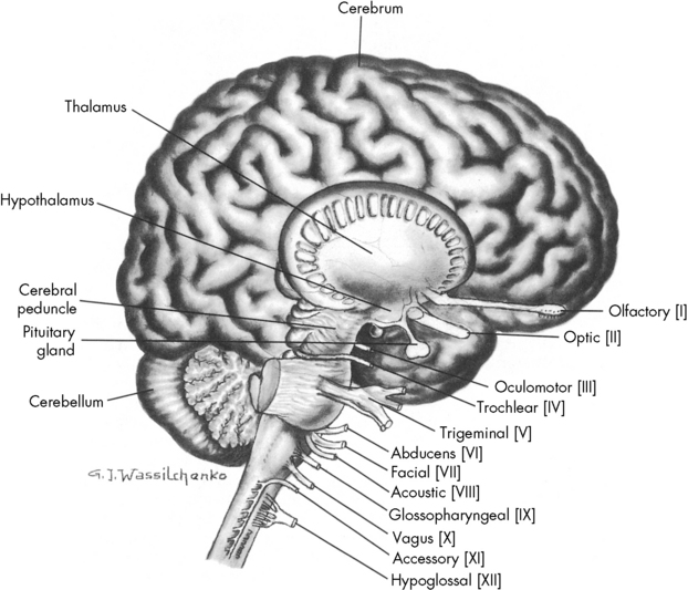
Fig. 11-1 Lateral view of the brain depicting origin of cranial nerves.
(From Rudy EB: Advanced neurological and neurosurgical nursing, St Louis, 1984, Mosby.)
At birth, the skull plates are not fused; they are separated by nonossified spaces called fontanelles. The anterior fontanelle is the junction of the coronal, sagittal, and frontal bones. The posterior fontanelle represents the junction of the parietal and occipital bones (see Evolve Fig. 11-1 in the Chapter 11 Supplement on the Evolve Website). Normally, the posterior fontanelle closes at approximately 2 months of age, and the anterior fontanelle closes at approximately 16 to 18 months of age. If the brain does not grow, such as in patients with microcephaly, the cranial bones can fuse early. Conversely, premature fusion of cranial bones, known as craniosynostosis, can result in microcephaly unless surgery is performed, because brain growth is inhibited by the restricted intracranial space.
At birth, the brain is approximately 25% of the adult volume.76 By 2 years of age, approximately 75% of adult brain volume has been achieved. The cranium itself continues to expand until approximately 7 years of age, when most brain differentiation is complete. This growth of the brain can be assessed indirectly through measurements of the head circumference. These measurements should always be plotted on a growth chart, because they can aid in the detection of excessive or inadequate head and brain growth that may reflect neurologic disease.
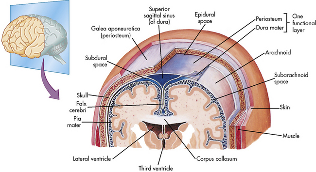
The Meninges
Three highly vascular membranes surround the brain and spinal column; the three membranes collectively are called the meninges. The outermost membrane, the dura mater, consists of tough connective tissue that lines the endocranial vault (Fig. 11-2). The dura mater is folded into tents of tissue immediately underneath the skull cap (the periosteum). The most familiar of the many dural folds is the fold that roofs the posterior fossa; this is called the tentorium cerebelli. This fold serves as an anatomic landmark; intracranial lesions are divided into those that occur above the tentorium cerebelli (supratentorial lesions) and those that occur below the tentorium cerebelli (infratentorial lesions). The dura not only lines the endocranium, but it also lines the vertebral column. It descends through the foramen magnum to the level of the second sacral vertebra and ends as a blind sac.

Fig. 11-2 Coverings of the brain. Frontal section of the superior portion of the head, as viewed from the front. The bony and the membranous coverings of the brain can be seen.
(From Patton KT, Thibodeau GA: Anatomy and physiology, ed 7, St Louis, 2010, Mosby.)
The middle membrane, the arachnoid, consists of spiderlike tissue from which it gains its descriptive name. The arachnoid membrane is separated from the dural membrane by the subdural space, which contains cerebral vessels. Because these vessels traverse the subdural space with relatively little support, serious head trauma can cause a rupture of these vessels and the development of a subdural hematoma. This space allows for some cerebral expansion or small hematoma without cerebral compression, but the critical capacity is low. Beneath the subdural space the arachnoid membrane follows the contour of the brain and spinal cord to the end of the spinal cord root.
The pia mater is the third and the innermost membrane. It consists of highly vascular tissue that is separated from the arachnoid membrane by a space called the subarachnoid space. This space contains CSF and provides for two major CSF-collecting chambers. The largest chamber, the cisterna magna (also called the cisterna cerebello-medullaris), is located between the cerebellum and the medulla. The smallest, the lumbar cistern, is located at the level of the sacrum. Because this space contains CSF, obstruction in this subarachnoid space can obstruct the flow of CSF. Head injury can result in the accumulation of blood in the subarachnoid space; this lesion is called a subarachnoid hemorrhage.
The Brain
The brain is contained within the cranial vault and extends through the foramen magnum. It is composed of distinct structures, each having a specific function. The brain can be considered in three major functional areas: the cerebrum, the brainstem, and the cerebellum (see Fig. 11-1). The cerebrum (from the embryologic forebrain) consists of the cerebral hemispheres, the thalamus, hypothalamus, basal ganglia, and the olfactory and optic nerves. The brainstem consists of the midbrain, pons, and medulla. The cerebellum is the final major division of the brain. The brainstem and cerebellum develop from the embryologic hindbrain. Table 11-1 lists the divisions of the brain and their major functions. Each of these brain divisions is presented separately in the following pages.
Table 11-1 Basic Brain Divisions and Functions
Structure | Division | Function |
Cerebrum | Cerebral hemispheres | Integration of sophisticated sensory and motor activities and thoughts |
Cerebral cortex | ||
Frontal lobes | Reception of smell, memory banks, and higher intellectual processes | |
Parietal lobes | Sensory discrimination, localization of body awareness (spatial relationships), and speech | |
Temporal lobes | Auditory functions and emotional equilibrium | |
Occipital lobes | Vision and memory of events | |
Limbic lobes | Primitive behavior, moods, and instincts | |
Basal ganglia | Transmission of motor tracts, linking pyramidal pathways | |
Corpus callosum | Provision of intricate connection between cerebral hemispheres | |
Brain stem | Midbrain | Hypothalamic response to neuroendocrine stimuli |
Pons | Origin of cranial nerves V, VI, VII, VIII | |
Medulla | Vital center activity (cardiac, vasomotor, respiratory centers); origin for cranial nerves IX, X, XI | |
Cerebellum | White and gray matter | Muscle and proprioceptive activity, balance, and dexterity |
The Cerebrum
The Cerebral Cortex
The cerebral cortex is the convoluted gray matter that forms the outermost layer of the brain. It consists largely of specialized neurons that process and respond to specific sensory stimuli. The cortex receives electrical discharges from other neurons and converts them into ideas or actions. The cortex is divided into five anatomic divisions: the frontal, parietal, temporal, occipital, and limbic divisions.
The cortical neurons are specialized so that within each major division of the brain, specific areas are devoted to specific functions. Fifty-two specialized areas were identified in the late 1800s by Korbinian Brodmann; these areas are numbered according to histologic appearances and functions. If brain injury is identified in one of these areas, it is possible to predict the resulting sensory or functional impairment. Conversely, a lesion often can be localized according to the motor functions or sensations that are impaired. It is important to note, however, that most functions can be performed through impulses from several areas of the brain.
The cerebral cortex performs the highest functions of the human brain. As a result, it continues to develop beyond infancy and childhood. The newborn responds to the environment with simple awareness and reflex behavior. During infancy, individual sensations, sights, and sounds can be stored in memory in the cerebral cortex, and the infant learns to associate these sights and sounds with events or feelings. As the infant develops into a toddler, higher cortical functions such as imagination and language become apparent.
There is tremendous growth of cortical function during the early years of life. Most developmental and neurologic assessment tools evaluate only basic reflexes and motor skills of the young infant and toddler, and it is not until the preschool and the early childhood years that cognitive functions and learning can be evaluated.
The cerebral hemispheres are two mirror image portions of the brain that consist largely of the cerebral cortex and fiber tracts. In general, each cerebral hemisphere governs the functions of and receives sensations from the contralateral side of the body. Therefore, the right cerebral hemisphere governs movement of and receives sensory input from the left side of the body. The left cerebral hemisphere governs movement of and receives sensory input from the right side of the body.
In most humans, one side of the brain is considered dominant; right-handed people are thought to have a dominant left side of the brain, and left-handed people are thought to have a dominant right side of the brain. Each hemisphere also has primary responsibility for some functions. In most people, the left hemisphere controls language and speech, and the right hemisphere helps interpret three-dimensional images and spaces. Other distinctions have been postulated. For example music understanding is thought to be predominantly controlled by the left hemisphere and arithmetic and design are thought to be controlled by the right hemisphere. To a certain extent, if one side of the brain is injured, the other side of the brain can be taught to assume the dominant functions (this compensation is called plasticity). This compensation is more likely when the injury occurs during infancy or early childhood, because cerebral dominance is not established fully until approximately 3 years of age.
The cerebral hemispheres are connected by nerve fibers called the corpus callosum. These nerve fibers allow the brain to function as a single unit despite its division into two hemispheres.
The Basal Ganglia
The basal ganglia are paired masses of gray matter deep within the cerebral hemispheres. They contain the nuclei of neurons and networks of tracts that control motor function. These basal ganglia send information to the motor cortex through the thalamus to inhibit unintentional movement. Thus, the basal ganglia regulate the extrapyramidal motor system. This system selects motor messages from lower pathways for interpretation up to the cerebral cortex; thus it influences motor activities, musculoskeletal control, rhythmic movement, and maintenance of an erect posture.
Interference with neurotransmission to the basal ganglia produces disturbances of intentional movement. The uptake of bilirubin by the brain during infancy, known as kernicterus, affects this area and can result in the development of neurologic dysfunction.
The Thalamus and Hypothalamus
The thalamus surrounds the third ventricle and is composed of tracts of gray matter. The thalamus is a major integrating center for afferent impulses from the body to the cerebral cortex.85 The thalamus integrates and modifies messages that come from the basal ganglia and cerebellum and then transmits information up to the cerebral cortex. All sensory impulses, with the exception of those from the olfactory nerve, are received by the thalamus. These impulses are then associated, synthesized, and relayed through thalamocortical tracts to specific cortical areas. The thalamus is the center for the primitive appreciation of pain, temperature, and tactile sensations.
The anterior pituitary gland, called the adenohypophysis, secretes hormones that control glands throughout the body; these hormones include growth hormone (somatotrophin), adrenocorticotropic hormone, thyroid-stimulating hormone, melanocyte-stimulating hormone, follicle-stimulating hormone, luteinizing hormone releasing factor, and prolactin.
Injury to or disease of the hypothalamus or the pituitary can produce a wide variety of neuroendocrine problems and can result in fluid and electrolyte imbalance and growth disturbances (see Chapter 12).
The Brainstem
The brainstem, located at the base of the skull, is the major nerve pathway between the cerebral cortex and the spinal cord. The three major divisions of the brainstem are the midbrain, the pons, and the medulla. Together they control many of the involuntary functions of the body.
The midbrain is a short segment between the hypothalamus and the pons. It contains the cerebral peduncles and the corpus quadrigemina. The midbrain consists of fibers that join the upper and lower brainstem; it is the origin of the oculomotor and trochlear cranial nerves. The midbrain is the center for reticular activity and assimilates all sensory input from the lower neurons before it is relayed to the cortex (see Evolve Fig. 11-2 in the Chapter 11 Supplement on the Evolve Website). It is because of this relay that the cortex can maintain consciousness, arousal, and sleep.
The medulla oblongata lies between the pons and the spinal cord at the level of the foramen magnum. It is the site of decussation (crossing) of many corticospinal motor neurons. In addition, it transmits messages to and from the spinal pathways for interpretation and reaction by the cortex. The medulla is the origin for the glossopharyngeal, vagus, spinal accessory, and hypoglossal cranial nerves. Critical regulatory centers for cardiovascular and respiratory functions are found within this portion of the brain. Severe intracranial injury can result in the loss of medullary control of respirations and cardiac output. A blow to the back of the head can result in respiratory arrest, labile blood pressure, and decreased cardiac output.
Although posture is controlled by the cortex, it is integrated in the medulla, so medullary injury can produce decorticate and decerebrate posturing. Loss of medullary function can lead to decreased gag reflex or swallowing difficulties, because the glossopharyngeal and vagus nerves originate in the medulla. Any disease or injury to the medulla can be life threatening.
The Cerebellum
The cerebellum is located in the posterior fossa, directly below the occipital lobe. It consists of two hemispheres that contain gray matter, and it joins with the basal ganglia and reticular system. This area of the brain integrates voluntary movement. Spatial orientation, fine motor movements, muscle tone, balance, and dexterity are controlled by visual, auditory, and proprioceptive stimuli that are processed by the cerebellum. The cerebral cortex can exert voluntary control over the cerebellum by virtue of cognitive nerve pathways that adjust movement.
The Ventricles
The four ventricles of the brain are cavities that contain CSF. The ventricles are joined to one another by foramina, and the ventricular system ultimately communicates with the subarachnoid spaces of the brain and the spinal column. The ventricles are designed specifically for the production and circulation of CSF.
The first two ventricles are called the lateral ventricles, and each is located in a cerebral hemisphere. These ventricles communicate with the third ventricle through the foramen of Monro at the level of the thalamus. The third ventricle joins the fourth ventricle through a channel known as the Sylvian aqueduct. This fourth ventricle is located at the level of the pons and medulla. From this last chamber rise three foramina that open into the subarachnoid space to allow distribution of the CSF into the subarachnoid spaces of the brain and spinal cord. Insult or injury to structures surrounding the CSF pathway can cause acute or chronic CSF flow obstruction and can result in the development of hydrocephalus. In addition, the presence of blood or inflammation within this system can obstruct the CSF pathway.
The Cranial Nerves
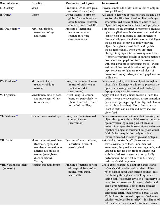
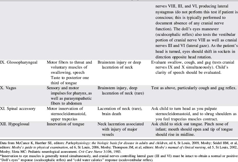
The cranial nerves are 12 pairs of peripheral nerves that arise from the brain; each has a specific motor or sensory function, and four cranial nerves have parasympathetic functions.111 Cranial nerve function can be lost as a result of lesions near the origin of the cranial nerve or following direct injury to the cranial nerve itself.
Identification of lost cranial nerve functions can help determine the location and severity of CNS disease or injury. For example, pupil inequality and a unilateral sluggish pupil response to light can develop with uncal herniation of the brain (lateral herniation of the temporal lobe through the tentorial notch) and with compression and stretching of the oculomotor nerve. Assessment of cranial nerve function becomes extremely important when the patient is comatose or unresponsive. (Table 11-2 lists cranial nerve origins and functions; see Fig. 11-1).
The Spinal Cord
The spinal cord is a cylindrical structure composed of neurons and nerve fibers. It joins the medulla at the foramen magnum and extends to the level of the second lumbar vertebra. There are 31 pairs of spinal nerves, which are distributed along the entire spinal cord (Fig. 11-3). These spinal nerves are all multifibered and transmit impulses between the CNS and the rest of the body. When a portion of the spinal cord is viewed in cross section, the cord fills only part of the vertebral column; it is surrounded by the pia mater, the CSF, the arachnoid, and the dura mater.

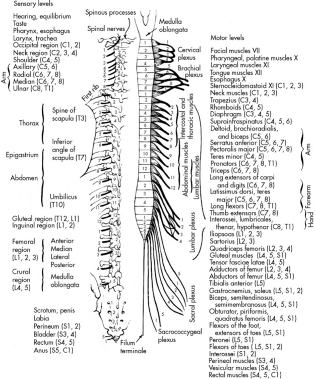
Fig. 11-3 Motor and sensory innervation from the spinal cord.
(From Chusid JG: The spinal nerves. In Feringa ER, editor, Correlative neuroanatomy and functional neurology, ed 18, Los Altos, CA, 1981, Lange Medical Publications.)
The spinal cord contains gray and white material, or matter. The gray matter consists of cell bodies and cell nuclei, and the white matter consists of nerve fibers that are grouped into tracts. The gray matter in the spinal cord is shaped like a butterfly, with anterior and posterior projections called the anterior and posterior horns or, respectively, the ventral or dorsal roots.
Peripheral sensory nerves carry impulses to the posterior horn (the dorsal root) of the spinal column where they synapse (i.e., connect or communicate) with other neurons that will carry information up the spinal column or to other neurons at the same level of the spinal column. Lower motor neurons are located in the anterior horn (the ventral root) of the spinal column. The lower motor neurons receive input from the brain and from other neurons within the spinal cord; they affect motor activity.
Spinal cord reflexes do not require any input from higher levels of the CNS. For example, when the lower leg hangs free and the patellar tendon is tapped with a reflex hammer, the rapid stretch of the muscle will produce a reflex contraction of the rectus femoris without the participation of higher CNS structures. As a result of the reflex, the lower leg swings upward. Occasionally, stimulus of a sensory neuron on one side of the body will result in movement on the opposite side of the body. For example, if the right hand is placed on something hot, that hand automatically will be withdrawn, and the left hand and left leg will extend to allow the body to move away from the painful stimulus. These behaviors can all occur at the spinal cord level, and they can continue despite injury to the cerebral cortex or even brain death. If damage to the brain or higher levels of the spinal cord occurs, however, it also can result in loss of inhibition to the lower motor neurons and cause flaccid or spastic paralysis.
Central Nervous System Circulation and Perfusion
The Cerebral Circulation
The brain requires a constant supply of oxygen and substrates (perfusion) to metabolize carbohydrates as an energy source. Adequate perfusion is also necessary to remove carbon dioxide and other metabolites from the brain. The brain requires approximately 20% of the child’s cardiac output. A healthy child’s brain consumes 5.5 mL of oxygen per 100 g of brain tissue per minute.37 As a result, if the brain is deprived of oxygen for even a few minutes, brain ischemia can develop and result in permanent neurologic dysfunction or brain death.32
The cerebral arterial blood flow is provided by the two vertebral arteries and the right and left internal carotid arteries. The internal carotid arteries enter the skull anteriorly and end in the anterior cerebral and the middle cerebral arteries; they supply approximately 85% of cerebral blood flow. The vertebral arteries enter the skull posteriorly and join to form the basilar artery. The basilar artery bifurcates to form two posterior communicating arteries (Fig. 11-4).

Fig. 11-4 The cerebral circulation. A, Major arteries of the head and neck. B, Arteries at the base of the brain. The arteries that compose the circle of Willis are the two anterior cerebral arteries, joined to each other by the anterior communicating two short segments of the internal carotids, from which the posterior communicating arteries connect to the posterior cerebral arteries.
(A, From Patton KT, Thibodeau GA: Anatomy and physiology, ed 7, St Louis, 2010, Mosby. B, Modified from Patton KT, Thibodeau GA: Anatomy and physiology, ed 7, St Louis, 2010, Mosby.)
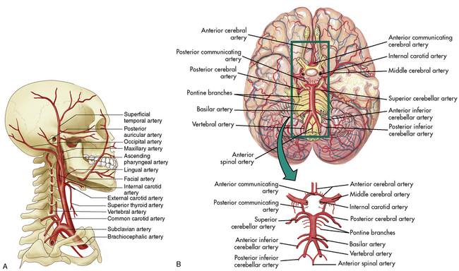
The circle of Willis at the base of the brain is formed by a junction of the two internal carotid arteries, the two anterior and two posterior cerebral arteries, and the posterior and anterior communicating arteries (see Fig. 11-4, B). This arterial configuration, present in approximately half of all adults,85 maintains effective cerebral perfusion despite a reduction in flow from any single contributory artery. Patients with an alternative form of arterial circulation are considered to have anomalous cerebral circulation, although their arterial circulation typically is not significantly different from that which is considered normal. Congenital anomalies of one or both carotid arteries or of the internal carotid system have been documented. In many of these patients, the development of collateral circulation early in life prevents any compromise in cerebral perfusion.29
The cerebral venous circulation is unique in that the cerebral veins have no valves, and they do not follow the course of the cerebral arteries.85 Venous drainage from the brain flows primarily into large vascular channels within the dura, known as dural sinuses, that ultimately drain into the internal jugular veins. Occlusion of the jugular vein can obstruct cerebral venous return.
Cerebral Blood Flow and Regulation
Cerebral Blood Flow
Cerebral blood flow (CBF) is the volume of blood that is in transit through the brain over a unit of time (e.g., per minute); this term is commonly used to describe the cerebral arterial flow perfusing the brain. Cerebral blood volume (CBV) is the total amount of blood in the intracranial vault at any one time, and it consists of both arterial and venous blood.
Normal CBF in adults is approximately 60 mL per 100 g brain tissue per minute. The normal quantity of CBF in children is unknown, but is thought to be approximately 50 to 100 mL per 100 g brain tissue per minute or more. The absolute quantity of CBF is not as important as the relationship between cerebral oxygen, substrate delivery, and cerebral metabolic requirements. It is essential that CBF is adequate to maintain effective cerebral oxygenation to sustain cerebral functions. Under normal circumstances, CBF, metabolism, and oxygen extraction are closely interrelated.133
Cerebral Venous Return
Cerebral venous return from superficial and deep cerebral veins flows into venous plexuses and dural sinuses, then into the internal jugular veins. If cerebral arterial flow is maintained in the face of obstructed cerebral venous return, CBV will increase and ICP may rise. Cerebral venous return can be obstructed by compression or thrombosis of the internal jugular vein, or by any condition that obstructs superior vena caval flow (e.g., mechanical ventilation with high inspiratory pressures, the development of a tension pneumothorax, or the Valsalva maneuver). Increased ICP can compress cerebral veins, impeding cerebral venous return. When increased ICP is present, turning the head to one side can obstruct internal jugular venous flow, increase CBV, and worsen ICP.
Autoregulation
CBF normally is maintained at a constant level by cerebral autoregulation, which is the constant adjustment of the tone and resistance in the cerebral arteries in response to local tissue biochemical changes.72 Autoregulation is essential to the maintenance of cerebral perfusion and function over a wide variety of clinical conditions. If systemic arterial pressure increases, cerebral arterial constriction will prevent a rise in the cerebral arterial pressure to maintain CBF at a constant level. Conversely, if systemic arterial pressures falls, cerebral vasodilation will minimize the effects on CBF. Severe alterations in systemic arterial blood pressure will exceed the limits of autoregulatory compensation, however, and will be associated with changes in CBF.
Autoregulation may be compromised or destroyed with severe traumatic or anoxic brain injury. If cerebral autoregulation is lost, CBF becomes related passively to the mean arterial pressure (MAP) so that a fall in MAP will result in a decrease in cerebral flow and perfusion. Recent studies suggest that impaired autoregulation in traumatic brain injury may correlate with severity of injury and young age.126
Alterations in Cerebral Blood Flow
In some conditions, including head injury and some encephalopathies, CBF (oxygen and substrate delivery) and cerebral metabolic demand may differ. This uncoupling of oxygen delivery and demand can lead to regional ischemia in some portions of the brain or to excessive CBV with subsequent increase in ICP.
CBF can increase with anemia, administration of vasodilators, and hyperthyroidism. It also can increase if the CSF pressure is abnormally low or if a hemangioma or arteriovenous malformation is present.
Seizures (particularly status epilepticus) increase CBF and can increase cerebral metabolic rate. If seizures develop in the patient with increased ICP, the additional CBF can produce a further rise in ICP.
CBF will likely be compromised if CSF pressure or ICP rise. The ultimate complication of an accelerating rise in ICP following head injury is the cessation of CBF, producing brain death. CBF also can decrease in the presence of coma or as a result of a rise in cerebral venous pressure, polycythemia, or hypothyroidism.
If CBF is reduced severely, local cerebral metabolism is compromised, and brain cell metabolic functions will be compromised and may cease. If ischemia continues, brain cell membranes will become more permeable, and water will enter the brain cells. Profound ischemia can result in permanent neurologic dysfunction or brain death.
Effects of Arterial Blood Gases on Cerebral Blood Flow
CBF is affected by significant changes in arterial oxygen and carbon dioxide tensions.
Oxygen Response
CBF is unchanged over the normal range of arterial oxygen tension (PaO2). However, severe hypoxemia (PaO2 <50-55 mm Hg) will produce cerebral vasodilation and an increase in CBF, in an attempt to maintain oxygen delivery to brain tissues. Tissue hypoxia and acidosis also will result in cerebral vasodilation.
Carbon Dioxide Response
Changes in the arterial carbon dioxide tension (PaCO2) can acutely affect CBF. When the PaCO2 is between 20 and 80 mm Hg, CBF is directly related to the arterial carbon dioxide tension; the higher the PaCO2 the greater is the cerebral vasodilation, and the greater is the CBF. A low PaCO2 causes cerebral vasoconstriction and a fall in CBF. When the PaCO2 is extremely high or low, the relationship is no longer linear, and the effect of the PaCO2 on CBF is blunted.
Normoventilation is the accepted method of management of patients with severe head injury and other causes of increased ICP. Mild hyperventilation to a PaCO2 of 30 to 35 mm Hg should be reserved for acute rises in ICP that are refractory to other medical management (e.g., sedation, hyperosmolar therapy) and that are thought to be associated with acute (impending) herniation syndrome. Although the reduction in CBF caused by hyperventilation can transiently reduce intracranial hypertension, it may worsen ischemia by decreasing oxygen delivery to an already compromised brain.9
When increased ICP is present, routine care such as suctioning must be performed skillfully to prevent the development of hypercarbia and cerebral vasodilation and an increase in ICP.9 The vasoconstrictive response to hypocarbia is unpredictable in patients after traumatic brain injury and the requires careful monitoring of clinical effects of any changes in PaCO2.9
Cerebral Perfusion Pressure
Cerebral perfusion pressure (CPP) is calculated as the difference between the systemic mean arterial pressure (MAP) and the ICP:
The normal range of CPP is thought to be approximately 50 to 150 mm Hg in healthy adults, with a goal of 70 mm Hg following traumatic brain injury. There is a paucity of information to identify the normal range of CPP in children; it is thought to be approximately 40 to 60 mm Hg, but normal ranges vary with age.9,48 A CPP of at least 40 mm Hg is thought to be necessary for effective cerebral perfusion; however, this number is not absolute because perfusion is determined by blood flow, not blood pressure. It is likely that a CPP of 40 mm Hg is acceptable in an infant, but a CPP of 50 to 65 mm Hg is likely to be necessary in older children and adolescents.
The calculated CPP will fall if the mean systemic arterial pressure falls, if the mean ICP rises, or if both occur simultaneously. The calculated CPP can be maintained despite a rise in ICP if the MAP rises commensurately with a rise in ICP. It is important to note that such compensation may or may not be associated with effective CBF and actual cerebral perfusion (for a Case Study of calculation of CPP, see the Chapter 11 Supplement on the Evolve Website); a normal CPP (40-50 mm Hg or more) has been recorded after brain death was pronounced.16
Evaluation of Cerebral Blood Flow
Qualitative radioisotope scans have been performed for a number of years to determine the presence or absence of CBF. However, quantitative CBF measurements cannot be readily performed at the bedside of the critically ill patient using standard pressure measuring devices. A variety of techniques can detect and monitor trends in CBF.
Jugular Venous Oxygen Saturation
The oxygen (actually oxyhemoglobin) saturation in the jugular venous bulb (SjO2) is normally 55% to 70%; this measurement reflects the saturation of the hemoglobin leaving the cranial vault, so it reflects trends in the amount of oxygen leaving the brain. Trends in the SjO2 can reflect changes in global cerebral perfusion in some clinical settings (discussed under Common Diagnostic Tests).
A fiberoptic catheter placed in the jugular bulb can be used to continuously monitor the SjO2. The jugular bulb fiberoptic catheter must be correctly positioned, calibrated, and functional and correct use requires the ability to troubleshoot its function and potential causes of misleading information. Use of continuous SjO2 monitoring has been reported for patients with head trauma, encephalopathy, status epilepticus, intracranial hemorrhage, and other conditions that may compromise cerebral perfusion and oxygen delivery.
Because the SjO2 reflects oxygen (specifically oxyhemoglobin) saturation of blood leaving the cranial vault, it can be affected by any factor influencing oxygen delivery to the brain or oxygen consumption by the brain. As a result, if the SjO2 changes, providers must attempt to evaluate each component affecting cerebral oxygen delivery and consumption to try to identify and treat the cause of the change. If the fiberoptic is not correctly positioned, calibrated and functional, trends in the SjO2 may not accurately reflect trends in CBF.
Oxygen delivery to the brain can be altered by cardiac output, arterial oxygen content (which is, in turn, affected chiefly by hemoglobin concentration and its saturation), and factors that affect regional or global CBF (e.g., ICP, arterial pressure, arterial oxygen tension and tissue oxygenation, hemoglobin concentration, arterial and tissue pH, arterial CO2, cerebral vasoconstriction, vasodilation, and tissue cytokines). Oxygen consumption by the brain can be increased by conditions such as fever and seizures and decreased by therapeutic hypothermia and by drugs such as barbiturates.
The SjO2 will likely fall if oxygen delivery to the brain falls; in this case, oxygen extraction in the brain will increase so that less oxygen is left in the venous system when it leaves the cranial vault. The SjO2 will fall if cerebral oxygen consumption increases (e.g., with fever or seizures) and oxygen delivery to the brain remains the same (i.e., it does not increase commensurately with increased consumption). Thus, a fall in the SjO2 can indicate a fall in oxygen delivery to the brain (caused by a fall in cardiac output or CBF or an uncompensated fall in arterial oxygen content), or a rise in oxygen consumption.
The SjO2 will rise, typically above 75%,127 if oxygen delivery to the brain rises in excess of cerebral oxygen consumption. This rise is unlikely to be caused by decreased cerebral oxygen consumption unless a drug such as a barbiturate is administered. The SjO2 will rise with the development of hypercarbia and associated cerebral vasodilation. An unexpected rise in SjO2 can indicate hyperemia (excessive CBF) that may signal a loss of cerebral autoregulation.
Several calculations can be made using the SjO2 (Box 11-1). Providers can calculate the cerebral extraction of oxygen (normally 20%-42%) and the cerebral arteriovenous oxygen content difference (normally 3.5-8.1 mL/dL of blood).127 A rise in these variables indicates a decrease in oxygen delivery versus demand, with increased oxygen extraction or uptake that may signal decreased CBF. A rise in the arterial-jugular lactate difference can indicate a compromise in CBF.
Box 11-1 Calculations Using Jugular Venous Bulb Oxyhemoglobin Saturation
Cerebral extraction of oxygen (normally 20%-42%):
Cerebral arteriovenous oxygen content difference (cerebral AVjDO2)*:
CEO2, Cerebral extraction of oxygen; SaO2, arterial oxygen saturation; SjO2, jugular venous oxygen saturation.
Note that calculations derived from SjO2 monitoring reflect only global brain oxygenation and cannot identify areas of regional ischemia. In addition, these calculations do not provide absolute values for cerebral metabolic rate and CBF. Sources of inaccurate values include a shift in the oxyhemoglobin dissociation curve—which will change the relationship between oxyhemoglobin saturation and partial pressure of oxygen, thus altering oxygen content at a given saturation—and technical errors related to positioning and calibration, especially in the pediatric population.96
Doppler Flow Velocity
Doppler flow velocity can be measured at the carotid artery or through an open fontanelle. In addition, transcranial Doppler flow velocity studies can be performed in older children and adolescents to evaluate cerebral flow velocity within the middle cerebral artery, anterior cerebral artery, and basal artery. Technical limitations of transcranial Doppler include inadequate visualization through available bone windows, the ability to view only medium to large vessels, variations in measurements between studies, and poor correlation with other indices of CBF.53 There are also limited data regarding pediatric norms. For further information regarding the interpretation of changes in mixed venous oxygen saturations and Doppler flow studies, see the final section of this chapter and Chapters 6 and 8.96
Partial Pressure of Oxygen in Brain Tissue
The cerebral tissue oxygen tension (PbtO2) can be monitored using a probe placed through the skull into brain tissue. The probe can be placed in healthy tissue or in an area of the brain in or near a lesion, to monitor trends in oxygenation. Normal PbtO2 is approximately 20 to 35 mm Hg, and critical values are those less than 15 mm Hg. The PbtO2 can be altered by factors that alter CBF and by those that shift the oxyhemoglobin dissociation curve and alter release of oxygen to the tissues.40
Noninvasive Near-Infrared Spectroscopy
Noninvasive techniques for detection of trends in CBF, including the near-infrared spectroscopy, have been used more frequently in recent years. These techniques require placement of a light source on the scalp to transmit light through the skin and skull into the brain, and then quantify light reflection from the brain. Light reflection will be affected by tissue or hemoglobin oxygenation. These devices can help to identify trends in cerebral perfusion (see Chapter 21).
The Blood-Brain Barrier
The blood-brain barrier is the name given to the cellular structures that filter (i.e., selectively inhibit) some circulating toxins or potentially harmful substances to prevent their entry into brain tissue and CSF. The cellular structures that are involved include the cerebral capillary wall and the brain cells, especially the glial astrocytes. The astrocytes occupy the space between the relatively impermeable cerebral capillaries and the tissues of the CNS. The low permeability of the capillaries and the surrounding brain cells can protect the cerebral tissue from exposure to wide fluctuations in blood acids or bases (e.g., hydrogen ion or bicarbonate) or ionic composition. Permeability can be altered by widening or narrowing of spaces between endothelial cells and by widening or narrowing of the junctions between the endothelial cells and surrounding brain cells.
Oxygen, carbon dioxide, and lipid-soluble drugs readily cross the blood-brain barrier. The blood-brain barrier is also freely permeable to water, so rapid changes in intravascular osmolality can affect cerebral function (see Chapter 12). The major factor affecting transport across the blood-brain barrier is lipid solubility; the more lipid-soluble the drug is, the more easily it will cross the blood-brain barrier. Many drugs, including some water-soluble contrast agents and some antibiotics, do not cross the blood-brain barrier.
The immature brain does not have adequate development of glial cells; therefore, the blood-brain barrier is incomplete in the preterm infant.64 This incomplete barrier is thought to contribute to increased risk of intracranial hemorrhage in preterm infants. It also makes the neonatal brain more vulnerable to some circulating drugs and toxins.
The Spinal Cord Circulation
The arterial supply of the spinal cord begins from paired spinal arteries that rise from the vertebral arteries at the level of the foramen magnum. In addition, the spinal cord is perfused from branches of the intercostal arteries that, in turn, branch from the thoracic aorta. This spinal cord circulation can be injured during thoracic surgery (e.g., during repair of coarctation of the aorta), resulting in spinal cord damage and paralysis.
Cerebrospinal Fluid and Its Circulation
CSF is a clear, colorless liquid that is produced in the ventricles and in specialized capillaries within the CNS. CSF circulates in the ventricles, the subarachnoid space, and the central canal of the spinal cord; it provides buoyancy to reduce the effective weight of the brain, and it cushions the CNS from injury.
CSF is not merely a filtrate of plasma. It contains water, oxygen, carbon dioxide, sodium, potassium, chloride, glucose, a small amount of protein, and an occasional lymphocyte (Table 11-3). The CSF glucose is normally approximately 75% of the serum glucose concentration, which is approximately 50 to 80 mg/dL. The normal protein concentration is in the range of 20 to 45 mg/dL (higher normal values, up to 125 mg/dL, are present in neonates), and there are usually less than five white blood cells per cubic millimeter present in children. Again, slightly higher numbers may be normal in the neonate.58
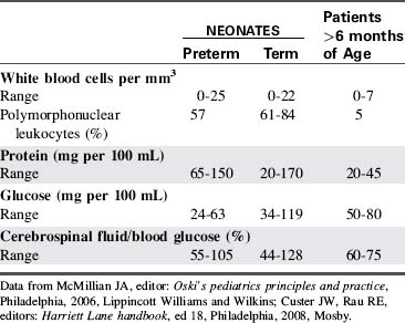
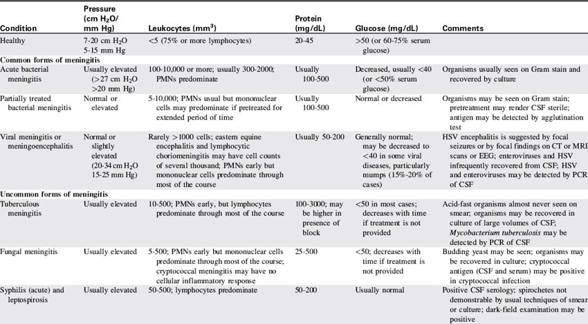
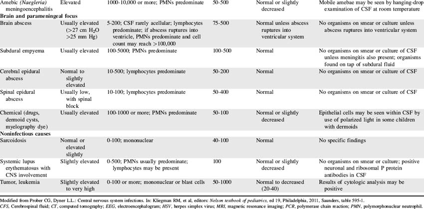
Red blood cells are present in a CSF sample only if a traumatic spinal tap was performed or if the patient has suffered a cerebral hemorrhage. Generally, CSF is hypertonic to blood, but changes in CSF osmolality will parallel those of blood (i.e., an increase in serum osmolality will soon be followed by an increase in CSF osmolality). Abnormalities of CSF composition can aid in the diagnosis of some CNS diseases (Table 11-4).
CSF is formed primarily by the choroid plexuses; these plexuses are collections of capillaries located on the floor of each lateral ventricle and in the third and fourth ventricles. Additional CSF is formed by ependymal cells lining the ventricles and meninges and by blood vessels of the brain and spinal cord. CSF formation requires both active transport and simple diffusion between the existing CSF and the secreting surfaces.
In healthy children, the rate of CSF production is approximately 20 mL per hour.58 The amount of CSF formed is affected by cerebral metabolism, CPP, blood pressure, and changes in the serum osmolality. An increase in the CPP or systemic arterial pressure usually results in an increase in CSF formation.
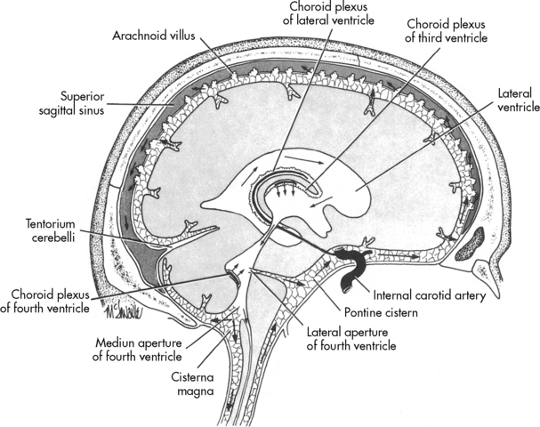
Once formed, the CSF flows from both lateral ventricles through the foramen of Monro into the third ventricle. From there the fluid passes through the cerebral aqueduct, known as the Sylvian aqueduct (or aqueduct of Sylvius), into the fourth ventricle. Some CSF then passes through the two lateral Luschka’s foramina into the subarachnoid space to bathe the brain. The remaining CSF passes through Magendie’s foramen and enters the subarachnoid space to circulate around the spinal cord. Most CSF ultimately is reabsorbed by venous sinuses that project into the subarachnoid space; these are known as the arachnoid villi (Fig. 11-5). Inflammation (e.g., meningitis) or blood in the ventricular system may obstruct CSF flow, often in the narrow aqueduct of Sylvius between the third and fourth ventricle. Subarachnoid hemorrhage can prevent normal reabsorption of CSF.

Fig. 11-5 Normal cerebrospinal fluid circulation. CSF is secreted from the floor of the lateral ventricles. After circulation though the ventricles and cisterns and around the spinal cord, the fluid is reabsorbed by the arachnoid villi in the subarachnoid space.
(From Nolte J: The human brain, St Louis, 1981, Mosby.)
An obstruction in the flow of CSF, an increase in its production, or a decrease in its reabsorption will result in a condition known as hydrocephalus. When hydrocephalus is caused by an obstruction to flow (e.g., with obstruction in the aqueduct of Sylvius or with intraventricular hemorrhage), it is referred to as obstructive or noncommunicating hydrocephalus. When hydrocephalus is caused by increased CSF production or decreased CSF reabsorption (e.g., with subarachnoid hemorrhage), it is known as communicating hydrocephalus. Hydrocephalus causes an increased head circumference in the infant and can produce increased ICP in a patient of any age.
The normal CSF pressure is 7 to 20 cm H2O or 5 to 15 mm Hg in the quiet, resting child; however, this pressure is not static. The CSF pressure normally varies during the cardiac and respiratory cycles and increases transiently during crying, sneezing, or a Valsalva maneuver (grunting or straining against a closed glottis), but it is normally <27 cm H2O or <20 mm Hg.
CSF pressure can be measured from the central canal of the spinal cord (during a lumbar puncture), through catheterization of a lateral ventricle, or by insertion of a catheter into the subarachnoid space. All of these techniques measure CSF pressure and are thought to represent the ICP. If the CSF pressure remains above 27 cm H2O (20 mm Hg), increased ICP is present.
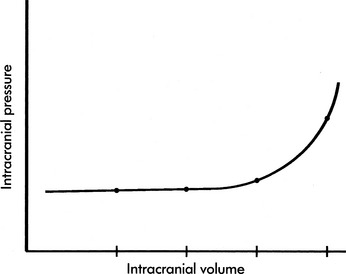
Intracranial Pressure and Volume Relationships
Monroe-Kellie Hypothesis
Monroe (in the eighteenth century) and Kellie (in the nineteenth century) made the observation that the total intracranial volume is relatively constant. In general, the skull is a rigid structure that contains a finite total intracranial volume. The ICP is determined by the total intracranial volume and the intracranial compliance (the change in pressure resulting from a change in volume).
The skull sutures are not fused during infancy, and the skull can expand to accommodate gradual increases in intracranial volume (e.g., hydrocephalus or a slow-growing brain tumor). However, the skull cannot expand rapidly to accommodate acute increases in intracranial volume; therefore, even during infancy the intracranial volume is relatively constant.
The intracranial contents include the brain, the blood, and CSF. Therefore, the intracranial volume is equal to the sum of the volumes of these substances:
If the volume of any of the intracranial contents increases without a commensurate and compensatory decrease in the volume of other intracranial content, the ICP will rise.
The Brain
The brain occupies the largest portion (80%) of the intracranial space; it is essentially not compressible, but it is somewhat movable within the cranium. If significant pressure gradients develop within the cranium or between the intracranial space and the spinal column, cerebral herniation can occur. A severe increase in ICP can cause herniation of the brainstem through the foramen magnum and brain death (cerebral circulation ceases).
Cerebral edema can increase brain volume. Cerebral edema is an increase in brain water content related to increased cellular membrane permeability or massive extravascular (intracellular) fluid shift. Cerebral edema can develop during some infections, metabolic derangements (e.g., treatment of diabetic ketoacidosis), and asphyxia. This edema may be further categorized as vasogenic, cytotoxic, osmotic, or interstitial edema.
Vasogenic cerebral edema may result from disruption of the blood-brain barrier that allows plasma proteins and fluid to enter the brain parenchyma. Cytotoxic edema results from metabolic derangements that alter sodium-potassium pump function and result in retention of sodium and water by the astrocytes. The blood-brain barrier remains intact in cytotoxic edema. Cytotoxic edema can develop following drug or alcohol intoxication, trauma, hypoxic-ischemic events such as cardiac arrest and in early stroke.
Osmotic cerebral edema occurs when the serum osmolality falls acutely, creating an acute difference between intravascular/extracellular osmolality and intracellular (brain) osmolality. This abnormal pressure gradient leads to movement of water into the brain cells (i.e., from the extracellular—including intravascular—space to the intracellular space) with resultant development of cerebral edema. Causes of osmotic edema include an acute fall in the serum sodium concentration, rapid lowering of blood glucose (such as in treatment of diabetic ketoacidosis) or rapid fall in blood urea nitrogen (BUN) during hemodialysis.
Interstitial edema is a consequence of hydrocephalus. The CSF-brain barrier is disrupted resulting in trans-ependymal flow of CSF into the extracellular space of the brain parenchyma.
Brain volume also can be increased as the result of an increase in CBF, such as occurs in some areas of the brain after head injury. Excessive blood flow is referred to as hyperemia.
Cerebral Blood Volume
CBV comprises approximately 7% to 10% of the total intracranial volume. CBF is influenced by the biochemical environment in brain tissue, and changes in CBF normally match changes in cerebral metabolic requirements. As noted previously, CBV can increase or decrease in some areas of the brain after head injury, and cerebral arterial flow may no longer match cerebral metabolic requirements in those areas.
Some cerebral blood is contained in venous capacitance vessels. This blood is dispensable and is shifted from the intracranial vault to reduce total CBV as a compensatory mechanism during periods of increased intracranial volume. Hyperventilation can produce cerebral vasoconstriction; it will decrease cerebral arterial flow and may contribute to displacement of this venous capacitance blood. However, hyperventilation and cerebral vasoconstriction can also produce cerebral ischemia.
If the volume of other intracranial contents increases significantly and ICP rises, CBF and oxygen delivery can be compromised. Cessation of CBF results in brain death.
Cerebrospinal Fluid
CSF normally comprises 7% to 10% of the total intracranial volume and this percentage remains constant if production is matched by absorption. CSF volume will increase if CSF flow pathways are obstructed (e.g., obstructive or noncommunicating hydrocephalus can develop with a brain tumor, after meningitis, or after head trauma associated with intraventricular bleeding), or if CSF reabsorption is diminished (e.g., following head injury with subarachnoid hemorrhage).
CSF is the material most easily displaced from the intracranial vault as compensation for an increase in brain volume or CBV. CSF can be removed from the intracranial vault by a shunt or the placement of a ventricular drain.
Normal Intracranial Pressure
The ICP is the pressure exerted by the intracranial contents. The normal ICP is approximately 5 to 15 mm Hg, but this pressure is not static. It can be increased transiently by anything that acutely increases cerebral venous pressure or by movement from an upright to a reclining position. Typically, the ICP varies by 0.5 to 1.3 mm Hg during respiration.
If the brain, cerebral blood, or CSF volume increases without a compensatory decrease in other intracranial components, the intracranial volume increases. Initially, however, the ICP does not rise (Fig. 11-6). This ability to tolerate an increase in the volume of one intracranial component results from the compensatory displacement of venous capacitance blood or CSF from the intracranial vault. In addition, intracranial compliance (including a small amount of brain compression) allows for some increase in intracranial volume without an increase in ICP. However, there is a limit to this compliance. If the brain, blood, or CSF volume continues to increase, ICP ultimately will rise. Once the limits of compliance have been reached, progressively smaller incremental increases in intracranial volume will be associated with progressively more significant increases in ICP (see Common Clinical Conditions, Increased Intracranial Pressure).

Fig. 11-6 Intracranial pressure (ICP)-volume curve. The change in ICP resulting from an increase in intracranial volume is not linear. Initially, an increase in volume can develop without any increase in pressure. However, once the limits of compliance have been reached, ICP will rise. Note that once the ICP begins to rise, small increases in intracranial volume will produce significant elevations in ICP.
Common clinical conditions
Nursing care of any child with an actual or potential neurologic problem requires careful and repeated assessments over time. For this reason, before presentation of common clinical conditions themselves, this section begins with a summary of critical bedside neurologic assessment (Box 11-2).
Box 11-2 Summary of Bedside Nursing Assessment of Neurologic Function
• Airway, Ventilation and Respiratory Pattern, Oxygenation


• Pupil Size and Response to Light: notify on-call provider immediately if pupils dilate or have decreased constriction to light


• Glasgow Coma Scale Score (see Table 11-6)


• Additional Motor Activity and Reflexes

Neurologic assessment and support includes assessment and support of airway, oxygenation, ventilation, and circulation, as well as evaluation of level of consciousness, pupil size and response to light, cranial nerve function, Glasgow Coma Scale score and additional evaluation of motor activity, reflexes, and movement. General neurologic assessment is summarized in Box 11-2. To ensure clear and consistent communication and to facilitate rapid identification of clinical changes, all members of the healthcare team must use consistent terminology and assessment tools and must apply them in a consistent fashion.
Evaluation of Respiratory Pattern
Patients with neurologic disease or dysfunction may demonstrate a wide variety of respiratory patterns. Regardless of the respiratory rate or pattern demonstrated by the patient, the nurse must ensure that the patient’s airway and arterial oxygen saturation and carbon dioxide removal are adequate, because hypercapnia, hypoxemia and hypoxia can contribute to cerebral vasodilation, increased CBF, increased ICP, and inadequate cerebral perfusion. If respiratory insufficiency develops, immediately notify the on-call provider and support airway, oxygenation and ventilation.
When intracranial injury or insult occurs, some characteristic breathing patterns may be noted that help identify the level of intracranial problem. Such breathing patterns include Cheyne-Stokes breathing, central neurogenic hyperventilation, apneusis, cluster breathing and ataxic breathing (Fig. 11-7). If the ICP rises to a point that brainstem compression occurs and cerebral herniation is imminent, the Cushing reflex is initiated, producing an abnormal breathing pattern that often includes apnea.

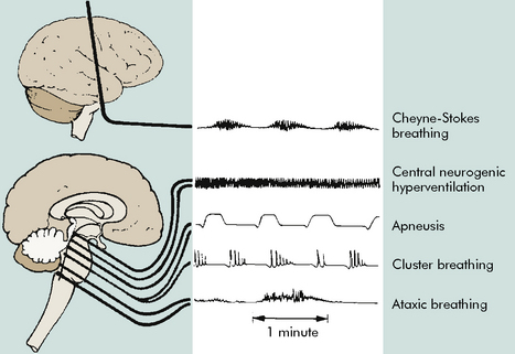
Fig. 11-7 Abnormal respiratory patterns with corresponding level of central nervous system activity.
(From Boss BJ: Alterations in cognitive systems, cerebral hemodynamics, and motor function. In McCance KE, Huether SE, editors: Pathophysiology: the biologic basis for disease in adults and children, ed 6, Philadelphia, 2010, Mosby, p. 531, fig. 16-1.)
Cheyne-Stokes respirations are defined as alternating hyperpnea and bradypnea, which means that the patient initially breathes faster and deeper, then more shallowly, and then demonstrates a long pause before beginning the cycle again. Cheyne-Stokes respirations can be observed in patients with encephalopathies or cerebrovascular disease and in patients with diabetic ketoacidosis.
Central neurogenic hyperventilation is present when the patient breathes deeply at a constant, rapid rate (hyperpnea) despite the presence of adequate arterial oxygenation and hypocapnia. This hyperventilation usually indicates the presence of cerebral hypoxia or ischemia or a midbrain or pontine lesion. Other abnormal breathing patterns include apneustic breathing (pauses after inspiration and possibly after expiration), cluster breathing (irregular breathing associated with irregular pauses), and ataxic breathing (very irregular rate, rhythm, and depth of breaths).
Evaluation of Systemic Perfusion
Careful monitoring of systemic perfusion is required for all critically ill or injured patients. If systemic perfusion is poor, cerebral perfusion may be compromised. This concept is especially true for trauma patients with head injury. Shock resuscitation is essential to optimizing cerebral perfusion.
Vital signs are evaluated in light of the patient’s clinical condition (see pages inside front cover for tables of normal heart rates, respiratory rates, and blood pressures in children). Tachycardia and tachypnea are usually more appropriate in critically ill children than are normal heart and respiratory rates. In children who are 1 to 10 years old and of average height, hypotension is present if the child’s systolic blood pressure is less than 70 mm Hg plus twice the patient’s age in years.31,56 Hypotension is also present if the MAP is less than 40 mm Hg plus one and one-half times the child’s age in years.56
The Cushing Reflex
The Cushing reflex is a late and ominous result of increased ICP and ischemia of the vasomotor center.83 The Cushing reflex indicates profound compromise in brainstem perfusion and may develop only when cerebral brainstem herniation is imminent. This reflex produces the clinical triad of bradycardia, an increase in systolic arterial blood pressure with widened pulse pressure, and abnormal breathing pattern. This clinical triad is referred to as the Cushing triad, a term often used interchangeably with the Cushing reflex. The abnormal breathing pattern that is part of the Cushing triad may consist of an abnormal or irregular respiratory effort or apnea.83
Evaluation of Level of Consciousness
Detection of neurologic deterioration requires careful monitoring of the patient’s level of consciousness, including assessment of behavior and responsiveness that is tailored to each patient. Excessive irritability is a common and nonspecific sign of pain, sleep deprivation, and cardiopulmonary or neurologic dysfunction in the critically ill child. Lethargy is almost always abnormal and is typically a more specific and crucial indicator of deterioration of neurologic function than irritability. However, evaluation of the child’s behavior and responsiveness is facilitated by knowledge of the patient’s normal behavior and condition.
An infant is expected to be irritable when hungry, tired, or overstimulated, so it is important to be aware of normal feeding times and sleep patterns and to attempt to reduce stimulation of the seriously ill or injured infant. It is normal for the infant to be comforted when swaddled or patted and to be quiet and sleepy after feeding. A healthy infant will not cry or sleep constantly, but a seriously ill infant will likely sleep much of the time. A high pitched cry is usually abnormal.
It is important to evaluate the activity of the infant or child in the context of surrounding events and environmental stimulation. The child is expected to be sleepy if the child was awake throughout the preceding night in the hospital. However, it would be extremely abnormal for the same child to sleep while a venipuncture is performed. A decreased response to frightening or painful procedures is abnormal and probably indicates cardiorespiratory or neurologic compromise.
Assessment of level of consciousness in the verbal child is facilitated if the nursing care plan includes information about the child’s normal activities and names of the child’s family members, pets, or favorite stuffed animals. This information will assist in assessment of the child’s short- and long-term memory and orientation to time and place. For example, a child stating that “Oscar was flying around my room at home” could be demonstrating confusion if Oscar is the child’s brother, but can be demonstrating accurate recall if Oscar is the child’s pet parakeet that frequently escapes from the cage. It is helpful to document the names of any imaginary friends that the child has if the child normally refers to them.
If acute coma is present, members of the healthcare team should use consistent terminology (Table 11-5) to describe the child’s level of response. Coma is present when the patient demonstrates no eye opening or verbal response to any stimuli, demonstrating only motor response to painful or noxious stimuli. Stupor is present when only vigorous and repeated stimulation produces arousal.
Table 11-5 Levels of Acute Coma
State | Definition |
Confusion | Loss of ability to think rapidly and clearly; impaired judgment and decision making |
Disorientation | Beginning loss of consciousness; disorientation to time followed by disorientation to place and impaired memory; self-recognition is last to be lost |
Lethargy | Limited spontaneous movement or speech; easy arousal with normal speech or touch; may not be oriented to time, place, or person |
Obtundation | Mild to moderate reduction in arousal (consciousness) with limited response to the environment; falls asleep unless stimulated verbally or tactilely; answers questions with minimal response |
Stupor | A condition of deep sleep or unresponsiveness from which the person may be aroused or caused to open eyes only by vigorous and repeated stimulation; response is often withdrawal or grabbing at stimulus |
Coma | No verbal response to the external environment or to any stimuli; noxious stimuli such as deep pain or suctioning yields motor movement |
Light coma | Associated with purposeful movement on stimulation |
Deep coma | Associated with unresponsiveness or no response to any stimulus |
From Boss BJ: Alterations in cognitive systems, cerebral hemodynamics, and motor function. In McCance KE, Huether SE, editors: Pathophysiology: the biologic basis for disease in adults and children, ed 6, Philadelphia, 2010, Mosby-Elsevier, p. 530, table 16-4.
When coma is present, careful assessment of the child’s motor function is extremely important. Such assessment includes the routine use of a scoring system of neurologic response (see Glasgow Coma Scale Scoring of Neurologic Function).

Stay updated, free articles. Join our Telegram channel

Full access? Get Clinical Tree

