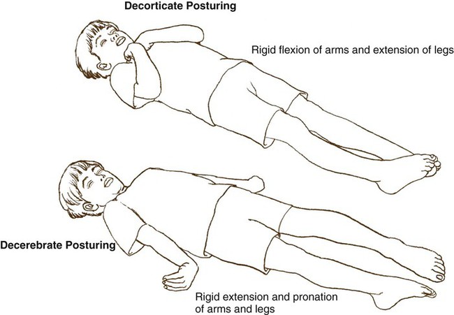
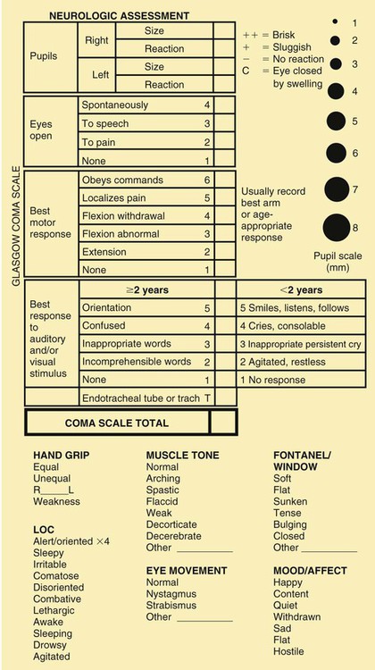
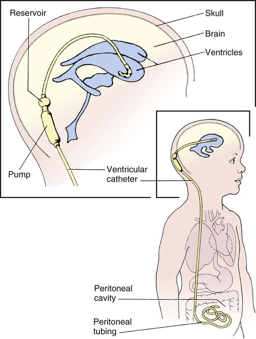
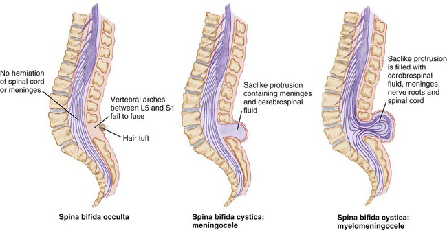
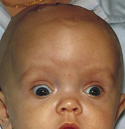
Neurologic and Sensory Disorders


Neurologic and Sensory Disorders


imagehttp://evolve.elsevier.com/Price/pediatric/


imageThe Nervous System


The nervous system is made up of the central nervous system (CNS), which is composed of the cerebrum, cerebellum, brainstem, and spinal cord; the peripheral nervous system, which is composed of the cranial nerves and spinal nerves; and the autonomic nervous system, which is composed of the sympathetic and parasympathetic systems. The focus of this chapter is primarily on the CNS and the disorders that occur in childhood.


The cerebellum is the center for consciousness, thought, memory, sensory input, and motor activity. The cerebellum coordinates all muscle movement including walking, talking, and balance. All cranial nerves (except 1) arise from the brainstem and control vital centers.


The nervous system grows rapidly before birth and well into the first year of life. It will slow to a more gradual rate in later childhood. Brain growth is measured with head circumference and plotted during the first 3 years of life. There is an increased cerebral blood flow and oxygen consumption in childhood that is almost twice that of adults (Hockenberry and Wilson, 2007). This is due to the increased metabolic requirement in early childhood. Myelinization of the nerves in the CNS progresses in a head-to-toe (cephalocaudal) and proximodistal sequence. This myelinization and maturation of the nervous system will lead to the development of motor skills in the growth and development process. Brain development will be influenced by environmental stimuli. Box 13-1 reviews additional physical differences.


Neurologic Disorders


Increased Intracranial Pressure

The brain, CSF, and blood occupy space within the cranium. A change in volume of any of these components must be compensated for. However, if the capacity increases to the point where compensation cannot occur any longer, there will be a rapid increase in intracranial pressure (ICP). Early signs and symptoms of increased ICP may be subtle; these signs and symptoms become more pronounced as the level of consciousness deteriorates. It is important to remember that as ICP increases, cerebral perfusion decreases or is at least compromised. A decrease in cerebral perfusion causes less oxygen to be delivered to the brain cells. Without oxygen, a cell dies, and brain cells do not replace themselves as other cells may do. Discussion of ICP will continue throughout this chapter.


Intracranial Hemorrhage

Intracranial hemorrhage, a common birth injury, may result from trauma or anoxia. It occurs more frequently in the preterm infant, in whom the blood vessels are fragile. Blood vessels within the skull are broken, and bleeding occurs into the brain. When the diagnosis is made, the specific location of the hemorrhage should be noted: subdural or subarachnoid, epidural or intraventricular (IVH is discussed in Chapter 5). This injury may also occur during precipitated delivery, prolonged labor, or when the newborn’s head is large in comparison with the mother’s pelvis.


Signs and Symptoms

The symptoms of intracranial hemorrhage may occur suddenly or gradually. Some or all may be present, depending on the severity. They include inability to move normally, lethargy, poor sucking reflex, irregular respirations, cyanosis, twitching, forceful vomiting, a high-pitched shrill cry, and convulsions. Opisthotonic (opistho, backward; tonos, tension) posturing may be observed. The fontanel may be tense and under pressure, rather than soft and compressible. The pupil of one eye is apt to be small and the other large. If the symptoms are mild, there is a good chance of complete recovery in most cases. Death results if there is a massive hemorrhage. The infant who survives an extensive hemorrhage may have residual defects such as mental retardation or cerebral palsy. The diagnosis is established by the history of the delivery, CT, MRI, evidence of an increase in CSF pressure, and the symptoms and course of the disease.


Treatment and Nursing Care

The newborn is placed in an isolette, which allows proper temperature control, ease in administering oxygen, and continuous observation. The baby is handled gently and as little as possible. The head is elevated. The doctor may prescribe medication to control bleeding and anticonvulsants if convulsions are apparent. The baby is fed carefully because the sucking reflex may be affected. The infant vomits easily. The nurse observes the baby for signs of increased ICP and convulsions. The nurse also assists the physician with such procedures as lumbar punctures and aspiration of subdural hemorrhage.


If a convulsion occurs, the nurse’s observation of its character aids the physician in determining the exact location of the bleeding. The following observations are of particular importance: Were the arms, legs, or face involved? Was the right or left side of the body involved? Was the convulsion mild or severe? How long did it last? What was the condition of the infant before and after the seizure? The nurse records observations in the nurses’ notes.


Head Injuries

Infants and children suffer from head injuries that may occasionally result in brain damage. Falls, shaken baby syndrome, motor vehicle injuries, and bicycle injuries account for a large number of these statistics. Toddlers especially are famous for the number of blows received to the head. Fortunately, most of these injuries are not serious, but they are alarming to parents. The skulls of infants and young toddlers are more pliable and absorb much of the impact to the head. By 2 years of age, both fontanels have completely closed, and the cranium no longer has the same pliability in response to force. Types of head injuries are discussed in Table 13-1.


Table 13-1


Types of Head Injuries


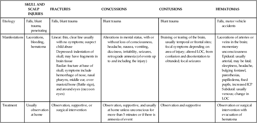
  SKULL AND SCALP INJURIES FRACTURES CONCUSSIONS CONTUSIONS HEMATOMAS
Etiology Falls, blunt trauma, penetrating Falls, blunt trauma Blunt trauma Blunt trauma Falls, motor vehicle accidents
Manifestations Lacerations, bleeding, hematoma Linear: thin, clear line usually with no symptoms; suspect child abuse
Depressed: indentation of skull; may have fragments in brain tissue
Basilar: fracture at base of skull; symptoms include hemorrhage of nose, nasal pharynx, middle ear, over mastoid bone (Battle sign), and around eyes (raccoon eyes)
Alterations in mental status, with or without loss of consciousness, headache, nausea, vomiting, dizziness, irritability, seizures, retrograde amnesia (of events up to and including the injury) Bruising or tearing of the brain, usually temporal or frontal sites; focal symptoms depending on area of injury; altered LOC, from confusion and disorientation to obtunded; focal seizures Lacerations of arteries or veins in the brain; momentary unconsciousness
Epidural: usually arterial, may be fatal; sleepiness, headache, bulging fontanel, paresthesias, papilledema, fixed pupils, increased ICP
Subdural: usually venous; change in LOC
Treatment Usually observation at home Observation, supportive, or surgical intervention Observation, supportive, and usually at home unless unconscious for more than 5 minutes or if there is amnesia of event Observation and supportive Observation or surgical intervention with evacuation of hematoma


image


ICP, Intracranial pressure; LOC, level of consciousness.


Treatment and Nursing Care

Frequently a child who has had a blow to the head is brought to the hospital for observation to rule out or confirm the diagnosis. Initial care of the child with a head injury includes assessment of the ABCs (airway, breathing, circulation), assessment for spinal cord injury, and documentation of baseline vital signs. The child may have all or some of the following symptoms: headache (manifested by fussiness in the toddler), drowsiness, blurred vision, vomiting, and dyspnea (see Did You Know?). In severe cases, the child may be completely unconscious or having seizures. Decerebrate (indicating injury to the midbrain) or decorticate (indicating injury to the cerebral cortex) posturing may be evident (Figure 13-1). A careful history is obtained to determine any preexisting conditions and to ascertain the exact circumstances of the accident. Of particular importance is the child’s state of consciousness immediately after the occurrence. Radiography, CT, and magnetic resonance imaging (MRI) can be used to diagnose the specific head injury. Should the child be alert, without significant symptoms, and the parents reliable, the child can be sent home with instructions for observation.


Further assessment includes the level of consciousness (LOC); changes are particularly meaningful and require immediate medical attention. Changes in behavior can be an early sign of increasing ICP. Response should be correlated with the developmental age of the child. Parents can be helpful in providing information about the child’s usual capabilities. In general, the child should be oriented to person, time, and place (may not be accessible in the toddler). Ask “What is your name?” and “Where are you?” Older children may know the day of the week. The child should recognize the parents. Point to the mother and ask “Who is this?” If there is no response to verbal stimuli, note the arousal to tactile stimulation. Determine the presence or absence of crying or speech. It is not unusual for the child to fall asleep, but he or she should be easily aroused. Record changes in sleeping pattern, posture, movements of extremities, and any signs of tremors or restlessness.


The Pediatric Coma Scale (Figure 13-2), based on the adult Glasgow Coma Scale, is valuable in determining various LOCs. It consists of three parts: eye opening, motor response, and verbal response. A numerical value is assigned to each part. The lower the score, the deeper the coma.


Evaluation of pupil and eye movement requires documentation of size, shape, and equality of pupils and their reaction to light and extraocular movements. (Have the child follow your finger from side to side and up and down to detect movement.) Strabismus, nystagmus, “sunset” eyes (eyes deviated downward), and inability to move eyes in all four quadrants indicate abnormality. If ICP is increasing, pupils become sluggish to light stimulus, dilated, and eventually fixed; this is an indication of a medical emergency.


Advanced ICP causes an increase in systolic blood pressure with a widening pulse pressure (the diastolic blood pressure usually decreases), a decrease in pulse, and altered respiratory pattern. This is referred to as the Cushing triad. Temperature is routinely monitored. Never take oral temperatures in children prone to seizures. Elevations may result from inflammation, systemic infection, or damage to the hypothalamus, which regulates body temperature. Body temperature may be reduced by administering antipyretics or by using a hypothermia blanket. Mild elevations in temperature are not uncommon in the first 2 days after trauma. Any seizure activity is treated with anticonvulsants.


The quality and strength of muscle tone should be observed in all four extremities. The child should be able to squeeze the nurse’s hands. The grip should be equal in both hands. The child should be able to move the legs and push against the examiner’s hands with both feet. The face should be symmetrical, and the child should be able to smile and frown. Drooping of the eyes, ptosis, inability to close the eyes tightly, and drooping of the corner of the mouth are considered adverse signs. The child should be able to raise the arms and turn the palms up and down. Abnormal posturing should be described and recorded.


Record the type and amount of any drainage from the ears and nose. Leakage of cerebrospinal fluid from a fractured skull is seen as clear drainage from the ears or nose. Head circumference (FOC) should be monitored in infants, as should tension of the fontanels and the presence of a high-pitched cry.


Fluids are carefully monitored to control cerebral edema. Overhydration increases the amount of cerebral fluid. Feeding difficulties should be noted as the child’s diet is increased. Children should be observed for signs of shock, which can also occur.


Children whose conditions have remained stable are discharged. Parents are instructed about any additional observations and follow-up care.


It is important to teach parents about preventing head injury. Teach parents of infants never to leave the child unattended on a changing table or bed. Childproofing the home with stairway gates decreases falls. Teaching the toddler and preschooler not to run out in the street is an important safety measure. As the child becomes older and more active, a helmet should be worn during activities that provide risk, such as biking, rollerblading, and contact sports.


Hydrocephalus

Hydrocephalus (hydro, water; cephalo, head) is a condition characterized by an increase in cerebrospinal fluid (CSF) in the ventricles of the brain, which causes an increase in the size of the head and pressure changes in the brain. It occurs as a result of an imbalance between production and absorption of CSF. Hydrocephalus may be congenital or acquired. It may occur in conjunction with a meningomyelocele or as a sequela of infections, including congenital TORCH infections (TORCH stands for toxoplasmosis, other, rubella, cytomegalovirus, and herpes simplex), encephalitis, or meningitis, or because of perinatal hemorrhage. The symptoms depend on the site of obstruction and the age at which it develops. Although there are many causes of hydrocephalus, all result in either an impairment of CSF absorption within the subarachnoid space (communicating hydrocephalus) or an obstruction of CSF flow within the ventricles (noncommunicating hydrocephalus). Hydrocephalus may proceed slowly or rapidly. Two forms of hydrocephalus are the Arnold-Chiari malformation and the Dandy-Walker syndrome. Because hydrocephalus can cause progressive cerebral damage, early recognition and treatment are important.


It should be recalled that the brain and spinal cord are surrounded by fluid, membranes, and bone. The three membranes, called meninges, are the dura mater, the arachnoid, and the pia mater. The arachnoid mater (also known as the middle membrane) resembles a cobweb, and its spaces are filled with fluid. CSF is also found in spaces of the brain called ventricles. The primary site of formation for CSF is believed to be the choroid plexus.


Signs and Symptoms

Signs and symptoms depend on the time of onset and the severity of the imbalance. The classic sign both in congenital hydrocephalus and in hydrocephalus with onset in infancy is an increase in head size (Figure 13-3). The direction of skull expansion depends on the site of obstruction. Transillumination (trans, across; illuminare, to enlighten), or the inspection of a cavity or organ by passing a light through its walls, is an older diagnostic procedure used to visualize fluid. (This method uses a flashlight with a sponge rubber collar held tightly against the infant’s head in a dark room to indicate areas of increased luminosity.) Another sign is a bulging anterior fontanel and separation of cranial sutures. The scalp is also shiny and the veins dilated. The infant appears helpless and lethargic. The body becomes thin, and the muscle tone of the extremities is often poor. In addition to the infant’s shrill and high-pitched cry, irritability, vomiting, and anorexia are present. Convulsions may also occur. In severe infantile hydrocephalus, the eyes may appear deviated downward, which is known as the “setting sun” sign (Figure 13-4). Children with an onset of hydrocephalus later in childhood may have minimal enlargement of the head and display the signs and symptoms of increased ICP.


Diagnosis and Treatment

The child’s head is measured daily. Echoencephalography, computed tomography (CT), or magnetic resonance imaging (MRI) is most frequently used to show the enlarged ventricles and to locate the level of obstruction. Sedation is required. A ventricular tap or puncture may be performed in a special procedures room with sterile technique. The equipment needed is the same as that for a lumbar puncture. The specimen is labeled and sent to the laboratory for analysis.


Treatment is directed toward relief of symptoms, treatment of the underlying problem (such as a tumor), and treatment of complications. If there is an obstruction, such as a tumor, it may be removed surgically. In other conditions, a shunt is placed by inserting special tubing that provides drainage of the CFS from the ventricles to another area of the body, where it is absorbed and eventually excreted. Two types of shunts are used: the ventriculoperitoneal (VP) shunt and the ventriculoatrial (VA) shunt. The VP shunt is the most commonly used (Figure 13-5). New shunt systems now allow for growth and have generally eliminated the necessity for shunt revisions. (Shunts still need to be replaced if a malfunction occurs). Shunts work with a one-way valve that opens at a predetermined intraventricular pressure and closes when the pressure falls below that level. This prevents CSF from flowing back upward. Nurses and parents may be taught how to pump the shunt.


The prognosis for hydrocephalus has improved with modern drugs and surgical techniques. If the brain has not been seriously damaged before the operation, mental function can be preserved. However, motor development may occur at a slower rate, if the child cannot lift the head normally. Complications associated with shunts are usually caused by mechanical flaws (kinking or plugging of tubing) or infection. Shunt malfunction may be identified by headaches, nausea, vomiting, irritability, and lethargy. The shunt is also subject to infection. The symptoms of shunt infections may include an unexplained fever and symptoms similar to those of shunt malfunction. If the infections persist, it may become necessary to remove the shunt.


Nursing Care

The general nursing care of an infant with hydrocephalus who has not undergone surgery presents several challenges. The child may be barely able to raise the head. Mental development is also delayed. Lack of appetite, a tendency to vomit easily, and poor resistance to infections pose additional problems.


The position of the infant must be changed frequently to prevent hypostatic pneumonia and pressure sores. Hypostatic pneumonia occurs when there is poor circulation of the blood in the lungs and when the child remains too long in one position. It is particularly prevalent in children who are poorly nourished, weak, or have a debilitating disease. Whenever the nurse turns the child with hydrocephalus, the head must always be supported. To turn the patient in bed, the weight of the head should be borne in the palm of one hand and the head and body should be rotated together to prevent a strain on the neck. When the child is lifted from the crib, the head must be supported by the nurse’s arm and chest. The head circumference (FOC) is measured daily. This measurement is critical, and it is often marked so that daily measurements will be performed in the same location. If there is a deviation in the findings, the charge nurse should be notified. Pressure sores may occur if the child’s position is not changed at least every 2 hours. The tissues of the head and ears and the bony prominences have a tendency to break down. A soft fluid-filled pad may be placed under the head to help prevent lesions. In most cases, the nurse may hold the infant for feeding. The nurse sits with the arm supported because the baby’s head is heavy. A calm, unhurried manner is necessary. The room should also be as quiet as possible to avoid unnecessary stimulation.


Observations that need to be made include vomiting, condition of skin, motor abilities, restlessness, irritability, lethargy, and changes in vital signs. Fontanels are palpated for size and bulging. Changes in vital signs associated with increased ICP are usually a sign later in infancy. They include elevated blood pressure and a decrease in pulse and respirations. Signs of a cold or other infection should be reported to the charge nurse immediately and recorded.


Many infants and children are treated surgically for hydrocephalus. Postoperative nursing care is complex, and in addition to routine postoperative care and observations, the nurse observes the child for signs of increased ICP and for infection at the operative site or along the shunt line. Pain issues should be included in the care of the postoperative child.


Bacterial infection is a life-threatening complication that sometimes makes it necessary to remove the shunt. Signs of infection include poor feeding, elevated vital signs, decreased level of consciousness, vomiting, and seizure activity. The nurse should also observe for signs of inflammation at the shunt insertion site. If the fontanels are sunken, the infant should be kept flat because too rapid a reduction in fluid may lead to seizures or cortical bleeding. If the fontanels are bulging, the child is usually placed in the semi-Fowler’s position to assist in drainage of the ventricles through the shunt. Usually the physician will indicate the position to be maintained and the extent of activity allowed. The child is always positioned so as to avoid pressure on the operative site. Head measurements are recorded. In children with peritoneal shunts, the abdomen should also be measured or observed to detect malabsorption of fluid. Skin care continues to remain a priority. As the child’s condition improves, parents are instructed regarding the care of the shunt.


Hydrocephalus, even with a shunt, is a chronic condition, and the child needs to be followed throughout life. Parents are taught how to recognize shunt malfunction and infection. It is also important that family issues and the child’s own growth and development be considered.


image
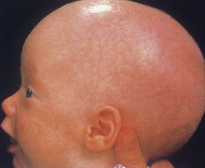
Community Considerations


The National Hydrocephalus Foundation (www.nhfonline.org) provides information and networking opportunities for families.


Myelodysplasia/Spina Bifida

Myelodysplasia refers to a group of CNS disorders characterized by abnormal development of the spinal cord and associated neural tube structures. These defects are categorized as neural tube defects (NTDs). One of these disorders is spina bifida.


Spina bifida (divided spine) is a congenital embryonic NTD in which there is imperfect closure of the spinal vertebrae. There are two forms: occulta (hidden) and cystica (sac or cyst). Spina bifida occulta is a relatively minor variation of the disorder in which the opening is small and there is no associated protrusion of structures. It is often undetected and occurs most commonly at L5 and S1 levels. There may be a tuft of hair, a dimple, a lipoma, or a port-wine birthmark at the site. Generally, treatment is not necessary unless neuromuscular symptoms appear. These consist of progressive disturbances of gait, footdrop, or disturbances of bowel and bladder sphincter function.


Spina bifida cystica consists of the development of a cystic mass in the midline of the spine (Figure 13-6). Meningocele and meningomyelocele (also called myelomeningocele) are two types of spina bifida cystica. A meningocele (meningo, membrane; cele, tumor) contains portions of the membranes and CSF. The size varies from that of a walnut to that of the head of a newborn infant.


Stay updated, free articles. Join our Telegram channel

Dec 22, 2016 | Posted by in NURSING | Comments Off on Neurologic and Sensory Disorders

Full access? Get Clinical Tree

Get Clinical Tree app for offline access