Introduction to Anatomy and Physiology


Introduction to Anatomy and Physiology


Learning Objectives


1. Explain why it is important for the medical assistant to be knowledgeable about anatomy and physiology.


2. Explain the relationship between anatomy and physiology.


3. State the six levels of organization within the human body.


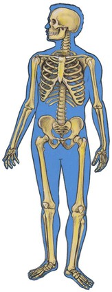
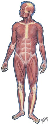
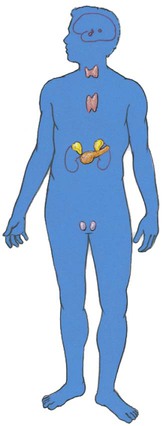
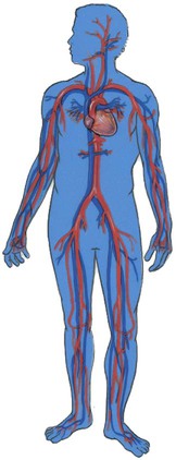
4. List the 11 organ systems of the body, and describe the function of each.


5. Describe homeostasis and its importance to the human body.


6. Explain how the body maintains homeostasis using a negative feedback system.


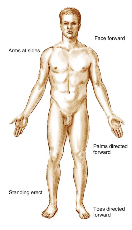
7. List the four criteria used to describe the anatomic position.


8. Identify body planes, body regions, and relative positions using anatomic terms.


9. Distinguish between the dorsal body cavity and the ventral body cavity, and list the subdivisions of each cavity.


10. Describe the cell membrane.


11. Describe the composition of the cytoplasm.


12. Describe the components of the nucleus, and state the function of each component.


13. Identify and describe each of the cytoplasmic organelles, and state the function of each organelle.


14. Explain how the cell membrane regulates the composition of the cytoplasm.


15. Describe the various mechanisms that result in the transport of substances across the cell membrane.


16. List the phases of a cell cycle, and describe the events that occur in each phase.


17. Explain the difference between mitosis and meiosis.


18. List the four main types of tissues found in the body.


19. Describe the various types of epithelial tissues in terms of structure, location, and function.


20. Describe the general characteristics of connective tissue.


21. List three types of connective tissue cells, and state the function of each.


22. Describe the features and location of the various types of connective tissue.


23. Explain the differences among skeletal muscle, smooth muscle, and cardiac muscle in terms of structure, location, and control.


24. State the two categories of cells in nerve tissue, and explain their function.


A Brief Summary of Medical History


Studies of illness and aging of the human body are major components in the field of medicine. The study of “modern” medicine began in the fifth century BC.


Hippocrates, who was born in approximately 460 BC on the island of Cos, Greece, is recognized as the “Father of Medicine.” After his death, all the existing writings on medicine were gathered into a work called the Hippocratic Collection and attributed to him whether he wrote them or not. The Hippocratic Oath, which is still in use today, is from the Collection. Hippocrates concluded that illness had rational explanations instead of being caused by evil spirits or disfavor of the gods. This freed medicine from superstition and allowed for scientific study.


In 1249, Roger Bacon invented eyeglasses, bringing better vision to many. Leonardo da Vinci advanced the understanding of human anatomy by carefully dissecting corpses and making detailed anatomic drawings during the fifteenth century. William Harvey published An Anatomical Essay on the Motion of the Heart and Blood in Animals in 1628, detailing how blood was pumped from the heart throughout the body and then returned to the heart and recirculated. This work showed that food was not converted into blood by the liver and then consumed as fuel by the body, as was widely alleged at that time.


A Dutch cloth merchant, Antony van Leeuwenhoek discovered blood cells in 1670. This discovery was made possible by his microscope. Although the microscope had been invented by Robert Hooke a few years earlier, van Leeuwenhoek, by grinding his own glass lenses, greatly improved the microscope’s design and achieved magnifications of greater than 270 diameters. He also observed bacteria, yeast cells, spermatozoa, and protozoa. In addition, his microscope allowed capillaries to be observed, thus showing the link between arteries and veins and confirming Harvey’s theory of blood circulation.


Edward Jenner, an English microbiologist, is known as the “Father of Immunology.” He observed that people who had contracted cowpox seemed immune to the deadly smallpox. He theorized that deliberately infecting people with cowpox would protect them from smallpox. He tested his theory on a young boy in 1796 and then demonstrated that the lad was indeed immune to smallpox.


William Beaumont, while serving as an army post surgeon, treated a patient who had been blasted by a musket at close range. The resulting large wound affected part of his lung, two ribs, and his stomach. Beaumont treated the wounds but was unable to get the hole in the stomach to completely close; repeated bandaging was required to prevent food and drink from coming out. He quickly realized this was an opportunity to study the digestion process. He tied small pieces of food with silk string and dangled them through the hole in the patient’s stomach, removing the items at 1-hour intervals. He published his observations in 1833.


Medicine truly came of age during the second half of the nineteenth century. Louis Pasteur and Robert Koch established the germ theory of disease. Florence Nightingale showed the importance of hygiene and sanitation to reduce hospital infections. Sir Humphry Davy discovered the anesthetic properties of nitrous oxide, and Joseph Lister pioneered the use of carbolic acid as an antiseptic to clean wounds and surgical instruments. His antiseptic technique reduced deaths from infection after surgery from about 60% to under 4%. Then in 1895 a German scientist named Wilhelm Roentgen discovered the x-ray. Medicine has never been the same since.


The twentieth century continued to build on all these discoveries. The pharmaceutical industry mushroomed with the development of thousands of new medicines. Vaccines were developed to prevent polio, measles, mumps, and many other diseases. Cardiac pacemakers and defibrillators were invented. Medical imaging advanced with the development of computed tomography (CT), magnetic resonance imaging (MRI), positron emission tomography (PET), ultrasound, and many other techniques. Numerous advances occurred in surgery, including open-heart surgery and organ transplants. Radiation therapy and chemotherapy for the treatment of cancer were developed. Kidney dialysis machines were invented, and hospital intensive care units (ICUs) were established to better treat very sick patients. The list of medical advances goes on and on and is added to every year.


The Human Body


The human body is an awesome masterpiece. Imagine billions of microscopic parts, each with its own identity, working together in an organized manner for the benefit of the total being. The human body is more complex than the greatest computer, yet it is personal. The study of the human body is as old as history itself because people have always had an interest in how the body is put together, how it works, why it becomes defective (illness), and why it wears out (aging).


The study of the human body is essential for those planning a career in health sciences, just as knowledge about automobiles is necessary for those planning to repair them. How can you fix an automobile if you do not know how it is put together or how it works? How can you help fix a human body if you do not know how it is put together or how it works?


Anatomy and Physiology


Human anatomy is the study of the shape and structure of the human body and its parts. It encompasses a wide range of study, including the development and microscopic organization of structures, the relationship between structures, and the interrelationship between structure and function. Gross human anatomy deals with the large structures of the human body that can be seen through normal dissection. Microscopic anatomy deals with the smaller structures and fine detail that can be seen only with the aid of a microscope.


Human physiology is the scientific study of the functions or processes of the human body. It answers how, what, and why anatomic parts work. Anatomy and physiology are interrelated because structure and function are always closely associated. The function of an organ, or how it works, depends on how it is put together. Conversely, the anatomy or structure provides clues to understanding how it works. The structure of the hand, with its long, jointed fingers, is related to its function of grasping things. The heart is designed as a muscular pump that can contract to force blood into the blood vessels. By contrast, lungs are made of a thin tissue and function to exchange oxygen and carbon dioxide between the outside environment and the blood. Imagine what would happen if the heart were made of thin tissue and the lungs were made of thick muscle. Structure and function are always related.


Levels of Organization


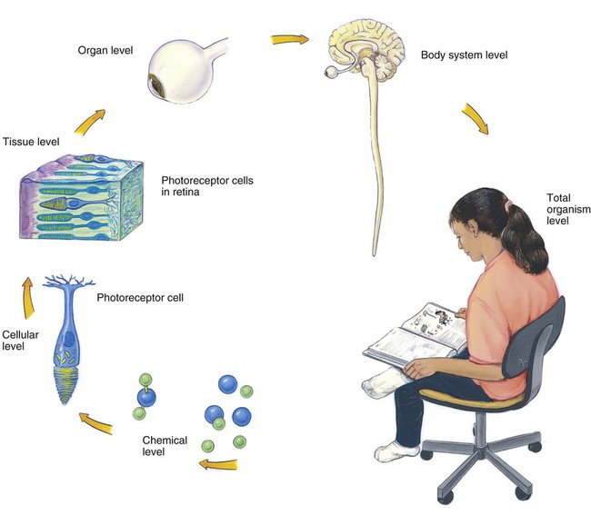
Among the most outstanding features of the complex human body are its order and organization—how all the parts, from tiny cells to visible organs, work together to make a functioning whole. The organizational scheme of the body has six levels (Figure 5-1).


Starting with the simplest and proceeding to the most complex, the six levels of organization are chemical, cellular, tissue, organ, body system, and total organism. The structural and functional characteristics of all organisms are determined by their chemical makeup.


Organ Systems


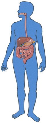
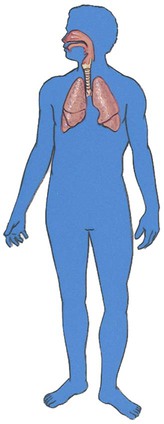
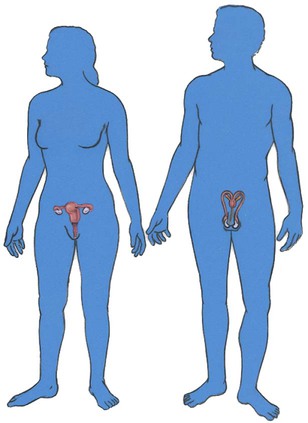
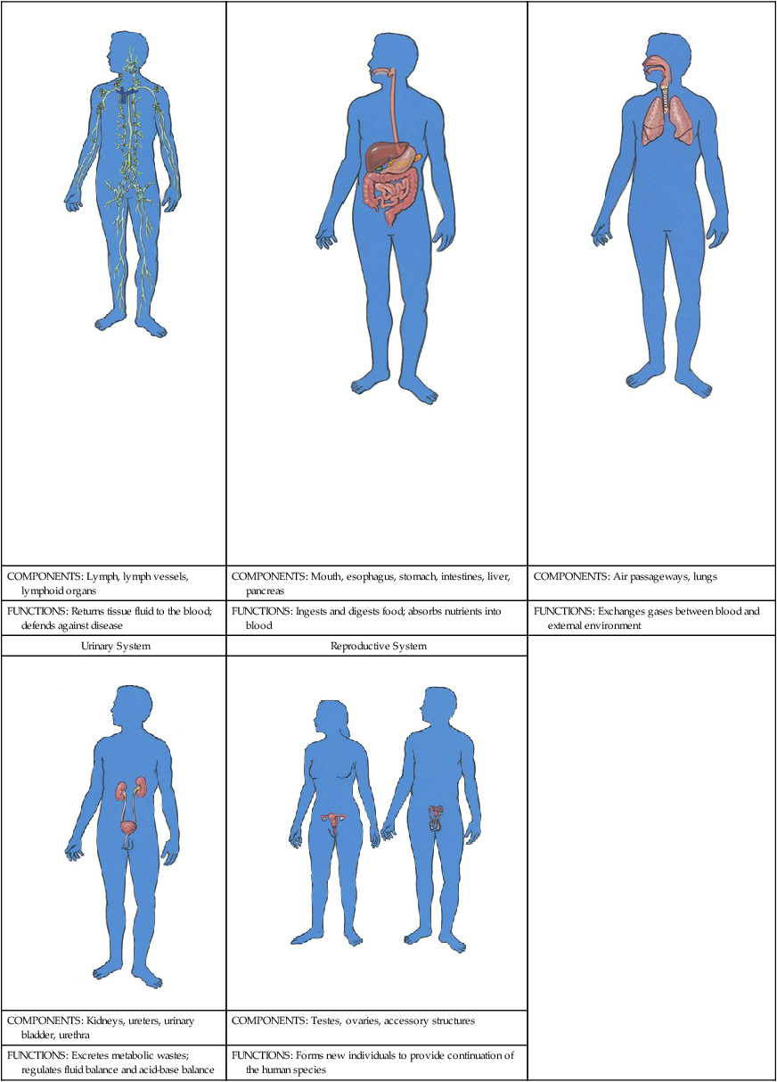
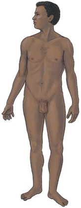
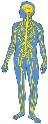
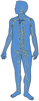
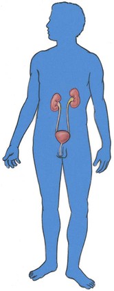
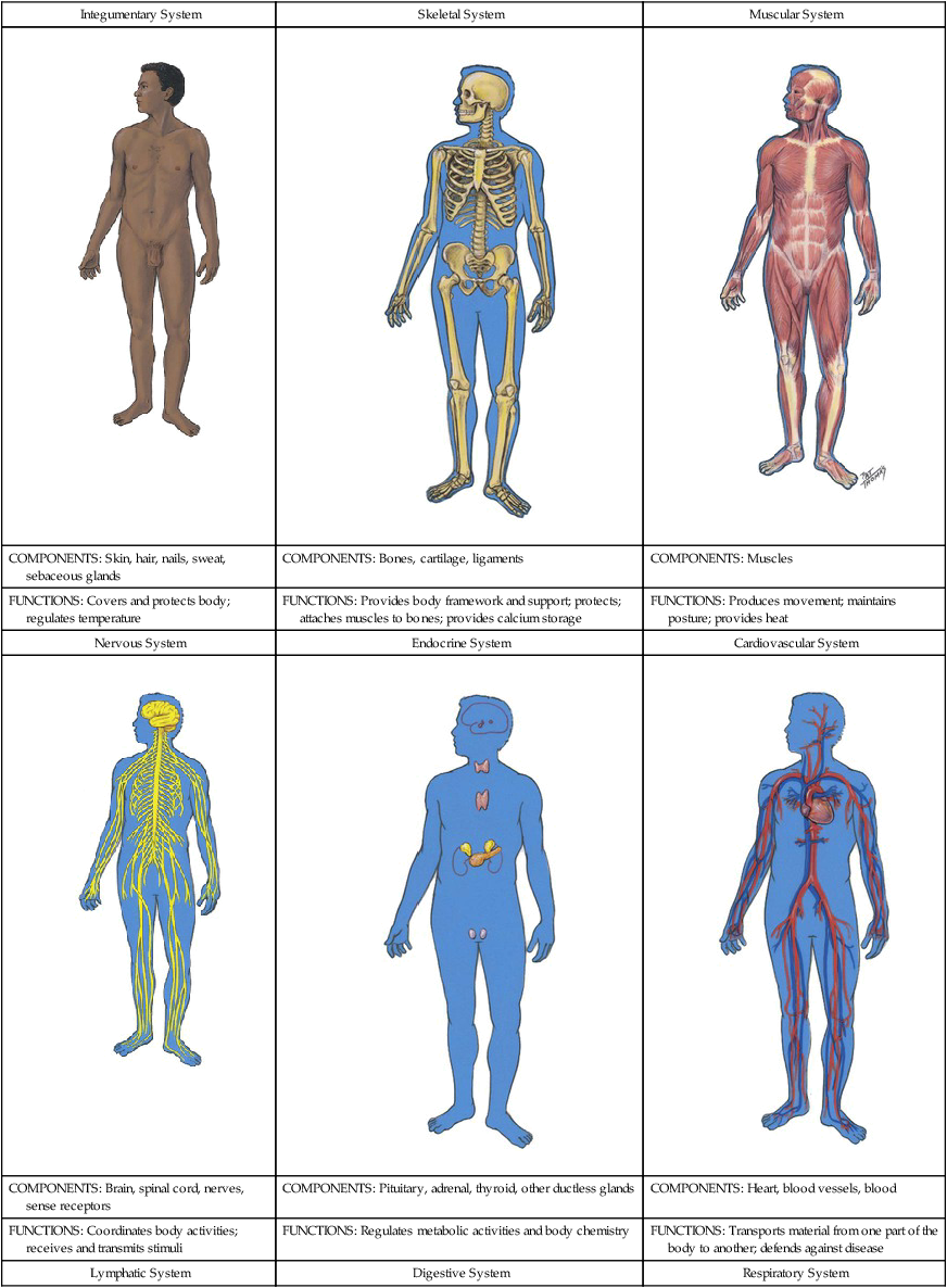
The human body has 11 major organ systems, each with specific functions, yet all are interrelated and work together to sustain life. Each system is described briefly here and then in more detail in later chapters. The organ systems are illustrated and summarized in Table 5-1.


Table 5-1


Organ Systems of the Body


Integumentary System Skeletal System Muscular System
image

image

image
COMPONENTS: Skin, hair, nails, sweat, sebaceous glands COMPONENTS: Bones, cartilage, ligaments COMPONENTS: Muscles
FUNCTIONS: Covers and protects body; regulates temperature FUNCTIONS: Provides body framework and support; protects; attaches muscles to bones; provides calcium storage FUNCTIONS: Produces movement; maintains posture; provides heat
Nervous System Endocrine System Cardiovascular System
image

image

image
COMPONENTS: Brain, spinal cord, nerves, sense receptors COMPONENTS: Pituitary, adrenal, thyroid, other ductless glands COMPONENTS: Heart, blood vessels, blood
FUNCTIONS: Coordinates body activities; receives and transmits stimuli FUNCTIONS: Regulates metabolic activities and body chemistry FUNCTIONS: Transports material from one part of the body to another; defends against disease
Lymphatic System Digestive System Respiratory System
image

image

image
COMPONENTS: Lymph, lymph vessels, lymphoid organs COMPONENTS: Mouth, esophagus, stomach, intestines, liver, pancreas COMPONENTS: Air passageways, lungs
FUNCTIONS: Returns tissue fluid to the blood; defends against disease FUNCTIONS: Ingests and digests food; absorbs nutrients into blood FUNCTIONS: Exchanges gases between blood and external environment
Urinary System Reproductive System  
image

image
COMPONENTS: Kidneys, ureters, urinary bladder, urethra COMPONENTS: Testes, ovaries, accessory structures
FUNCTIONS: Excretes metabolic wastes; regulates fluid balance and acid-base balance FUNCTIONS: Forms new individuals to provide continuation of the human species

image


image


From Applegate E: The anatomy and physiology learning system, ed 4, St Louis, 2011, Saunders.


Homeostasis


Homeostasis refers to the constant internal environment that must be maintained for the cells of the body. The word is derived from two Greek words: homeo, which means “alike” or “the same,” and stasis, which means “always” or “staying.” Putting these together, the word homeostasis means “staying the same.” When the body is healthy, the internal environment always stays the same. It remains stable within limited normal ranges.


Everyone is familiar with aspects of the external environment—whether it is cold or hot, humid or dry, smoggy or clear. The internal environment is not quite as obvious. It involves the tissue fluid that surrounds and bathes every cell of the body. Normal functional activities of the cell depend on the internal environment being maintained within limited normal ranges. The chemical content, volume, temperature, and pressure of the fluid must stay the same (homeostasis), regardless of external conditions, so that the cell can function properly. If the conditions in the tissue fluid deviate from normal, mechanisms respond that try to restore conditions to normal. If the mechanisms are unsuccessful, the cell malfunctions and dies. This leads to illness and disease. Ultimately the goal of medical treatment is to restore homeostasis.


Negative and Positive Feedback

Any condition or stimulus that disrupts the homeostatic balance in the body is a stressor. When a stressor causes internal conditions to deviate from normal, all the body systems work to bring conditions back to the normal range. This is usually accomplished by a negative feedback mechanism in which a stimulus initiates reactions that reduce the stimulus. This mechanism works similarly to a thermostat connected to a furnace and an air conditioner. When the temperature in the room decreases (stressor) below the thermostat setting (normal), the sensing device in the thermostat detects the change and causes the furnace to add heat to the room. When the room becomes too warm, the furnace stops and the air conditioner begins to cool the room. Negative feedback mechanisms do not prevent variation, but they keep variation within a normal range.


An example of a physiologic negative feedback mechanism in the human body involves blood pressure. When blood pressure decreases below normal, body sensors detect the deviation and initiate changes that bring the pressure back within the normal range. When the pressure increases above normal, changes occur to decrease the pressure to normal. Variations in blood pressure occur, but homeostatic mechanisms keep them within the limits of a normal range.


The nervous and endocrine systems work together to control homeostasis, but all the organ systems in the body help maintain the normal conditions of the internal environment. The brain contains centers that monitor temperature, pressure, volume, and the chemical conditions of body fluids. Endocrine glands secrete hormones in response to deviations from normal conditions, and these hormones affect other organs. The changes required to bring conditions back to the normal range are mediated by various organ systems. Good health depends on homeostasis. Illness results when the negative feedback mechanisms that maintain homeostasis are disrupted. Medical therapy attempts to assist the negative feedback process to restore balance, or homeostasis.


Anatomic Terms


Certain basic terms need to be understood to communicate effectively in the health care profession. In other words, you have to speak the language. This section explains some basic terms that relate to the anatomy of the body. They are used to describe directions and regions of the body.


Directions in the Body

Directional terms are used to describe the relative position of one part to another. Note that in the following list of directional terms, the two items in each pair of terms are opposites.


Superior means that a part is above another part, or closer to the head. The nose is superior to the mouth. Inferior means that a part is below another part, or closer to the feet. The heart is inferior to the neck.


Anterior (or ventral) means toward the front surface. The heart is anterior to the vertebral column. Posterior means that a part is toward the back. The heart is posterior to the sternum.


Medial means toward, or nearer, the midline of the body. The nose is medial to the ears. Lateral means toward, or nearer, the side, away from the midline. The ears are lateral to the eyes.


Proximal means that a part is closer to a point of attachment, or closer to the trunk of the body, than another part. The elbow is proximal to the wrist. The opposite of proximal is distal, which means that a part is farther away from a point of attachment than is another part. The fingers are distal to the wrist.


Superficial means that a part is located on or near the surface. The superficial (or outermost) layer of the skin is the epidermis. The opposite of superficial is deep, which means that a part is away from the surface. Muscles are deep to the skin.


Visceral pertains to internal organs or the covering of the organs. The visceral pericardium covers the heart. Parietal refers to the wall of a body cavity. The parietal peritoneum lines the wall of the abdominal cavity.


Planes and Sections of the Body

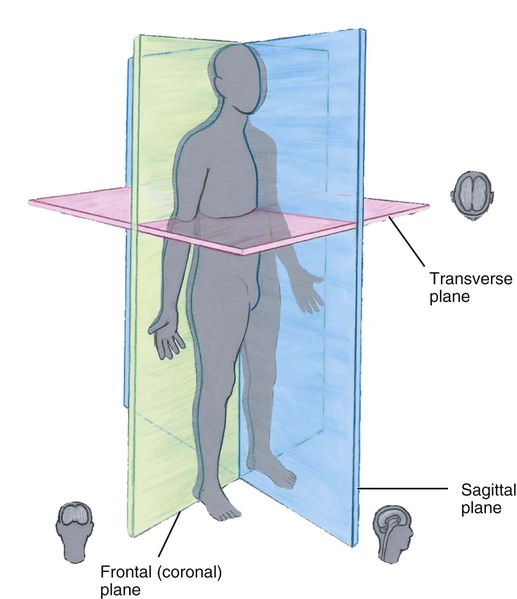
To aid in visualizing the spatial relationships of internal body parts, anatomists use three imaginary planes, each of which is cut through the body in a different direction. Figure 5-3 illustrates these three planes.


Body Cavities

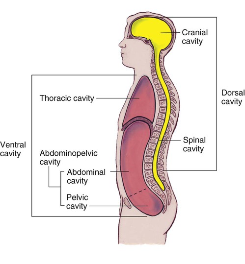
Spaces within the body that contain the internal organs or viscera are called body cavities. The two main cavities are the dorsal cavity and the larger ventral cavity, which are illustrated in Figure 5-4. The dorsal cavity is divided into the cranial cavity, which contains the brain, and the spinal cavity, which contains the spinal cord. The cranial and spinal cavities join with each other to form a continuous space.


The ventral cavity is much larger than the dorsal cavity and is subdivided into the thoracic (tho-RAS-ik) cavity and the abdominopelvic (ab-dahm-ih-noh-PEL-vik) cavity. The thoracic cavity is superior to the abdominopelvic cavity and contains the heart, lungs, esophagus, and trachea. It is separated from the abdominopelvic cavity by the muscular diaphragm. Although there is no clear-cut partition to divide it, the abdominopelvic cavity is separated into the superior abdominal cavity and the inferior pelvic cavity. The stomach, liver, gallbladder, spleen, and most of the intestines are in the abdominal cavity. The pelvic cavity contains portions of the small and large intestines, the rectum, the urinary bladder, and the internal reproductive organs.


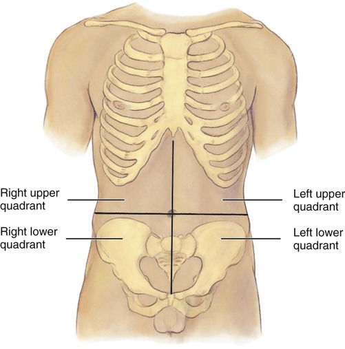
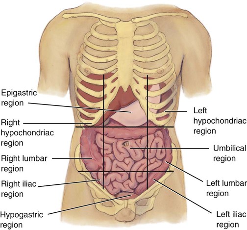
To help describe the location of body organs or pain, health care professionals frequently divide the abdominopelvic cavity into regions using imaginary lines. One such method uses the midsagittal plane and a transverse plane that passes through the umbilicus. This divides the abdominopelvic area into four quadrants, illustrated in Figure 5-5. Another system uses two sagittal planes and two transverse planes to divide the abdominopelvic area into the nine regions illustrated in Figure 5-6. The three central regions are, from superior to inferior, the epigastric (ep-ih-GAS-trik), umbilical (um-BIL-ih-kal), and hypogastric (hye-poh-GAS-trik) regions. Lateral to these, from superior to inferior, are the right and left hypochondriac (hye-poh-KAHN-dree-ak), right and left lumbar, and right and left iliac (ILL-ee-ak) or inguinal (IN-gwih-nal) regions.


Regions of the Body

The body may be divided into the axial (AK-see-al) portion, which consists of the head, neck, and trunk, and the appendicular (ap-pen-DIK-yoo-lar) portion, which consists of the limbs. The trunk, or torso, includes the thorax, abdomen, and pelvis. In addition to these terms and the nine abdominopelvic regions identified in the previous section, there are numerous other terms that apply to specific body areas. Some of these are listed in Table 5-2 and are identified in Figure 5-7.


Table 5-2


Body Area Terms


Abdominal (ab-DAHM-ih-nal) Portion of the trunk between the thorax and pelvis; celiac region
Antebrachial (an-te-BRAY-kee-al) Region between the elbow and wrist; forearm; cubital region
Antecubital (an-te-KYOO-bih-tal) Space in front of elbow
Axillary (AK-sih-lair-ee) Armpit area
Brachial (BRAY-kee-al) Arm; proximal portion of upper limb
Buccal (BUK-al) Region of cheek
Buttock (BUT-tuck) Posterior aspect of lower trunk; gluteal region
Carpal (KAR-pal) Wrist
Celiac (SEE-lee-ak) Abdomen
Cephalic (seh-FAL-ik) Head
Cervical (SER-vih-kal) Neck region
Costal (KAHS-tal) Ribs
Cranial (KRAY-nee-al) Skull
Crural (KROO-rahl) Portion of lower extremity between knee and foot; leg
Cubital (KYOO-bih-tal) Forearm; region between elbow and wrist; antebrachial
Cutaneous (kyoo-TAY-nee-us) Skin
Femoral (FEM-or-al) Thigh; part of lower extremity between hip and knee
Frontal (FRUN-tal) Forehead
Gluteal (GLOO-tee-al) Buttock region
Groin (GROYN) Depressed region between abdomen and thigh; inguinal
Inguinal (IN-gwih-nal) Depressed region between abdomen and thigh; groin
Leg (LEG) Portion of lower extremity between knee and foot; also called crural region
Lumbar (LUM-bar) Region of lower back and side between lowest rib and pelvis
Mammary (MAM-ah-ree) Pertaining to the breast
Navel (NAY-vel) Middle region of abdomen; umbilical region
Occipital (ahk-SIP-ih-tal) Lower portion of the back of the head
Ophthalmic (off-THAL-mik) Pertaining to the eyes
Oral (OH-ral) or (AW-ral) Pertaining to the mouth
Otic (OH-tik) Ears
Palmar (PAWL-mar) Palm of hand
Pectoral (PEK-toh-ral) Chest region
Pedal (PED-al) Foot
Pelvic (PEL-vik) Inferior region of abdominopelvic cavity
Perineal (pair-ih-NEE-al) Region between anus and pubic symphysis; includes region of external reproductive organs
Plantar (PLAN-tar) Sole of foot
Popliteal (pop-LIT-ee-al or pop-lih-TEE-al) Area behind knee
Sacral (SAY-kral) Posterior region between hip bones
Sternal (STIR-nal) Anterior midline of the thorax
Tarsal (TAHR-sal) Ankle and instep of foot
Thigh (THIGH) Part of lower extremity between hip and knee; femoral region
Thoracic (tho-RAS-ik) Chest; part of trunk inferior to neck and superior to diaphragm
Umbilical (um-BIL-ih-kal) Navel; middle region of abdomen
Vertebral (ver-TEE-bral or VER-teh-bral) Pertaining to spinal column; backbone

From Applegate E: The anatomy and physiology learning system, ed 4, St Louis, 2011, Saunders.

Stay updated, free articles. Join our Telegram channel

Apr 16, 2017 | Posted by in NURSING | Comments Off on Introduction to Anatomy and Physiology

Full access? Get Clinical Tree
Get Clinical Tree app for offline access