Gastrointestinal Disorders


Gastrointestinal Disorders


Gastrointestinal System


imagehttp://evolve.elsevier.com/Price/pediatric/


The gastrointestinal system includes the mouth; esophagus; stomach; small and large intestine; and accessory organs such as the gall bladder, liver, and pancreas. In the gastrointestinal tract, foods and fluids are ingested, absorbed, and eliminated. The absorption of essential nutrients is necessary for growth and development.


The gastrointestinal system is completely developed at birth but is not fully mature until after the second year of life. The infant’s sucking and swallowing are automatic reflexes until about 6 weeks of age when nerve and voluntary muscle control has developed. The stomach capacity of the child increases as the child grows (Table 14-1).


Intestinal motility (peristalsis) is quicker with infants and becomes slower in older children. The emptying time of the stomach is 2 to 3 hours for a newborn and increases to 4 to 6 hours by 2 months of age. These factors explain the necessity for small, frequent feedings with newborns and infants. Infants can have issues with regurgitation of feedings as a result of a relaxed and immature cardiac sphincter.


The infant’s small intestine is proportionally greater than an adult’s. This allows for more absorption of nutrients to help with meeting the higher caloric demands needed for growth. The infant’s large intestine is proportionally shorter than an adult’s. With less surface area, less water absorption occurs resulting in the frequent loose stools characteristic of a young infant. With a decreased ability to recapture large amounts of water, the infant and young child are at risk for developing dehydration.


Several digestive enzymes are deficient until about 4 to 6 months of age. Amylase (digests carbohydrates), lipase (fat absorption), and lactase (carbohydrate digestion) are insufficient to aid with digestion. Infants who are given solid foods such as cereal before 4 to 6 months of age may develop gas and diarrhea. Introduction of solid foods should not be started until 6 months of age due to the infant’s inability to digest them. It also exposes them to food allergens that may cause food allergies. Despite this recommendation, many parents begin the introduction as early as 2 weeks of age to help the infant sleep better at night or gain weight (Morin, 2004). Iron-fortified infant cereal is usually the first. Rice is suggested as it has low allergy potential. It can be mixed with both breast milk and commercial formula. Fruits and vegetables are next, with meats and eggs by 1 year of age. Either fruits or vegetables can be given first. Whichever, it is highly recommended that only one new food is introduced in a limited amount every 5 to 7 days while monitoring for any reactions. While commercial baby food is very popular, making baby food at home is easy and inexpensive. Steamed fruits and vegetables without seeds can be pureed with water from cooking. Placing small amounts of pureed food in ice cube trays for freezing and storing in freezer bags offers an economical alternative.


Liver function is also immature, but after the first few weeks the infant can conjugate bilirubin and excrete bile. The immaturity of the liver affects the infant’s ability to detoxify substances, effectively process medications, and break down vitamins and minerals.


Cleft Lip


A cleft lip is characterized by a fissure or opening in the upper lip and is a result of the failure of the embryonic structures of the face to unite. In many cases, the condition seems to be caused by hereditary predisposition, coupled with a minor deviation in the intrauterine environment. This disorder appears more frequently in boys than in girls and may occur on one or both sides of the lip. The extent of the defect may vary from slight to severe. Sometimes it is accompanied by a cleft palate, a fissure in the midline of the roof of the mouth. Cleft lip and cleft palate are common congenital anomalies, occurring in about 1 in approximately 700 births.


Treatment and Nursing Care


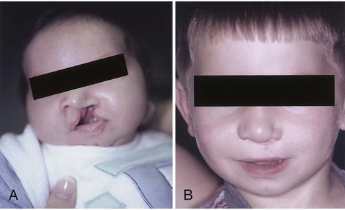
The initial treatment is surgical repair. The cleft lip is usually repaired first because it interferes with the infant’s ability to eat. If the infant cannot create a vacuum in the mouth, there can be difficulty with sucking. Surgery not only improves the infant’s sucking, it also greatly improves the appearance. Currently, it is performed any time after birth if the infant’s general health is good and there is no infection. Most infants undergo repair at around 10 to 12 weeks of age (Figure 14-1).


Feeding Method for Neonates with Cleft Lip, Cleft Palate, or Both.

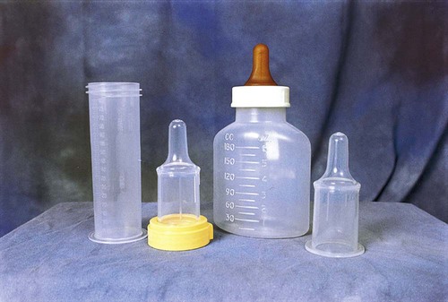
Babies with cleft lip and/or cleft palate can be fed by bottle or breast. Several specialized feeders (e.g., the Haberman Feeder, Pigeon Bottle, and Mead-Johnson Cleft Palate Feeder) are used to feed the infant before and after surgery. These feeders are designed to provide easy but limited flow of the liquid. Sometimes a soft, cross-cut nipple can be used.


Breastfeeding is possible for these babies but may require the assistance of a lactation specialist. Infants with cleft lip are usually more successful with breastfeeding than the infant with cleft palate or a cleft lip and cleft palate. Mothers who wish to breastfeed to gain all the benefits from breastfeeding should be supported. Skill 14-1 describes mechanical feeding methods that may be used for infants with both cleft lip and cleft palate. After the infant has established a feeding routine, the infant should be able to complete the feeding in 18 to 30 minutes. An infant who requires a longer feeding period could be working too hard and expending too many calories. This would not promote growth, which is a goal for these infants. It is important for the nurse and the caregiver to remain flexible and patient in feeding these infants. It may require trying several different systems and techniques before the best one is found (Figure 14-2).


Skill 14-1


Oral Feeding for Infants with Cleft Lip or Cleft Palate


image image image image image image image image


Equipment Description
imageSoft, thin-walled nipple (preemie nipple) •Compresses easily; readily available
imageNUK orthodontic nipple •Large surface for compression
imageCross-cut nipple •Allows easy flow of milk with compression
imageRoss Cleft Palate Nurser •For infants with weak suck; has soft tubelike nipple that delivers past the cleft
imageMead Johnson Cleft Palate Nurser •Soft, long cross-cut nipple; soft bottle for squeezing and monitoring milk flow; can use cross-cut or NUK nipples with this system
imageHaberman Feeder •Large, squeezable nipple with a slit cut; has one-way valve to reduce amount of air ingested; two nipple lengths available: regular and short
imagePigeon Cleft Palate Nurser •Larger, more bulbous Y-cut nipple; firm on top with soft bottom; has air valve to prevent collapse and air flow; has flow setting in bottle collar
imageAsepto syringe, rubber tip •Readily available; places milk beyond cleft


Method


1. Use a nipple or feeding system that provides a controllable flow rate and is energy-efficient for the infant.


2. Hold infant in upright position to assist in reducing the amount of nasal regurgitation.


3. Use a pillow for additional support for infant to assist with longer feeding times.


4. Keep chin tucked because neck extension inhibits swallowing.


5. If regurgitation occurs (milk coming out through the nose), stop feeding and allow infant to cough/sneeze to clear nasal airway.


6. Place nipple on top of tongue. Nipple insertion may push tongue to the back of mouth.


7. Burp the infant frequently because of increased air ingestion.


8. Monitor for distress and fatigue during feeding.


9. Limit feeding time to approximately 30 minutes to avoid fatigue.


10. Follow feeding with sterile water to clean any trapped food in the cleft.


11. Clean mouth and nose.


Adapted from Cleft Palate Foundation. (2002). Feeding an infant with a cleft. Chapel Hill, NC: Author.


Postoperative Care


Postoperative nursing goals for a child with cleft lip/palate repair include (1) preventing the child from excessive crying, which could cause tension on the suture line, (2) careful positioning (never on the abdomen) to avoid injury to the operative site, (3) cleaning the suture line to prevent crusts from forming, which could cause scarring, (4) applying restraints to prevent injury to the operative site and using a butterfly strip or a Logan bow (bar) (to reduce tension on the suture line), and (5) cuddling and other forms of affection to provide for the infant’s emotional needs. This last is of particular importance because the infant is unable to obtain the usual satisfactions from sucking.


Suture line care may involve application of an antibacterial ointment, depending on the surgeon’s instruction. Cotton-tipped applicators should be avoided to prevent injuring the surgical site. The surgical site may be rinsed with sterile saline solution. The infant receives feedings by special feeder until the wound is completely healed (from 1 to 2 weeks).


Cleft Palate


A cleft palate is more serious than a cleft lip. It forms a passageway between the nasopharynx and the nose, which not only complicates feeding but easily leads to infections of the respiratory tract and middle ear and is generally responsible for speech difficulties in later life. Unlike cleft lip, cleft palate is more common in girls than in boys.


Treatment


Most surgeons prefer to operate between 7 to 15 months of age, so that speech patterns are not affected. Even with the advances in surgical techniques, many centers are electing to repair the cleft palate during these times (Campbell et al., 2010). There are several approaches to repair depending on the severity of the defect. While a single surgery may be all that is necessary, future surgical interventions may be required as the child grows.


The management of the child with a cleft lip and cleft palate requires expert teamwork over a long period of time. The emotional problems that sometimes occur with this condition may require more extensive attention than the repair itself. A child born with a facial deformity encounters many problems. It is difficult to be unattractive when society places such importance on good looks. The parent’s first reaction to a disfigured newborn infant may be one of shock, hurt, disappointment, and guilt. Some parents may regard the deformity as a result of their inadequacies. It should be strongly stressed that they are not responsible for causing the defect. They may desire to hide the child from relatives and friends. Feedings are difficult. As the child grows, irregular tooth eruptions, drooling, delayed speech, and intermittent hospitalizations and frequent clinic appointments can be frustrating. The developing child senses the feelings of the parents and acquires either a positive or a negative self-attitude.


In large cities, special cleft palate clinics are available where several specialists can work together in convenient consultation. The parents should be informed of the resources available in the state in which they live. Financial assistance is usually indicated because of the length of treatment required.


Postoperative Management and Nursing Care


Nutrition.

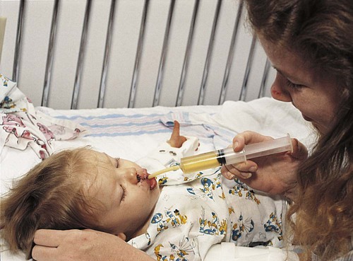
Fluids are best taken by a cup, although an Asepto syringe with a rubber tip (gravity feeder) may be used (Figure 14-3). Hot foods and liquids should be avoided to prevent injury to the surgical site. All objects should be kept out of the mouth. This includes straws, tongue blades, spoons, forks, and pacifiers. A wide-bodied spoon may be used if the food is fed from the side of the spoon and does not come in contact with the roof of the mouth. The diet progresses from a clear to a full liquid diet. The older child may go home on a soft diet (nothing harder than mashed potatoes).


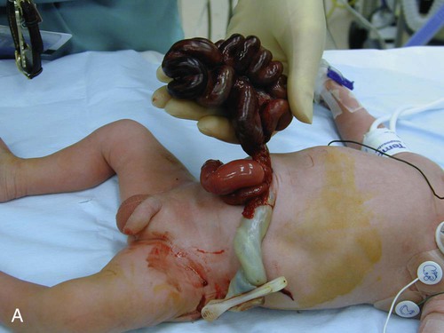
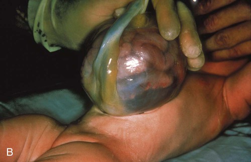
Gastroschisis and Omphalocele


These two defects allow abdominal contents to herniate outside the abdominal cavity (Figure 14-4). The gastroschisis usually occurs to the right of the umbilical cord. It is usually a small defect and involves only the bowel. It does not have a sac covering the defect. An omphalocele is a herniation of the gut into the umbilical cord. It is generally a large defect involving the bowel, liver, spleen, bladder, uterus, or ovaries. It is contained in a translucent sac with amniotic fluid. Improvement with survival rates has been helped by prenatal detection and cesarean delivery in a medical facility able to handle the care of this infant.


Treatment and Nursing Care


After delivery, care is given to prevent rupture of the sac with the omphalocele. Any exposed viscera are covered with warm saline-soaked gauze covered with plastic dressing to prevent radiant heat loss. The infant requires monitoring of respirations, temperature, and hydration status. Surgical repair is necessary for both defects. Small defects may be repaired by replacing the exposed viscera back into the abdominal cavity. For large defects, the surgical repair may require a staged repair. A Silastic silo or tubelike material is sutured around the defect. The abdominal contents are within the silo and are slowly pushed into the abdominal cavity. This procedure may take 7 to 10 days, which allows for the abdominal cavity to expand accommodating the bowel.


During the reduction period, the infant is at risk for infection, hypothermia, dehydration and shock, and decreased lower extremity circulation. When the bowel is completely reduced into the abdominal cavity, the infant is ready for complete closure. Postoperative nursing care requires pain control. Nursing priorities include monitoring respiratory and circulatory status and bowel function. Total parenteral nutrition (TPN) provides nutritional needs until bowel function has returned. Feeding is introduced slowly as tolerance is monitored. With the improvement of parenteral nutrition and neonatal care, survival rates are 90% to 95% (Durken and Shaaban, 2009).


Esophageal Atresia and Tracheoesophageal Fistula Atresia


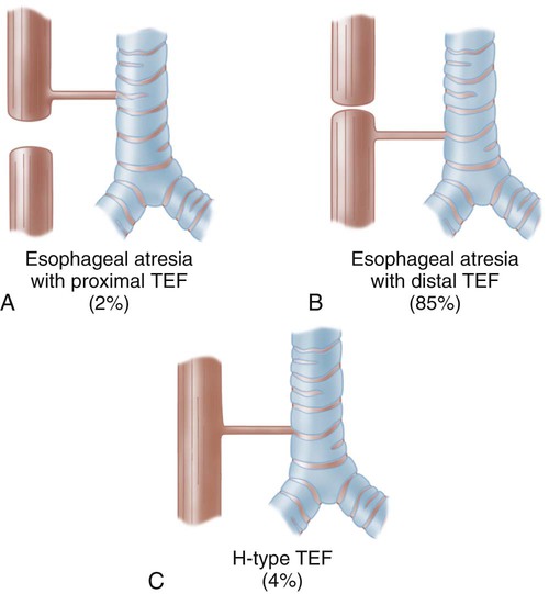
Esophageal atresia (EA) is a congenital defect in which the esophagus fails to connect to the stomach. It ends in a blind pouch. Most cases are associated with tracheoesophageal fistula (TEF), which is characterized by an abnormal connection or fistula between the esophagus and the trachea (Figure 14-5). There may be a maternal history of polyhydramnios (excess of amniotic fluid in pregnancy).


Treatment and Nursing Care


At birth, the infant presents with excessive oral secretions and accompanying coughing, choking, and cyanosis. These symptoms may worsen when feeding is attempted. Respiratory distress can present when the excessive secretions are aspirated or when the secretions pass into the trachea through a fistula. If an EA is not present, diagnosis may be more difficult. Diagnosis is determined by attempting to pass a small-bore NG tube into the esophagus. If passage is not possible, radiograph is used to diagnosis the defect.


A low-suction catheter is placed in the blind pouch to control secretions. The infant is maintained in an upright position or reverse Trendelenburg position to reduce the risk of aspiration. Priority nursing interventions include monitoring respiratory status, maintaining NPO (nothing by mouth) status, and administering oxygen. Antimicrobials are given for possible infection related to aspiration pneumonia. A gastrostomy tube or button is placed to provide nutrition and gastric decompression.


Surgical intervention is necessary. It involves reattaching the ends of the esophagus and ligation of the fistula. If the distance between the two ends of the esophagus is too far, surgery to connect these may be delayed. The esophagus may need to be lengthened before the ends can be attached.


Postoperative care involves care of a chest tube, a gastrostomy tube, and an NG/orogastric tube connected to low suction. As with any postoperative child, the nurse needs to monitor respiratory status, hydration status, thermoregulation, pain, and infection and assist in providing bonding between parent and infant. The infant may be prescribed peptic acid blockers to help with any reflux. As the infant progresses toward discharge, parents need education regarding home care and feedings. This may be more relevant for infants who require further surgical interventions.


Biliary Atresia


Biliary atresia is a disorder in which an obstruction of the flow of bile results from the destruction or absence of extrahepatic bile ducts. It is more common in girls than in boys. Infants are generally born with a normal birth weight.


Signs and Symptoms


There is persistent jaundice continuing after the period of newborn physiologic hyperbilirubinemia (beyond 14 days of age). The liver becomes enlarged (hepatomegaly), and the abdomen is distended. Lab values for bilirubin are elevated. The body tries to eliminate the excessive bilirubin by excretion through the kidneys, which turns the urine a dark tea color. Bile is deposited in the tissue, causing jaundice and pruritus. Eventually the spleen becomes enlarged (splenomegaly) as the disorder progresses. Early stools may be normal color, but as the disorder progresses, stools become acholic. Lacking bile, the infant is unable to digest fats and absorb A, D, E, and K fat-soluble vitamins. The infant becomes malnourished. The lack of bile flow results in destruction of liver tissue and development of cirrhosis.


Diagnostic tools include liver biopsy, hepatobiliary excretory exam, and ultrasound.


Treatment


Early diagnosis is critical to the course of the disorder. A palliative surgery (hepatoportoenterostomy or Kasai procedure) has the best results when done within 60 days of birth. Untreated biliary atresia has a poor prognosis, with death within 2 years (Feldman, 2006). Most of these children will require a liver transplant. The success rate for transplantation has improved. The availability of small donor livers remains an issue. Advances using partial liver transplantation are promising.


Nursing Care


The diagnosis of biliary atresia is devastating to the family. Parents will need emotional and educational support. Special formulas may be needed to maximize absorption. Monitoring the infant’s intake and weight is important. Itching as a result of the deposit of bile salts in the tissue is a frustrating side effect. Medications such as ursodeoxycholic acid (Ursodiol, Actigall) help to increase the bile flow and decrease itching. Diphenhydramine (Benadryl) can be used to help alleviate itching. Skin integrity should be closely monitored. Measures to reduce scratching, such as covering hands and keeping the nails short, are helpful. Tepid baths may also help relieve itching and patting rather than rubbing the skin can help prevent further skin irritation. Children can become irritable with the intense itching. The nurse can assist the family in coping with a child who is extremely irritable.

Stay updated, free articles. Join our Telegram channel

Dec 22, 2016 | Posted by in NURSING | Comments Off on Gastrointestinal Disorders

Full access? Get Clinical Tree

Get Clinical Tree app for offline access