Chapter 30 The fetal skull
After reading this chapter, you will be able to:
Development of the fetal skull
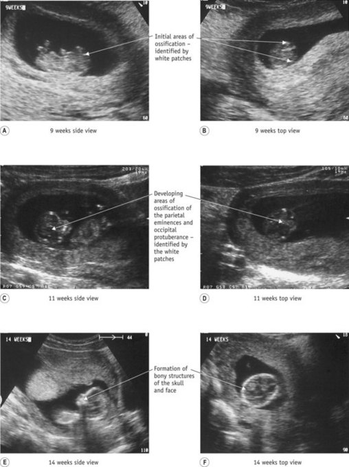
The earliest visible signs of development can be seen on ultrasonography at about 4 weeks’ gestation with calcification of the membranes and the development of the occiput. This becomes easier to determine from approximately 8 weeks, when intramembranous ossification is more prominent. At 12 weeks, the outline of the individual bones become evident (Moore & Persaud 2007, Sadler 2009) (see Fig. 30.1). Ossification of the bones continues throughout pregnancy with individual bones ossifying from their centre. At term, the bones of the skull are thin and pliable, enabling some movement of bones to take place during labour. The two frontal bones have usually united by term.
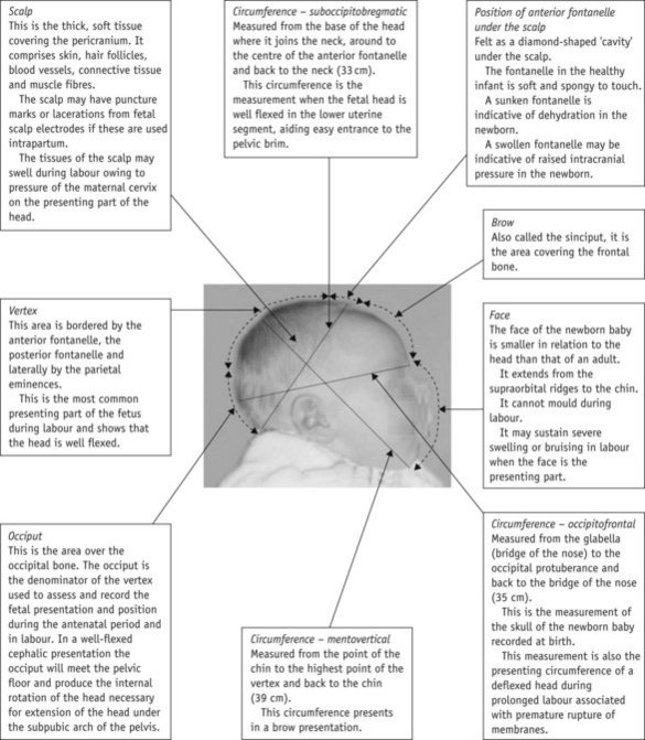
The external structures of the newborn skull (Fig. 30.2)
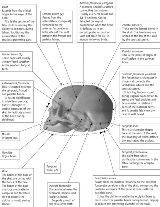
The skull (Fig. 30.3)
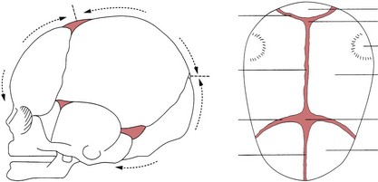
The fetal skull is a complex structure consisting of 29 irregular flat bones with 22 of these paired symmetrically: 8 bones form the cranium, 14 the face, and 7 the base. Knowledge of the fetal skull in the antenatal period enables a midwife to assess the size of the fetal head in relation to the size of the pelvis, assess engagement of the fetal skull in the pelvis (Dietz & Lanzarone 2005). It also helps inform reviews of ultrasonography and pelvimetry reports.
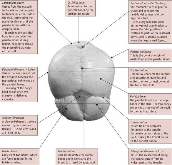
Fontanelles
A fontanelle is a membranous, non-ossified area of the skull where three or more sutures meet.
The significant fontanelles of the skull are:
Sinuses
Reflective activity 30.1
Use the diagrams of the fetal skull in Figure 30.19 to revise the information outlined within this chapter. Photocopy the diagrams and revise the structures until you are confident that you know all of the components of the fetal skull and their application to midwifery practice.
The bones and regions of the skull (Figs 30.3 and 30.4)
The skull is divided into three main regions:
The vault of the skull comprises:


