Describe the etiology, pathophysiology, and clinical manifestations of parasitic infections.
 Identify the prototype and describe the action, use, adverse effects, contraindications, and nursing implications for the amebicides.
 Identify the prototype and describe the action, use, adverse effects, contraindications, and nursing implications for the antimalarial drugs.
 Identify the prototype and describe the action, use, adverse effects, contraindications, and nursing implications for the anthelmintic drugs.
 Identify the prototype and describe the action, use, adverse effects, contraindications, and nursing implications for the scabicides and pediculicides.
 Implement the nursing process in the care of the patient being treated with antiparasitic agents.
Clinical Application Case Study
Lacy Michelson, a 35-year-old woman, is a missionary. She has just returned from a 2-year stay in a developing country. She comes to the clinic with severe diarrhea, fever, chills, headache, and myalgia. Ms. Michelson receives a diagnosis of giardiasis and malaria as well as a prescription for chloroquine 500 mg daily by mouth for 3 weeks and metronidazole 250 mg three times a day by mouth for 7 days.
KEY TERMS
Amebicides: drugs that destroy amebae and are classified according to the site of action
Anthelmintics: drugs used for treatment of helminthiasis, which is an infestation of parasitic worms
Larva: developmental form of a parasite
Parasite: organism that lives in or on the host and gains nutrition from the host
Pediculicides: drugs that have the ability to destroy lice
Plasmodium: genus of ameboid parasites that causes malaria
Introduction
A parasite is a living organism that survives at the expense of another organism, called the host. Parasitic infestations are common human ailments worldwide. The effects of parasitic diseases on human hosts vary from minor to major and can be life-threatening. Parasitic diseases discussed in this chapter are those caused by protozoa, helminths (worms), scabies, and pediculi (lice).
Overview of Parasitic Infections
Protozoa can infect the digestive tract and other body tissues, and resulting infections include amebiasis, giardiasis, malaria, toxoplasmosis, and trichomoniasis. Helminths can also infect these sites, causing a number of infections. Scabies and pediculi affect the skin.
Etiology and Pathophysiology
Protozoal Infections
Amebiasis
Amebiasis is a common disease in Africa, Asia, and Latin America, but it can occur in any geographic region. In the United States, it is most likely to occur in residents of institutions for the mentally challenged, in men who have sex with men, and in travelers from countries with poor sanitation.
Amebiasis is caused by the pathogenic protozoan Entamoeba histolytica, which exists in two forms, cysts and trophozoites. Cysts are inactive; resistant to drugs, heat, cold, and drying; and can survive outside the body for long periods. Transmission occurs by ingestion of food or water contaminated with human feces containing amebic cysts. After people ingest the cysts, some cysts remain intact to be expelled in feces and continue the chain of infection, and some open in the ileum to release amebae, which produce trophozoites. Trophozoites are active amebae that feed, multiply, move about, and produce clinical manifestations of amebiasis.
Trophozoites produce an enzyme that allows them to invade body tissues. They may form erosions and ulcerations in the intestinal wall with resultant diarrhea (this form of the disease is called intestinal amebiasis or amebic dysentery), or they may penetrate blood vessels and be carried to other organs, where they form abscesses. These abscesses are usually found in the liver (hepatic amebiasis) but also may occur in the lungs or brain.
Giardiasis
Giardiasis is a protozoal disease that may affect children more than adults and may cause community outbreaks of diarrhea. It also occurs in people who camp or hike in wilderness areas or who drink untreated well water in areas where sanitation is poor.
Giardiasis is caused by Giardia lamblia, a common intestinal parasite. The disease spreads by ingestion of food or water contaminated with human feces containing encysted forms of the organism or by contact with infected people or animals. Infections occur 1 to 2 weeks after ingestion of the cysts. Person-to-person spread often occurs in children in day care centers, in people in institutions, and in men who have sex with men.
Malaria
Malaria is a common cause of morbidity and mortality in many parts of the world, especially in tropical regions. In the United States, malaria is rare and affects travelers or immigrants from malarious areas.
Malaria is caused by four species of protozoa of the genus Plasmodium. The human being is the only natural reservoir of these parasites. Only Anopheles mosquitoes transmit the malarial plasmodia. Plasmodium vivax, Plasmodium malariae, and Plasmodium ovale cause recurrent malaria by forming reservoirs in the human host. In these types of malaria, signs and symptoms may occur months or years after the initial attack. Plasmodium falciparum causes the most life-threatening type of malaria but does not lead to formation of a reservoir. This type of malaria may be cured and prevented from recurring.
The plasmodia protozoa have a life cycle in which one stage of development occurs within the human body. When an uninfected Anopheles mosquito bites a person with malaria, it ingests blood that contains gametocytes (male and female forms of the protozoan parasite). These forms produce sporozoites, which are transported to the mosquito’s salivary glands. When the mosquito bites the next person, the sporozoites are injected into that person’s bloodstream. From the bloodstream, the organisms lodge in the liver and other tissues, where they reproduce and form merozoites. The liver cells containing the parasite eventually rupture and release the merozoites into the bloodstream, where they invade red blood cells. After a period of growth and reproduction, merozoites rupture red blood cells, invade other erythrocytes, form gametocytes, and continue the cycle.
Trichomoniasis
Trichomoniasis is caused by Trichomonas vaginalis, a single-cell protozoan. The disease is usually spread by men who have no signs and symptoms of infection who engage in sexual intercourse. Women are more likely than men to become infected.
Helminthiasis
Helminthiasis, or infestation with parasitic worms, often occurs in many parts of the world. It affects about 1 billion people, making it one of the most common of all diseases. Although helminthiasis is quite frequent in topical areas, it is also found in other regions, including countries such as the United States and Canada. Box 23.1 describes the etiology and pathophysiology of some helminthic infections.
BOX 23.1 Helminthic Infections
Hookworm infections are caused by N. americanus, a species found in the United States, and A. duodenale, a species found in Europe, the Middle East, and North Africa. Hookworm is spread by ova-containing feces from infected people. Ova develop into larvae when deposited on the soil. Larvae burrow through the skin (e.g., if the person walks on the soil with bare feet), enter blood vessels, and migrate through the lungs to the pharynx, where they are swallowed. Larvae develop into adult hookworms in the small intestine and attach themselves to the intestinal mucosa.
Pinworm infections (enterobiasis), caused by E. vermicularis, are the most common parasitic worm infections in the United States. They are highly communicable and often involve schoolchildren and household contacts. Infection occurs from contact with ova in food or water or on bed linens. The female pinworm migrates from the bowel to the perianal area to deposit eggs, especially at night. Touching or scratching the perianal area deposits ova on hands and any objects touched by the contaminated hands.
Roundworm infections (ascariasis), caused by A. lumbricoides, are the most common parasitic worm infections in the world. They occur most often in tropical regions but may occur wherever sanitation is poor. The infection is transmitted by ingesting food or water contaminated with feces from infected people. Ova are swallowed and hatch into larvae in the intestine. The larvae penetrate blood vessels and migrate through the lungs before returning to the intestines, where they develop into adult worms.
Tapeworms attach themselves to the intestinal wall and may grow as long as several yards. Segments called proglottids, which contain tapeworm eggs, are expelled in feces. Tapeworms are transmitted by ingestion of contaminated, raw, or improperly cooked beef, pork, or fish. Beef and fish tapeworm infections are not usually considered serious illnesses. Pork tapeworm, which is uncommon in the United States, is more serious because it produces larvae that enter the bloodstream and migrate to other body tissues (i.e., muscles, liver, lungs, and brain).
Threadworm infections (strongyloidiasis), caused by S. stercoralis, are potentially serious infections. This worm burrows into the mucosa of the small intestine, where the female lays eggs. The eggs hatch into larvae that can penetrate all body tissues.
Trichinosis, a parasitic worm infection caused by Trichinella spiralis, occurs worldwide. It is caused by ingestion of inadequately cooked meat, especially pork. Encysted larvae are ingested in infected pork. In the intestine, the larvae excyst, mature, and produce eggs that hatch into new larvae. The larvae enter blood and lymphatic vessels and are transported throughout the body. They penetrate various body tissues (e.g., muscles and brain) and evoke inflammatory reactions. Eventually, the larvae are reencysted or walled off in the tissues and may remain for 10 years or longer.
Whipworm infections (trichuriasis) are caused by T. trichiura. Whipworms attach themselves to the wall of the colon.
Scabies and Pediculosis
Scabies and pediculosis are parasitic infestations of the skin. Scabies is caused by the “itch mite” (Sarcoptes scabiei), which burrows into the skin and lays eggs that hatch in 4 to 8 days.
Pediculosis may be caused by one of three types of lice. Pediculosis capitis (head lice) is the most common type in the United States. It is diagnosed by finding louse eggs (nits) attached to hair shafts close to the scalp. Pediculosis corporis (body lice) is diagnosed by finding lice in clothing, especially in seams. Pediculosis pubis (pubic or crab lice) is diagnosed by the presence of nits in the pubic and genital areas. Although scabies and pediculosis are caused by different parasites, the conditions have two common characteristics:
• They are more likely to occur in areas of poverty, overcrowding, and poor sanitation. However, they may occur in people from any socioeconomic group, in any geographic area.
• They are highly communicable and transmitted by direct contact with an infected person or the person’s personal effects (e.g., clothing, combs and hairbrushes, bed linens).
Clinical Manifestations
Amebiasis may be asymptomatic. Affected people may have nausea, vomiting, diarrhea, abdominal cramping, and weakness. If the disease is severe, prolonged, and untreated, these people may experience symptoms from ulcerations of the colon or abscesses of the liver (hepatic amebiasis).
Giardiasis may be asymptomatic or produce diarrhea, abdominal cramping, and distention. If the infection is untreated, it may resolve spontaneously or progress to a chronic disease with anorexia, nausea, malaise, weight loss, and continued diarrhea with large, foul-smelling stools. Deficiencies of vitamin B12 and fat-soluble vitamins (see Chapter 33) may occur.
Malaria initially seems to resemble influenza in terms of its symptoms (e.g., headache, myalgia). The characteristic paroxysms of chills, fever, and copious perspiration may not be present at the early stage of disease. During acute malarial attacks, the cycles occur every 36 to 72 hours. Clinical symptoms occur because of the large parasite burden. The characteristic cycles correspond to the release of merozoites from erythrocytes. Additional manifestations include nausea and vomiting, splenomegaly, hepatomegaly, anemia, leukopenia, thrombocytopenia, and hyperbilirubinemia.
Trichomoniasis affects women and men differently. Women usually have vaginal burning, itching, and foul-smelling yellow–gray, frothy discharge. Men may be asymptomatic or have symptoms of urethritis.
Helminthiasis involves worm infestations; helminths are most often found in the gastrointestinal (GI) tract. However, several types penetrate body tissues or produce larvae (developmental forms of parasites) that migrate to the blood, lymph channels, lungs, liver, and other body tissues. Hookworm, roundworm, and threadworm larvae migrate through the lungs and may cause symptoms of pulmonary congestion. The hookworm may cause anemia by feeding on blood from the intestinal mucosa; the fish tapeworm may cause megaloblastic or pernicious anemia by absorbing folic acid and vitamin B12. Large masses of roundworms or tapeworms may cause intestinal obstruction. The major symptom usually associated with pinworms is intense itching in the perianal area (pruritus ani).
Scabies is characterized by burrows produced by the mite that create visible skin lesions, most often between the fingers, around the nails, and on the elbows and wrists. The lesions may also involve skin that is usually covered by clothing; the buttocks, belt line, penis, and skin around the nipples are likely places for mites to burrow.
Pediculosis leads to pruritus, which is usually the major symptom of the disease. This symptom results from an allergic reaction to parasite secretions. In addition to the intense discomfort associated with pruritus, scratching is likely to cause skin excoriation with secondary bacterial infection and formation of vesicles, pustules, and crusts.
NCLEX Success
1. During the second week of July, the emergency department of a local hospital treats 45 residents of one neighborhood for diarrhea. The neighborhood has a swimming pool, and in the past 10 days, 42 of the 45 affected people swam in the pool. What should the emergency department staff consider to be the cause of this outbreak of diarrhea?
A. Salmonella
B. Clostridium difficile
C. Pseudomonas
D. Giardia
2. A woman has received a diagnosis of Trichomonas vaginalis. What should the nurse teach the patient?
A. Avoid yeast-containing foods.
B. Wear a feminine pad to absorb the drainage.
C. Use acetic acid douches.
D. Avoid sexual intercourse.
Clinical Application 23-1
 Ms. Michelson has received a diagnosis of a Giardia infection. What vitamin deficiency is she at risk for developing?
 What are the initial symptoms of giardiasis?
Drug Therapy
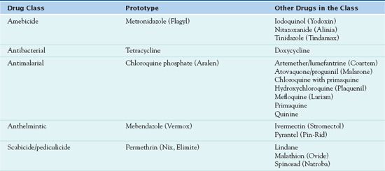
To combat parasitic diseases, health care professionals use antiparasitic drugs, including amebicides, antimalarials, other antiprotozoal agents, anthelmintics, scabicides, and pediculicides. Table 23.1 lists these drugs.

Amebicides
Amebicides, or drugs used to treat amebiasis, are classified according to their site of action. The prototype  metronidazole (Flagyl), a synthetic compound with amebicidal and trichomonacidal activity, is effective in both intestinal and extraintestinal amebiasis. Chloroquine is a tissue or extraintestinal amebicide because it acts in the bowel wall, liver, and other tissues. Chloroquine is discussed as an antimalarial agent because it is most commonly used for the prevention and treatment of malaria.
 Tetracycline and doxycycline, which are antibacterial drugs (see Chapter 18), act against amebae in the intestinal lumen by altering the bacterial flora required for amebic viability. One of these drugs may be used with other amebicides in the treatment of all forms of amebiasis except asymptomatic intestinal amebiasis.
Pharmacokinetics
Metronidazole is 80% absorbed by the GI tract, reaching a peak of action in 1 to 3 hours. The drug is widely distributed to the cerebrospinal fluid, bone, and cerebral and hepatic abscesses. Its half-life is 6 to 8 hours. Metronidazole is metabolized in the liver (30%–60%), with most excreted in the urine (77%) and some in the feces (14%). The drug readily crosses the placenta and enters the breast milk.
Action and Use
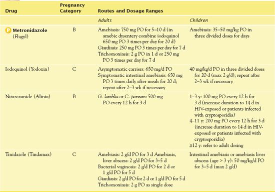
Metronidazole diffuses across the cell membrane of anaerobic and aerobic microorganisms to cause cell death. The biochemical mechanism of action of the drug is unknown. Indications include intestinal amebiasis, amebic liver abscess, trichomoniasis, and bacterial vaginosis. The U.S. Food and Drug Administration (FDA) has not approved this drug or any other amebicide for the prophylaxis of amebiasis. Table 23.2 gives route and dosage information for metronidazole and other amebicides.
 TABLE 23.2
Adverse Effects and Contraindications
The adverse effects of metronidazole include headache, dizziness, ataxia, darkening of urine, diarrhea, nausea, vomiting, and an unpleasant metallic taste. Contraindications include a known hypersensitivity to the drug. Pregnancy is also a contraindication.
Nursing Implications
Preventing Interactions
Concomitant use of metronidazole with barbiturates increases the metabolism of the antiamebic drug, thus decreasing its therapeutic effect. Administration of metronidazole with anticoagulants (e.g., warfarin) increases bleeding tendencies due to decreased vitamin K metabolism. Also, disulfiram and alcohol combined with metronidazole may lead to a disulfiram-like reaction, with tachycardia, nausea, flushing, and vomiting. No herbal interactions with metronidazole reportedly occur.
Administering the Drug
Patients may take metronidazole with food to improve medication adsorption. It is important not to crush extended-release preparations. If the patient is unable to swallow regular metronidazole pills, it may be necessary to crush them.
Assessing for Therapeutic Effects
The nurse assesses patients with intestinal amebiasis for decreased abdominal pain and diarrhea. It is necessary to assess stool specimens for amebic cysts and trophozoites periodically for 6 months. Also, the nurse checks the feces for increase in form that is indicative of diminished diarrhea stools and improved hydration.
It is important to assess women with trichomoniasis for decreased vaginal drainage and odor.
Assessing for Adverse Effects
It is necessary to assess for headache; diminished muscular coordination; GI upset, including metallic taste; and hypersensitivity reactions such as rash and bronchospasm.
Patient Teaching
Box 23.2 identifies patient teaching guidelines for antiparasitic drugs, including metronidazole.
BOX 23.2  Patient Teaching Guidelines for Antiparasitic Drugs


