Digestive and Endocrine Disorders
Objectives
• Define the key terms and key abbreviations in this chapter.
• Describe the care required for diverticular disease.
• Describe the care required for gallstones.
• Describe the care required for hepatitis and cirrhosis.
• Describe the care required for diabetes.
• Explain how to promote PRIDE in the person, the family, and yourself.
Key Terms
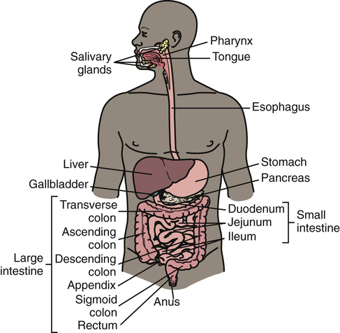
Problems can develop in any part of the digestive system. This includes the accessory organs of digestion—liver, gallbladder, and pancreas. The pancreas also is part of the endocrine system.
Digestive Disorders
The digestive system breaks down food for the body to absorb. Solid wastes are eliminated. See Chapter 26 for diarrhea, constipation, flatulence, fecal incontinence, and ostomy care.
See Body Structure and Function Review: The Digestive System, p. 740.
Gastro-Esophageal Reflux Disease
Gastro-esophageal reflux disease (GERD) occurs when stomach (gastro) contents flow back up (reflux) into the esophagus (esophageal). Stomach contents contain acid that can irritate and inflame the esophagus lining. This is called esophagitis—inflammation (itis) of the esophagus.
Heartburn is the most common symptom. Heartburn is a burning sensation in the chest or throat. The person may taste stomach fluid in the back of the mouth. Besides heartburn, other signs and symptoms of GERD include:
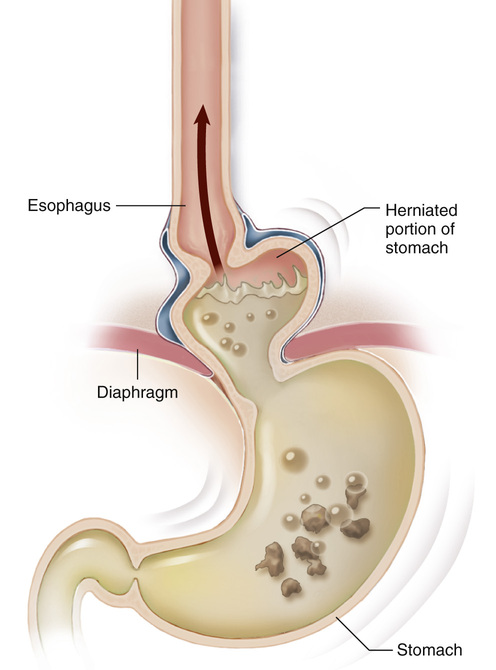
Risk factors include being over-weight, alcohol use, pregnancy, and smoking. Hiatal hernia is a risk factor. With hiatal hernia, the upper part of the stomach is above the diaphragm (Fig. 46-2). Large meals and lying down after eating can cause gastric reflux. So can chocolate, caffeine drinks, fried and fatty foods, garlic, onions, spicy foods, and tomato sauce.
The doctor may order drugs to prevent stomach acid production or to promote stomach emptying. Surgery may be needed. Life-style changes include:
Vomiting
Vomitus (emesis) is the food and fluids expelled from the stomach through the mouth. Vomiting signals illness or injury. Aspirated vomitus can obstruct the airway. Vomiting large amounts of blood can lead to shock (Chapter 54). These measures are needed.
• Follow Standard Precautions and the Bloodborne Pathogen Standard.
• Turn the person’s head well to 1 side if the person is supine. This prevents aspiration.
• Place a kidney basin under the person’s chin.
• Move vomitus away from the person.
• Provide oral hygiene. This helps remove the bitter taste of vomitus.
• Measure, report, and record the amount of vomitus.
• Save a specimen for laboratory study.
• Dispose of vomitus after the nurse observes it.
Diverticular Disease
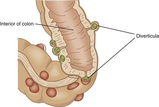
Small pouches can develop in the colon. The pouches bulge outward through weak spots in the colon wall (Fig. 46-3). A pouch is called a diverticulum. (Diverticulare means to turn inside out). Diverticulosis is the condition of having these pouches. (Osis means condition of.) The pouches can become infected or inflamed—diverticulitis. (Itis means inflammation.)
Many people over 50 years of age have diverticulosis. Aging, obesity, smoking, lack of exercise, low-fiber diet, a diet high in animal fat, and some drugs are risk factors.
When feces enter the pouches, they can become inflamed and infected. The person has abdominal pain and tenderness in the lower left abdomen. Fever, nausea and vomiting, chills, cramping, and constipation or diarrhea are likely.
A ruptured pouch is rare. Feces spill into the abdomen. This causes a severe, life-threatening infection. A pouch also can cause a blockage in the intestine (intestinal obstruction). Feces and gas cannot move past the blocked part.
Diet changes are ordered. Sometimes antibiotics and probiotics are ordered. Probiotics—found in dietary supplements and some foods—are live bacteria normally found in the colon. Surgery is done for severe disease, obstruction, and ruptured pouches. The diseased part of the bowel is removed. A colostomy may be needed (Chapter 26).
Inflammatory Bowel Disease
Thought to be an autoimmune disorder (Chapter 43), inflammatory bowel disease (IBD) involves a chronic inflammation of the digestive tract. Two types of IBD are:
• Crohn’s disease. The lining of the large intestine, small intestine, or both is inflamed.
• Ulcerative colitis. The lining of the large intestine and rectum is inflamed and has ulcers.
Signs and symptoms include:
• Diarrhea
• Fever
• Bleeding—bright red blood in the toilet, dark blood in the stools, or occult blood (Chapter 34)
IBD is often diagnosed before 30 years of age. Risk factors include a family history of IBD, cigarette smoking, some drugs, and a diet high in fat and refined foods.
Serious complications can result. They include bowel obstruction, ulcers in the GI tract (including the mouth and anus), colon cancer, osteoporosis, and liver disease. Treatment involves diet changes and drug therapy for inflammation, infection, diarrhea, pain, and nutrition. Surgery may be needed to remove damaged parts of the small intestine or colon. A colostomy or ileostomy (Chapter 26) may be necessary.
Gallstones
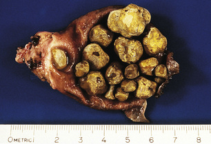
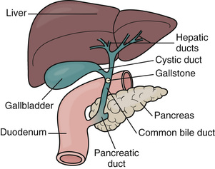
Bile is a liquid made in the liver. It is stored in the gallbladder until needed to digest fat. Gallstones form when the bile hardens into stone-like pieces (Fig. 46-4).
Bile is carried from the liver to the gallbladder through ducts (tubes). From the gallbladder, bile enters the small intestine through the common bile duct. Gallstones can lodge in any duct (Fig. 46-5). Bile flow is blocked. The gallbladder and ducts become inflamed. The liver and pancreas may be involved. Severe infections or damage can cause death.
Gallstones can be as small as a grain of sand or as big as a golf ball. A person may have 1 large stone or several that vary in size. Persons at risk include those who are:
• Use hormone replacement therapy
• American Indians or Mexican Americans
• Over-weight or obese or have had rapid weight loss
Stay updated, free articles. Join our Telegram channel

Full access? Get Clinical Tree


