Digestion, Absorption, and Metabolism


Digestion, Absorption, and Metabolism


imagehttp://evolve.elsevier.com/Grodner/foundations/ imageNutrition Concepts Online


Role in Wellness


Gulping down breakfast on the way to class or work, skipping lunch, and then eating dinner late may not seem to affect the health status of adults. However, if this kind of eating becomes routine, it characterizes an individual’s lifestyle and may negatively influence health status.


The body’s health is based on the nutrients available to support growth, maintenance, and energy needs. Inadequate nutritional intake can affect the body’s ability to use the foods consumed. The digestive system, which is responsible for processing foods, depends on nutrient intake for its maintenance. Although the body is resilient, we stress our physical limits when we adopt habits that do not support optimal health. A primary way to decrease the risk of future disease and achieve wellness is to use lifestyle choices that support positive health behaviors.


Physical health begins with the gastrointestinal (GI) tract as the first step to maintain body functioning; unless nutrients in foods are digested and absorbed, life cannot continue. The decision and follow-through to change lifestyle behaviors to positively improve health in relation to digestive disorders is an aspect of intellectual health. An individual’s emotional state and ability to handle stress may increase the risk of several disorders of the GI tract. Consequently, the emotional health effects of lifestyle behaviors may be related to constipation, diarrhea, and heartburn. Reducing the causes of intestinal gas helps guard against socially embarrassing moments. Our food choices and styles of eating may affect the level of flatus experienced. Negativity associated with body smells is defined by society and thus affects our social health dimension. Respecting the sanctity of the human body, thereby acknowledging our spiritual health dimension, may include one’s willingness to follow dietary and lifestyle changes to enhance the functioning of the GI tract (see the Cultural Considerations box, Wholeness of Body, Mind, and Self). This chapter presents a brief orientation to the processes of digestion, absorption, and metabolism. These processes work together to provide all body cells with energy and nutrients.


image Cultural Considerations


Wholeness of Body, Mind, and Self


This text’s presentation of digestion and absorption is based on Western perspectives. To most Westerners, body organs tend to be viewed separately from mind and spiritual influences. In contrast, Ayurveda, traditional Indian medicine, meaning “the science of life,” is based on living a balanced life. Consequently, Ayurveda treats physical disorders as the body (organs) or life being out of balance. Treatment works to bring balance or harmony back to the individual’s life. The wholeness of life is represented by body (shira), mind (manas), and self (atman). All three require attention to achieve and maintain health. Because each component is important, Ayurveda is a holistic approach recognizing the interdependent roles of body, mind, and self. A person is viewed as a combination of three forces or humors called doshas. Each person is a different combination of these forces, which are vata, pitta, and kapha. Vata is a force similar to air; pitta, a force similar to fire; and kapha, a force like mucus and water. Health occurs when these doshas are in balance; otherwise disease occurs. If pitta is too strong, fever, ulcers, and liver disorders may occur. An individual would need to strengthen the other doshas through (1) changes in behaviors and food choices, (2) use of natural medicines, and (3) yoga and meditation to decrease pitta and regain balance.


Application to nursing: This concept may assist clients to understand that their illnesses may be affected by other components of their lives. Sometimes illnesses force us to confront factors that may influence our ability to maintain health or to achieve balance in our lives.


Data from Ninivaggi, FJ: Ayurveda: A comprehensive guide to traditional Indian medicine for the West, Westport, Conn, 2008, Praeger Press.


Digestion


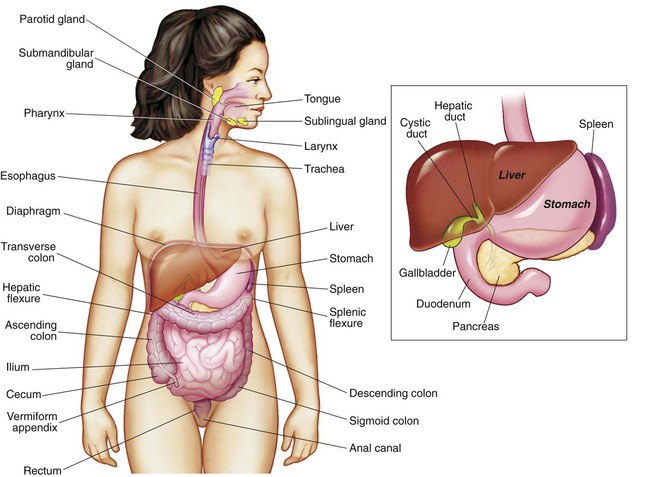
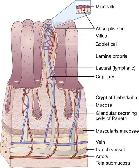
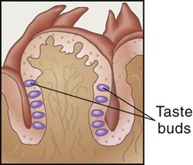
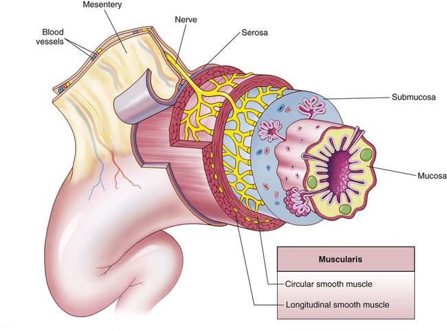
The main organs of the digestion system (Figure 3-1 and Box 3-1) form the gastrointestinal (GI) tract, or alimentary canal, which creates an open tube that runs from the mouth to the anus. Everything we eat is processed through the GI tract. The digestive system consists of a series of organs that prepare ingested nutrients for digestion and absorption and protect against consumed microorganisms and toxic substances. To do this, several processes take place. These processes of ingestion, digestion, absorption, and elimination depend on the motility or movement of the GI wall and the secretions of digestive juices and enzymes.1


imageThe Mouth


Are you hungry? Are you thinking about your favorite food? Is your mouth watering? Our mouths really do “water” when we think about or begin to eat foods. However, it is not actually water we sense but a thin mucous-like fluid called saliva. Saliva is the term for the secretions of the three salivary glands of the mouth. As exocrine glands, each set of salivary glands produces a different type of secretion that is released into the mouth. The parotid glands create watery saliva that supplies enzymes; the submandibular glands produce mucus and enzyme components; and the sublingual glands, the smallest, create a mucous type of saliva. A reflex mechanism controls these secretions.


Food in the mouth stimulates chemical and mechanical digestion. Chemical digestion occurs through the action of saliva that not only moistens the foods we chew but also contains amylase, an enzyme that begins the digestive process of starches.


Another digestive process that occurs in the mouth is mechanical digestion, which depends on teeth. Teeth rhythmically tear and pulverize food. The enamel covering teeth is the hardest substance in the body and therefore protects teeth from the harsh effects of chewing. The tongue assists with mechanical digestion by guiding food into chewing positions and then leading the pulverized food into the esophagus. Another function of the tongue is that of taste. More than 2000 taste buds are responsible for our sensations of sweet, bitter, sour, and salty when tasting foods (Figure 3-2).


As toddlers, we have the highest number of taste buds and a higher degree of taste sensitivity, so bland foods are more appealing. The number of taste buds declines as we grow older, which explains why older adults have diminished taste sensitivity. Older adults may need to be encouraged to avoid the use of too much salt, particularly if they have hypertension or cardiac disorders.


Our sense of smell works along with our taste bud sensations. These two combined senses actually account for the perception (and enjoyment) of the flavors of different foods. Our positive or negative response to specific foods based on our sensory perception affects our food choices.2


Portions of the pulverized or masticated food are formed into the shape of a ball called a bolus. The tongue effortlessly forms the bolus, which is then swallowed and passed by the epiglottis into the esophagus within about 5 to 7 seconds. The epiglottis is a flap of tissue that closes over the trachea to prevent the bolus from entering the lungs.


The Esophagus


The esophagus is a muscular tube through which the bolus travels from the mouth to the stomach. The process begins at the top of the esophagus when peristalsis, the involuntary movements of circular and longitudinal muscles, begins and draws the bolus farther into the GI tract.


This mechanical action further breaks down the size of foodstuff and increases exposure to digestive secretions. Muscular actions depend on the four layers of tissues that form the tube of the GI tract (Figure 3-3). The mucosa is composed of mucous membrane and forms the inside layer. Under the mucosa is the submucosa, a layer of connective tissue. Digestion depends on the blood vessels and nerves of the submucosa to regulate digestion. Surrounding the submucosa is a thick layer of muscle tissue called the muscularis. The outermost layer of the GI wall is made of serous membrane called serosa, which is actually the visceral layer of the peritoneum lining the abdominal pelvic cavity, and covers organs.1


The coordination of these layers provides the varied movements required for digestion. Essentially, muscular action controls the movement of the food mass through the GI tract. Churning action within a segment of the GI tract allows secretions to mix with food mass. Circular muscles surround the GI tube. Rhythmic contractions of these muscles cause the wavelike motions of peristalsis, moving food downward. Longitudinal muscles run parallel along the GI tube. The combined effect of the circular and longitudinal muscles causes segmentation as a forward and backward movement that assists in controlling food mass movement through the GI tract.


Sphincter muscles are stronger, circular muscles that act as valves to control the movement of the food mass in a forward direction. In effect, sphincter muscles prevent reflux by forming an opening when relaxed and closing completely when contracted. At the bottom of the esophagus the cardiac sphincter controls the movement of the bolus from the esophagus into the stomach. It also prevents the acidic contents of the stomach from moving upward back through the esophagus.


The Stomach


Functions of the stomach include the following:


When the bolus passes through the cardiac sphincter, it enters the fundus, the upper portion of the stomach that connects with the esophagus. The other divisions of the stomach include the body, or center portion, and the pylorus, the lower portion. The stomach wall contains gastric mucosa with gastric pits. At the base of the pits are the gastric glands whose chief cells create gastric juice, a mucous fluid containing digestive enzymes, and parietal cells, which secrete stomach acid called hydrochloric acid.


Gastric secretions occur in three phases: cephalic, gastric, and intestinal.1 The cephalic phase is called the “psychic phase” because mental factors can stimulate gastrin, a hormone secreted by stomach mucosa. In the gastric phase, gastrin increases the release of gastric juices when the stomach is distended by food. The third phase is the intestinal phase in which the gastric secretions change as chyme, a semiliquid mixture of food mass, passes through to the duodenum. Gastric secretions are inhibited by exocrine and nervous reflexes of gastric inhibitory peptides, secretin, and cholecystokinin (CCK) (also called pancreozymin), a hormone secreted by intestinal mucosa.


Some gastric juices provide acidity in the stomach to assist the effective function of certain enzymes. As agents of chemical digestion, enzymes are specific in action, working only on individual classes of nutrients and changing substances from one form to a simpler form. Enzymes are “organic catalysts” formed from protein structures. They function at specific pH and are continually created and destroyed. Specific enzymes are required for energy release and digestion.


Hormones regulate the release of gastric juices and enzymes, acting as messengers between organs to cause the release of needed secretions. In digestion, hormones affect the secretions from the stomach, intestines, and gallbladder. These secretions may slow or speed digestion and affect the pH levels of gastric juice. Overall, the mechanical and chemical actions work together to complete the process of digestion.


Gastric motility, or movement of food mass through the stomach, requires 2 to 6 hours. The churning and mixing of the food mass with gastric juices create a semiliquid mixture called chyme. When chyme enters the pylorus section of the stomach, it causes distention and the release of the hormone gastrin. Gastrin sends a message that hydrochloric acid (HCl) is needed to continue the breakdown of chyme. As HCl is released from the stomach lining, thick mucus is also secreted to protect the stomach walls from the harsh HCl.


Every 20 seconds chyme is released into the duodenum, the upper portion of the small intestine; this action is controlled by the hormonal and nervous system mechanism of enterogastric reflex. This consists of duodenal receptors in the mucosa that are sensitive to the presence of acid and distention. The impulses over sensory and motor fiber in the vagus nerve cause a reflex restriction of gastric peristalsis. For example, the gastric inhibitory peptide released in response to fats in the duodenum decreases peristalsis of stomach muscles and slows chyme passage. These result in decreased motility, which is why the stomach empties more slowly when a person eats a high-fat diet.


The combined action of mechanical digestion (the strong muscular movements of peristalsis) and chemical digestion (the effects of the gastric juices) work to prepare nutrients for the process of absorption. Chyme is kept in the stomach by the actions of the pyloric sphincter, which slowly releases it into the duodenum.


The Small Intestine


The chyme entering the duodenum soon moves through to the jejunum and ileum of the small intestine. It takes about 5 hours for chyme to pass through the small intestine because of the action of segmentation and peristalsis. Segmentation in the duodenum and upper jejunum mixes chyme with digestive juices from the pancreas, liver, and intestinal mucosa. Peristalsis is controlled by intrinsic stretch reflexes and is initiated by cholecystokinin (CCK), the hormone secreted by intestinal mucosa.


In the small intestine, the nutrients in chyme are prepared for absorption. The small intestine is the major organ of digestion, and the final stages of the digestive process occur here. Because it is also the site of almost all of the absorption of nutrients, the intestinal lining must be able to accommodate the actions of both digestion and absorption. The intestinal walls are covered with a thin layer of mucus, protecting the walls from digestive juices. The walls are also adapted to enhance the absorption process. Finger-like projections called villi greatly increase the amount of mucosal layer available for the absorption of nutrients (Figure 3-4). On the villi are hairlike projections called microvilli that also enhance absorption by their structure and movements.


As chyme enters the small intestine, hormones begin sending messages that regulate the release of digestive juices to continue the process of chyme digestion. Some hormones are provided by the small intestine; several are released by other organs into the small intestine. These secretions include enzymes from the small intestines, bile produced in the liver, and digestive juices from the pancreas.


One of the first hormones released by the small intestine is secretin. This hormone causes the pancreas to send bicarbonate to the small intestine to reduce the acidic content of the chyme. As the acidic level decreases, other pancreatic juices enter and begin their work. Another hormone secreted by the small intestine is CCK, or pancreozymin, which initiates pancreatic exocrine secretions; acts against gastrin by inhibiting gastric HCl secretion; and activates the gallbladder to contract, causing bile to be released into the duodenum.


Bile, which is secreted by the liver and stored in the gallbladder, is released to emulsify fats, which aids in the digestion of lipids. The emulsification creates more surface area, allowing lipid enzymes to digest fats to their component parts. The liver continuously secretes bile, and CCK and secretin spur the gallbladder to release bile for the digestion of fats. In addition, the small intestine produces enzymes to assist in the digestive process. Although much of the chyme is absorbed, the rest—which usually consists of fiber, minerals, and water—passes through the next sphincter (ileocecal valve) and into the large intestine (ascending colon).


The Large Intestine


The large intestine consists of the cecum, colon, and rectum. The cecum is a blind pocket; therefore, the mass bypasses it and enters the ascending colon, which leads into the transverse colon running across the abdomen over the small intestine to the descending colon. The descending colon extends down the left of the abdomen into the sigmoid colon and leads into the descending colon, on to the rectum, and into the anal canal. Finally, any remaining mass passes out through the anus. The journey through the large intestine takes about 9 to 16 hours.


In the large intestine or colon, final absorption of any available nutrients, usually water and some minerals, occurs. Bacteria residing in the large intestine produce several vitamins, which are then absorbed. Water is withdrawn from the fibrous mass, forming solidified feces. Mucous glands in the intestinal wall create mucus that lubricates and covers feces as it forms. Again, peristalsis continues to move substances through the GI tract, resulting in the excretion of feces from the colon through the anus, the last sphincter muscle of the GI tract.


The movement of the food mass through the GI tract is controlled to enhance digestion and absorption. During passage through the GI tract, more than 95% of the carbohydrates, fats, and proteins ingested are absorbed. Some minerals, vitamins, and trace elements may be less absorbed.1 Table 3-1 summarizes the primary mechanisms of the digestive system. Details of carbohydrate, protein, and lipid digestion follow in specific chapters.


Stay updated, free articles. Join our Telegram channel

Feb 9, 2017 | Posted by in NURSING | Comments Off on Digestion, Absorption, and Metabolism

Full access? Get Clinical Tree

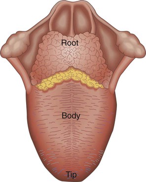
Get Clinical Tree app for offline access