Cardiovascular, Respiratory, and Lymphatic Disorders
Objectives
• Define the key terms and key abbreviations in this chapter.
• Describe congenital heart defects.
• Identify cardiovascular disorder risk factors and complications.
• Describe the care required for Ebola.
• Describe the care required for chronic obstructive pulmonary disease, asthma, and sleep apnea.
• Explain the differences between a cold and influenza and the care required.
• Describe the care required for enterovirus D68.
• Describe the care required for pneumonia and tuberculosis.
• Describe the care required for lymphedema and lymphoma.
• Explain how to promote PRIDE in the person, the family, and yourself.
Key Terms
Key Abbreviations
| CAD | Coronary artery disease |
| CDC | Centers for Disease Control and Prevention |
| CHF | Congestive heart failure |
| CO2 | Carbon dioxide |
| COPD | Chronic obstructive pulmonary disease |
| EV-D68 | Enterovirus D68 |
| IV | Intravenous |
| MI | Myocardial infarction |
| mm Hg | Millimeters of mercury |
| O2 | Oxygen |
| RBC | Red blood cell |
| TB | Tuberculosis |
| VHF | Viral hemorrhagic fever |
| WBC | White blood cell |
Cardiovascular and respiratory system disorders are leading causes of death in the United States. Many people have these disorders. Disorders also occur in the lymphatic system. Understanding these disorders gives meaning to the care you give.
Cardiovascular Disorders
The circulatory (cardiovascular) system delivers blood to the body’s cells. Problems occur in the heart or blood vessels. See Chapter 36 for circulatory ulcers.
See Body Structure and Function Review: The Circulatory System.
See Focus on Children and Older Persons: Cardiovascular Disorders, p. 726.
 Body Structure and Function Review
The Circulatory System
The circulatory system is made up of the blood, heart, and blood vessels. The heart pumps blood through the blood vessels.
The Blood
The blood consists of blood cells and plasma. Plasma is mostly water. It carries blood cells to other body cells. Plasma also carries substances (food, hormones, and chemicals) that cells need to function.
Red blood cells (RBCs) are called erythrocytes. Hemoglobin in the RBCs gives blood its red color. As RBCs circulate through the lungs, hemoglobin picks up oxygen (O2). Hemoglobin carries O2 to the cells. When blood is bright red, hemoglobin in the RBCs is filled with O2. As blood circulates through the body, O2 is given to the cells. Cells release carbon dioxide (CO2) (a waste product). It is picked up by the hemoglobin. RBCs filled with CO2 make the blood look dark red.
Blood also contains white blood cells (WBCs) and platelets (thrombocytes). WBCs are called leukocytes. They protect the body against infection. Platelets are needed for blood clotting.
The Heart
The heart is a muscle. It pumps blood through the blood vessels to the tissues and cells. The heart has 4 chambers (Fig. 45-1). Upper chambers receive blood and are called atria. The right atrium receives blood from body tissues. The left atrium receives blood from the lungs. Lower chambers are called ventricles. Ventricles pump blood. The right ventricle pumps blood to the lungs for O2. The left ventricle pumps blood to all parts of the body.
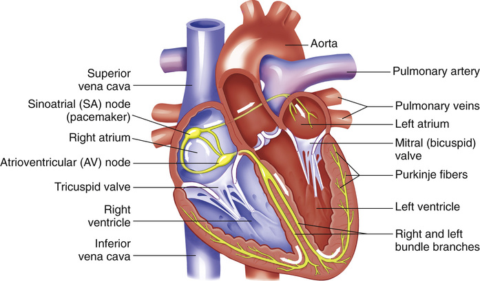
Valves are between the atria and ventricles (see Fig. 45-1). The valves allow blood flow in 1 direction. They prevent blood from flowing back into the atria from the ventricles. The tricuspid valve is between the right atrium and the right ventricle. The mitral valve (bicuspid valve) is between the left atrium and the left ventricle.
Heart action has 2 phases.
The heart has its own electrical system that stimulates the heart to contract. The electrical signal begins in the sinoatrial (SA) node (see Fig. 45-1). The SA node sets the pace of the heart. It stimulates the heart to beat at 60 to 100 beats per minute. The electrical signal spreads through the heart, causing the heart to contract.
The Blood Vessels
Blood flows to body tissues and cells through the blood vessels. There are 3 groups of blood vessels: arteries, capillaries, and veins (Fig. 45-2).
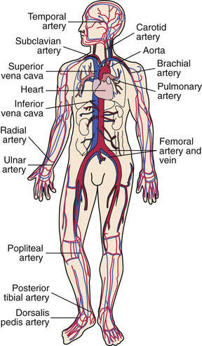
Arteries carry blood away from the heart. Arterial blood is rich in O2. The aorta (see Fig. 45-2) is the largest artery. It receives blood directly from the left ventricle. The aorta branches into other arteries that carry blood to all parts of the body. These arteries branch into smaller parts within the tissues. The smallest branch of an artery is an arteriole.
Arterioles connect to capillaries. Capillaries are very tiny blood vessels. Food, O2, and other substances pass from capillaries into the cells. The capillaries pick up waste products (including CO2) from the cells. Veins carry waste products back to the heart.
Veins return blood to the heart. They connect to the capillaries by venules. Venules are small veins. Venules branch together to form veins. The many veins also branch together as they near the heart to form 2 main veins—the inferior vena cava and the superior vena cava (see Fig. 45-2). Both empty into the right atrium. The inferior vena cava carries blood from the legs and trunk. The superior vena cava carries blood from the head and arms. Venous blood is dark red. It has little O2 and a lot of CO2.
Hypertension
With hypertension (high blood pressure), the systolic pressure is 140 mm Hg (millimeters of mercury) or higher (hyper). Or the diastolic pressure is 90 mm Hg or higher. The resting blood pressure is too high. Such measurements must occur several times. Pre-hypertension is when the systolic pressure is between 120 and 139 mm Hg or the diastolic pressure is between 80 and 89 mm Hg. Pre-hypertension eventually develops into hypertension. Most people have hypertension some time during their lives. See Box 45-1 for risk factors.
Narrowed blood vessels are a common cause. The heart pumps with more force to move blood through narrowed vessels. Kidney disorders, head injuries, some pregnancy problems, and adrenal gland tumors are causes.
Known as the “silent killer,” hypertension can go unnoticed for many years. Signs and symptoms develop over time. Headache, blurred vision, dizziness, and nose bleeds occur. Hypertension can lead to stroke, hardening of the arteries, heart attack, heart failure, kidney failure, and blindness.
Life-style changes can lower blood pressure. A diet low in fat and salt, a healthy weight, and regular exercise are needed. No smoking is allowed. Alcohol and caffeine are limited. Managing stress and sleeping well also lower blood pressure. Certain drugs lower blood pressure.
Coronary Artery Disease
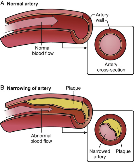
The coronary arteries are the arteries that supply the heart muscle with blood. In coronary artery disease (CAD) (coronary heart disease, heart disease), the coronary arteries become hardened and narrow. One or all are affected. The heart muscle gets less blood and O2.
The most common cause is atherosclerosis (Fig. 45-3). Plaque—made up of cholesterol, fat, and other substances—collects on artery walls. The narrowed arteries block some or all of blood flow. Blood clots can form along the plaque and block blood flow.
Major complications of CAD are angina, myocardial infarction (heart attack), irregular heartbeats, and sudden death. The more risk factors (see Box 45-1), the greater the chance of CAD and its complications.
CAD can be treated. Treatment goals are to:
• Relieve symptoms (see “Angina,” p. 728)
• Slow or stop atherosclerosis
• Lower the risk of blood clots
• Widen or bypass clogged arteries
• Reduce cardiac events (see “Angina” and “Myocardial Infarction,” p. 729)
CAD requires life-style changes. The person must quit smoking, exercise, and reduce stress. A healthy diet is needed to lower blood pressure, lower blood cholesterol, and maintain a healthy weight. If over-weight, the person must lose weight.
Some drugs decrease the heart’s workload and relieve symptoms. Other drugs prevent a heart attack or sudden death. Drugs can delay medical and surgical procedures that open or bypass diseased arteries (Fig. 45-4, p. 728).

Cardiac Rehabilitation.
CAD complications may require cardiac rehabilitation (cardiac rehab). The cardiac rehab team includes doctors (the person’s doctor, a heart specialist, a heart surgeon), nurses, exercise specialists, physical and occupational therapists, dietitians, and mental health professionals.
Cardiac rehab has 2 parts.
Angina
Angina is chest pain from reduced blood flow to part of the heart muscle (myocardium). (Angina comes from the Latin word angor that means strangling.) It occurs when the heart needs more O2. Normally blood flow to the heart increases when O2 needs increase. Exertion, a heavy meal, stress, and excitement increase the heart’s need for O2. So does smoking and very hot or cold temperatures. In CAD, narrowed vessels prevent increased blood flow.
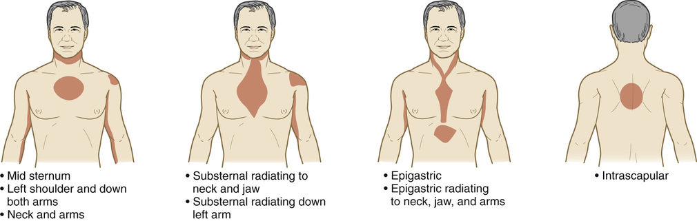
Chest pain is described as a tightness, pressure, squeezing, or burning in the chest. Pain can occur in the shoulders, arms, neck, jaw, or back (Fig. 45-5). Pain in the jaw, neck, and down 1 or both arms is common. The person may be pale, feel faint, and perspire. Dyspnea is common. Nausea, fatigue, and weakness may occur. Some persons complain of “gas” or indigestion. Rest often relieves symptoms in 3 to 15 minutes. Because rest reduces the heart’s need for O2, normal blood flow is achieved. Heart damage is prevented.
Besides rest, a nitroglycerin tablet is taken when angina occurs. A tablet is placed under the tongue, where it dissolves and is rapidly absorbed into the bloodstream. Kept within the person’s reach, the person takes a tablet and then tells the nurse. For some persons, the nurse applies and removes nitroglycerin patches.
Things that cause angina are avoided—over-exertion, heavy meals and over-eating, emotional stress, cold weather, hot and humid weather. Doctor-supervised programs are helpful.
See “Coronary Artery Disease” (p. 727) for the treatment of angina. The goal is increased blood flow to the heart. This prevents or lowers the risk of heart attack and death. Chest pain lasting longer than a few minutes and not relieved by rest and nitroglycerin may signal a heart attack. Emergency care is needed.
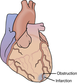
Myocardial Infarction
Myocardial refers to the heart muscle. Infarction means tissue death. With myocardial infarction (MI) part of the heart muscle dies from sudden blockage of blood flow in a coronary artery. A thrombus (blood clot) in an artery with atherosclerosis blocks blood flow. The damaged area may be small or large (Fig. 45-6).
MI also is called:
CAD, angina, and previous MI are risk factors. See Box 45-2 for signs and symptoms. MI is an emergency. Efforts are made to:
Box 45-2
Myocardial Infarction—
Signs and Symptoms
• Sudden, severe; usually in the center or on the left side
• Described as pressure, tightness, fullness, squeezing, or aching
• More severe and lasts longer than angina
• Not relieved by rest or nitroglycerin
• Pain or numbness in 1 or both arms, the back, neck, jaw, or stomach
• Dyspnea
• Fainting
• Perspiration and cold, clammy skin
• Pallor (pale skin) or cyanosis (bluish color)
Stay updated, free articles. Join our Telegram channel

Full access? Get Clinical Tree


