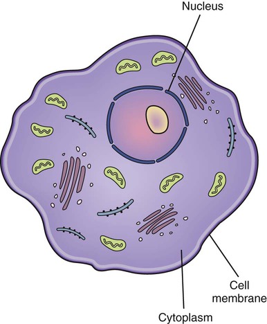
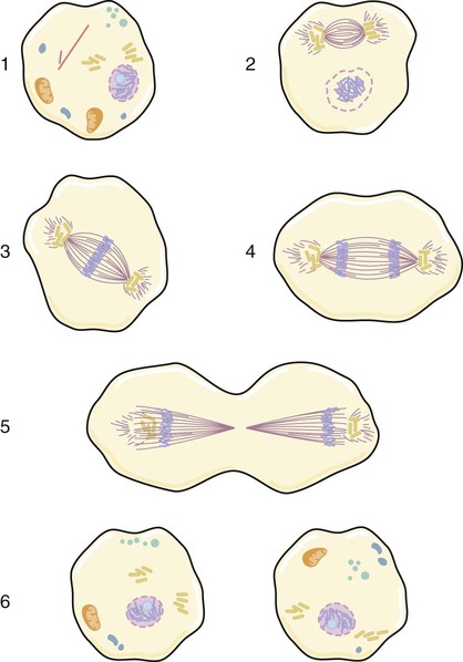
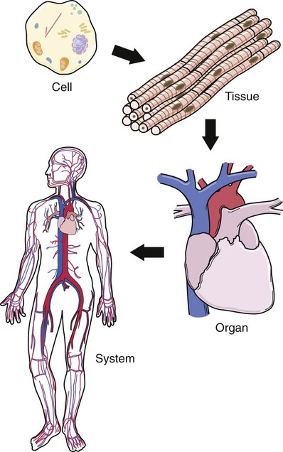
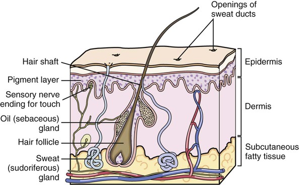
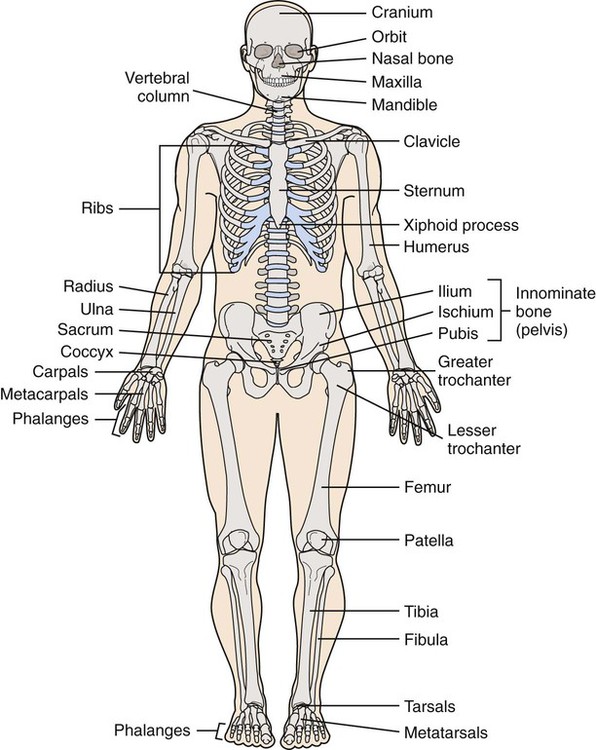
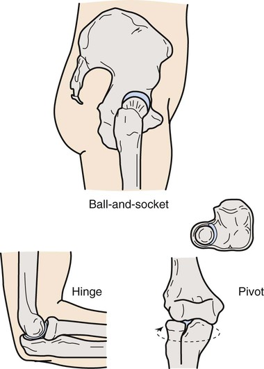
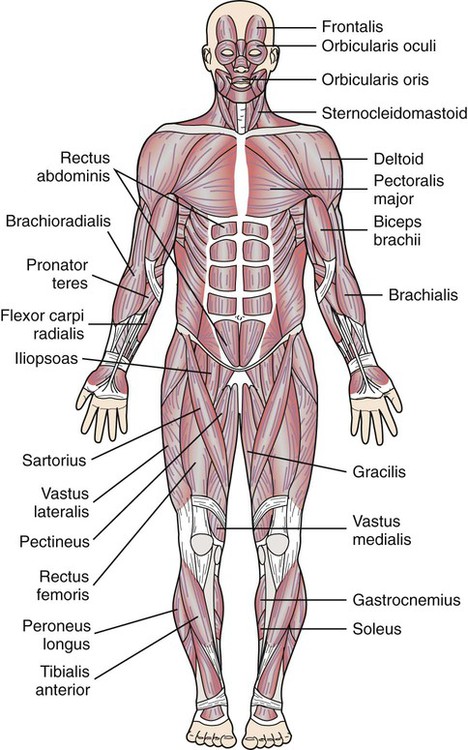
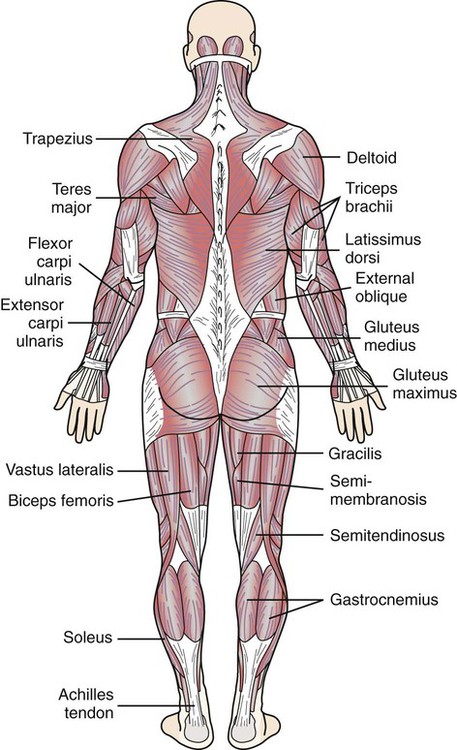
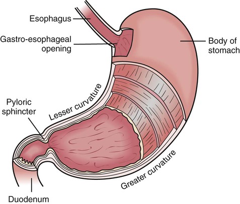
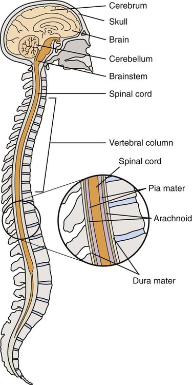
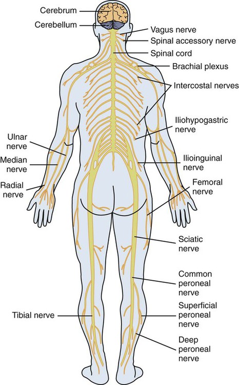
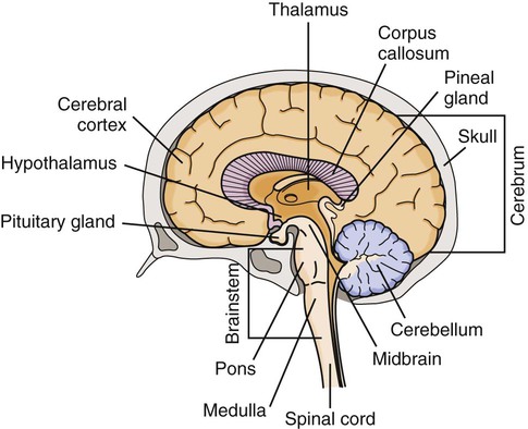
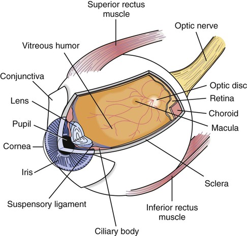
• Define the key terms and key abbreviations listed in this chapter. • Identify the basic structures of the cell. • Describe 4 types of tissues. • Identify the structures of each body system. • Identify the functions of each body system. • Explain how to promote PRIDE in the person, the family, and yourself. See Chapter 8 for the changes in body structure and function that occur with aging. Figure 7-1 shows the cell and its structures. The cell membrane is the outer covering. It encloses the cell and helps to hold its shape. The nucleus is the control center of the cell. It directs the cell’s activities. The nucleus is in the center of the cell. The cytoplasm surrounds the nucleus. Cytoplasm contains smaller structures that perform cell functions. Protoplasm means “living substance.” It refers to all structures, substances, and water within the cell. Protoplasm is a semi-liquid substance much like an egg white. The nucleus controls cell reproduction. Cells reproduce by dividing in half. The process of cell division is called mitosis. It is needed for tissue growth and repair. During mitosis, the 46 chromosomes arrange themselves in 23 pairs. As the cell divides, the 23 pairs are pulled in half. The 2 new cells are identical. Each has 46 chromosomes (Fig. 7-2). • Epithelial tissue covers internal and external body surfaces. Tissue lining the nose, mouth, respiratory tract, stomach, and intestines is epithelial tissue. So are the skin, hair, nails, and glands. • Connective tissue anchors, connects, and supports other tissues. It is in every part of the body. Bones, tendons, ligaments, and cartilage are connective tissue. Blood is a form of connective tissue. • Muscle tissue stretches and contracts to let the body move. • Nerve tissue receives and carries impulses to the brain and back to body parts. Groups of tissue with the same function form organs. An organ has 1 or more functions. Examples of organs are the heart, brain, liver, lungs, and kidneys. Systems are formed by organs that work together to perform special functions (Fig. 7-3). The integumentary system, or skin, is the largest system. Integument means covering. The skin covers the body. It has epithelial, connective, and nerve tissue. It also has oil glands and sweat glands. There are 2 skin layers (Fig. 7-4). • The epidermis is the outer layer. It has living cells and dead cells. The dead cells were once deeper in the epidermis. They were pushed upward as the cells divided. Dead cells constantly flake off. They are replaced by living cells. Living cells die and flake off. Living cells of the epidermis contain pigment. Pigment gives skin its color. The epidermis has no blood vessels and few nerve endings. • The dermis is the inner layer. It is made up of connective tissue. Blood vessels, nerves, sweat glands, and oil glands are found in the dermis. So are hair roots. Oil glands and sweat glands, hair, and nails are skin appendages. • Hair—covers the entire body, except the palms of the hands and the soles of the feet. Hair in the nose and ears and around the eyes protects these organs from dust, insects, and other foreign objects. • Nails—protect the tips of the fingers and toes. Nails help fingers pick up and handle small objects. • Sweat glands (sudoriferous glands)—help the body regulate temperature. Sweat consists of water, salt, and a small amount of wastes. Sweat is secreted through pores in the skin. The body is cooled as sweat evaporates. • Oil glands (sebaceous glands)—lie near the hair shafts. They secrete an oily substance into the space near the hair shaft. Oil travels to the skin surface. This helps keep the hair and skin soft and shiny. • It is the body’s protective covering. • It prevents microorganisms and other substances from entering the body. • It prevents excess amounts of water from leaving the body. • It protects organs from injury. • Nerve endings in the skin sense both pleasant and unpleasant stimulation. Nerve endings are over the entire body. They sense cold, pain, touch, and pressure to protect the body from injury. • It helps regulate body temperature. Blood vessels dilate (widen) when temperature outside the body is high. More blood is brought to the body surface for cooling during evaporation. When blood vessels constrict (narrow), the body retains heat. This is because less blood reaches the skin. The human body has 206 bones (Fig. 7-5). There are 4 types of bones. • Long bones bear the body’s weight. Leg bones are long bones. • Short bones allow skill and ease in movement. Bones in the wrists, fingers, ankles, and toes are short bones. • Flat bones protect the organs. They include the ribs, skull, pelvic bones, and shoulder blades. • Irregular bones are the vertebrae in the spinal column. They allow various degrees of movement and flexibility. A joint is the point at which 2 or more bones meet. Joints allow movement (Chapter 23). Cartilage is connective tissue at the end of the long bones. It cushions the joint so that the bone ends do not rub together. The synovial membrane lines the joints. It secretes synovial fluid. Synovial fluid acts as a lubricant so the joint can move smoothly. Bones are held together at the joint by strong bands of connective tissue called ligaments. There are 3 major types of joints (Fig. 7-6). • A ball-and-socket joint allows movement in all directions. It is made of the rounded end of 1 bone and the hollow end of another bone. The rounded end of 1 fits into the hollow end of the other. The joints of the hips and shoulders are ball-and-socket joints. • A hinge joint allows movement in 1 direction. The elbow is a hinge joint. • A pivot joint allows turning from side to side. A pivot joint connects the skull to the spine. Some joints are immovable. They connect the bones of the skull. The human body has more than 500 muscles (Figs. 7-7 and 7-8). Some are voluntary. Others are involuntary. • Voluntary muscles can be consciously controlled. Muscles attached to bones (skeletal muscles) are voluntary. Arm muscles do not work unless you move your arm; likewise for leg muscles. Skeletal muscles are striated. That is, they look striped or streaked. • Involuntary muscles work automatically. You cannot control them. They control the action of the stomach, intestines, blood vessels, and other body organs. Involuntary muscles also are called smooth muscles. They look smooth, not streaked or striped. • Cardiac muscle is in the heart. It is an involuntary muscle. However, it appears striated like skeletal muscle. Strong, tough connective tissues called tendons connect muscles to bones. When muscles contract (shorten), tendons at each end of the muscle cause the bone to move. The body has many tendons. See the Achilles tendon in Figure 7-8. Some muscles constantly contract to maintain the body’s posture. When muscles contract, they burn food for energy. Heat is produced. The more muscle activity, the greater the amount of heat produced. Shivering is how the body produces heat when exposed to cold. Shivering is from rapid, general muscle contractions. • The pyloric sphincter (Fig. 7-9) is an opening from the stomach into the small intestine. Closed, it holds food in the stomach for partial digestion. It opens to allow partially digested food to enter the small intestine. • The anal sphincter keeps the anus closed. It opens for a bowel movement. • Urethral sphincters seal off the bladder. This allows urine to collect in the bladder. The sphincters open for urination. The nervous system controls, directs, and coordinates body functions. Its 2 main divisions are: • The central nervous system (CNS). It consists of the brain and spinal cord (Fig. 7-10). • The peripheral nervous system. It involves the nerves throughout the body (Fig. 7-11). The brain and spinal cord make up the central nervous system. The brain is covered by the skull. The 3 main parts of the brain are the cerebrum, the cerebellum, and the brainstem (Fig. 7-12). • The outer layer lies next to the skull. It is a tough covering called the dura mater. • The middle layer is the arachnoid. Receptors for vision are in the eyes (Fig. 7-13). The eye is easily injured. Bones of the skull, eyelids and eyelashes, and tears protect the eyes from injury. The eye has 3 layers. • The sclera, the white of the eye, is the outer layer. It is made of tough connective tissue. • The choroid is the second layer. Blood vessels, the ciliary muscle, and the iris make up the choroid. The iris gives the eye its color. The opening in the middle of the iris is the pupil. Pupil size varies with the amount of light entering the eye. The pupil constricts (narrows) in bright light. It dilates (widens) in dim or dark places. • The retina is the inner layer. It has receptors for vision and the nerve fibers of the optic nerve.
Body Structure and Function
Cells, Tissues, and Organs
The Integumentary System
The Musculo-Skeletal System
Bones
Joints
Muscles
The Nervous System
The Central Nervous System
The Sense Organs
The Eye.
![]()
Stay updated, free articles. Join our Telegram channel

Full access? Get Clinical Tree


Body Structure and Function
Get Clinical Tree app for offline access
