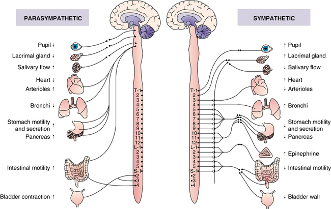
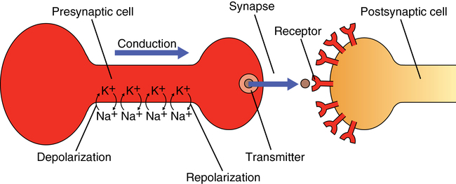
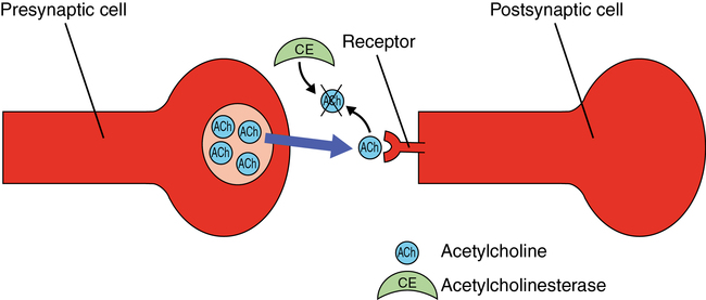
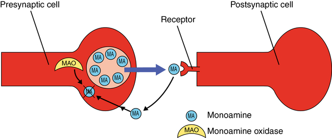
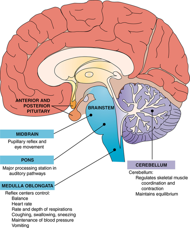
Biological basis for understanding psychiatric disorders and treatments

CHAPTER 3


Biological basis for understanding psychiatric disorders and treatments


Mary A. Gutierrez, Jerika T. Lam and Mary Ann Schaepper


image


Visit the Evolve website for a pretest on the content in this chapter: http://evolve.elsevier.com/Varcarolis


image


Although the origin of a psychiatric illness may be related to a number of factors, such as genetics, neurodevelopmental factors, drugs, infection, and bad experiences, there is ultimately a physiological alteration in brain function that accounts for the disturbances in the patient’s behavior and mental experiences. These physiological alterations are the targets of the psychotropic (Greek for psyche, or mind, + trepein, to turn) drugs used to treat mental disease. From a holistic point of view, mental disorders have psychobiological components that support the efficacy of treating these disorders both pharmacologically and with appropriate psychotherapy, or “talk” therapy that provides social and psychological support and actually alters brain function (Karlsson, 2011).


The treatment of mental illness with psychotropic drugs extends back more than half a century, yet a full understanding of how these drugs improve the symptoms of these illnesses continues to elude investigators. Early biological theories associated a single neurotransmitter with a specific disorder. The dopamine theory of schizophrenia and the monoamine theory of depression are now viewed as overly simplistic because a large number of other neurotransmitters, hormones, and co-regulators are now thought to play important and complex roles. The focus of research on neurotransmitters is now on how they are released from presynaptic cells and then act on postsynaptic cells and on how psychotropic drugs interact with these substances to make changes in brain functioning.


Recent discoveries have influenced the direction of research and treatment. While scientists have long known about basic receptors for neurotransmitters, many subtypes of receptors for the various neurotransmitters have been discovered in recent years.


The overall purpose of this chapter is to relate psychiatric disturbances and the psychotropic drugs used to treat them to normal brain structure and function. First, this chapter looks at the normal functions of the brain and how these functions are carried out from an anatomical and physiological perspective. Then, it reviews current theories of the psychobiological basis of various types of emotional and physiological dysfunctions. Finally, the chapter reviews the major drugs used to treat mental disorders, explains how they work, and identifies how both the beneficial and the problematic effects of psychiatric drugs relate to their interaction with various neurotransmitter-receptor systems.


Despite new information about the complex brain functions and neurotransmitters, there is still much to be clarified in understanding the complex ways in which the brain carries out its normal functions, is altered during disease, and is improved upon by pharmacological intervention. After reading this chapter, you should have a neurobiological framework into which you can place existing, as well as future, information about mental illnesses and their treatments. Additional, detailed information regarding adverse and toxic effects, dosage, nursing implications, and teaching tools is presented in the appropriate clinical chapters (Chapters 11-24).


Structure and function of the brain


Functions and activities of the brain


Regulating behavior and carrying out mental processes are important, but far from the only, responsibilities of the brain. Box 3-1 summarizes some of the major functions and activities of the brain. Because all of these brain functions are carried out by similar mechanisms (interactions of neurons) often in similar locations, it is not surprising that mental disturbances are often associated with alterations in other brain functions and that the drugs used to treat mental disturbances can also interfere with other activities of the brain.


Maintenance of homeostasis


The brain serves as the coordinator and director of the body’s response to both internal and external changes. Appropriate responses require a constant monitoring of the environment, interpretation and integration of the incoming information, and control over the appropriate organs of response. The goal of these responses is to maintain homeostasis and thus to maintain life.


Information about the external world is relayed from various sense organs to the brain by the peripheral nerves. This information, which is at first received as a gross sensation, such as light, sound, or touch, must ultimately be interpreted as a key, a train whistle, or a hand on the back, respectively. Interestingly, a component of major psychiatric disturbance (e.g., schizophrenia) is an alteration of sensory experience; thus, the patient may experience a sensation that does not originate in the external world. For example, people with schizophrenia may hear voices talking to them (i.e., auditory hallucination).


The brain not only monitors the external world but also keeps a close watch on the internal functions; thus, the brain continuously receives information about blood pressure, body temperature, blood gases, and the chemical composition of the body fluids so that it can direct the appropriate responses required to maintain homeostasis.


To respond to external changes, the brain has control over the skeletal muscles. This control involves the ability to initiate contraction (i.e., to contract the biceps and flex the arm) but also to fine-tune and coordinate contraction so that a person can, for example, guide the fingers to the correct keys on a piano. Unfortunately, both psychiatric disease and the treatment of psychiatric disease with psychotropic drugs are often associated with disturbance of movement.


It is important to remember that the skeletal muscles controlled by the brain include the diaphragm, which is essential for breathing, and the muscles of the throat, tongue, and mouth, which are essential for speech; thus, drugs that affect brain function can stimulate or depress respiration or lead to slurred speech.


Adjustments to changes within the body require that the brain exert control over the various internal organs. For example, if blood pressure drops, the brain must direct the heart to pump more blood and the smooth muscles of the arterioles to constrict. This increase in cardiac output and vasoconstriction allows the body to return blood pressure to its normal level.


Regulation of the autonomic nervous system and hormones


The autonomic nervous system and the endocrine system serve as the communication links between the brain and the cardiac muscle, smooth muscle, and glands of which the internal organs are composed (Figure 3-1). If the brain needs to stimulate the heart, it must activate the sympathetic nerves to the sinoatrial node and the ventricular myocardium. If the brain needs to bring about vasoconstriction, it must activate the sympathetic nerves to the smooth muscles of the arterioles.


The linkage between the brain and the internal organs that allows for the maintenance of homeostasis may also serve to translate mental disturbances, such as anxiety, into alterations of internal function. For example, anxiety can activate the sympathetic nervous system, leading to symptoms such as increased heart rate and blood pressure, shortness of breath, and sweating.


The brain also exerts influence over the internal organs by regulating hormonal secretions of the pituitary gland, which in turn regulates other glands. A specific area of the brain, the hypothalamus, secretes hormones called releasing factors. These hormones act on the pituitary gland to stimulate or inhibit the synthesis and release of pituitary hormones. Once in the general circulation they influence various internal activities. An example of this linkage is the release of gonadotropin-releasing hormone by the hypothalamus at the time of puberty. This hormone stimulates the release of two gonadotropins—follicle-stimulating hormone and luteinizing hormone—by the pituitary gland, which consequently activates the ovaries or testes. This linkage may explain why anxiety or depression in some women may lead to disturbances of the menstrual cycle.


The relationship between the brain, the pituitary gland, and the adrenal glands is particularly important in normal and abnormal mental function. Specifically, the hypothalamus secretes corticotropin-releasing hormone (CRH), which stimulates the pituitary to release corticotropin, which in turn stimulates the cortex of each adrenal gland to secrete the hormone cortisol. This system is activated as part of the normal response to a variety of mental and physical stresses. Among many other actions, all three hormones—CRH, corticotropin, and cortisol—influence the functions of the nerve cells of the brain. There is considerable evidence that this system is overactive in anxiety and that the normal negative feedback mechanism that is supposed to bring the hormone levels back down does not respond properly.


Control of biological drives and behavior


To understand the neurobiological basis of mental disease and its treatment, it is helpful to distinguish between the various types of brain activity that serve as the basis of mental experience and behavior. An understanding of these activities shows where to look for disturbed function and what to hope for in treatment. The brain, for example, is responsible for the basic drives, such as sex and hunger, that play a strong role in molding behavior. Disturbances of these drives (e.g., overeating or undereating, loss of sexual interest) can be an indication of an underlying psychiatric disorder such as depression.


Cycle of sleep and wakefulness.

The entire cycle of sleep and wakefulness, as well as the intensity of alertness while the person is awake, is regulated and coordinated by various regions of the brain. Although we do not fully understand the true homeostatic function of sleep, we know that it is essential for both physiological and psychological well-being. Assessment of sleep patterns is part of what is required to determine a psychiatric diagnosis.


Unfortunately, many of the drugs used to treat psychiatric problems interfere with the normal regulation of sleep and alertness. Drugs with a sedative-hypnotic effect can blunt the degree to which a person feels alert and focused and can cause drowsiness. The sedative-hypnotic effect requires caution in using these drugs while engaging in activities that require a great deal of attention, such as driving a car or operating machinery. One way of minimizing the danger is to take drugs at night just before bedtime.


Conscious mental activity


All aspects of conscious mental experience and sense of self originate from the neurophysiological activity of the brain. Conscious mental activity can be a basic, meandering stream of consciousness that can flow from thoughts of future responsibilities, memories, fantasies, and so on. Conscious mental activity can also be much more complex when it is applied to problem solving and the interpretation of the external world. Both the basic stream of consciousness and the complex problem solving and environment interpretation can become distorted in psychiatric illness. A person with schizophrenia may have chaotic and incoherent speech and thought patterns (e.g., a jumble of unrelated words known as word salad and unconnected phrases and topics known as looseness of association) and delusional interpretations of personal interactions, such as beliefs about people or events that are not supported by data or reality.


Cellular composition of the brain
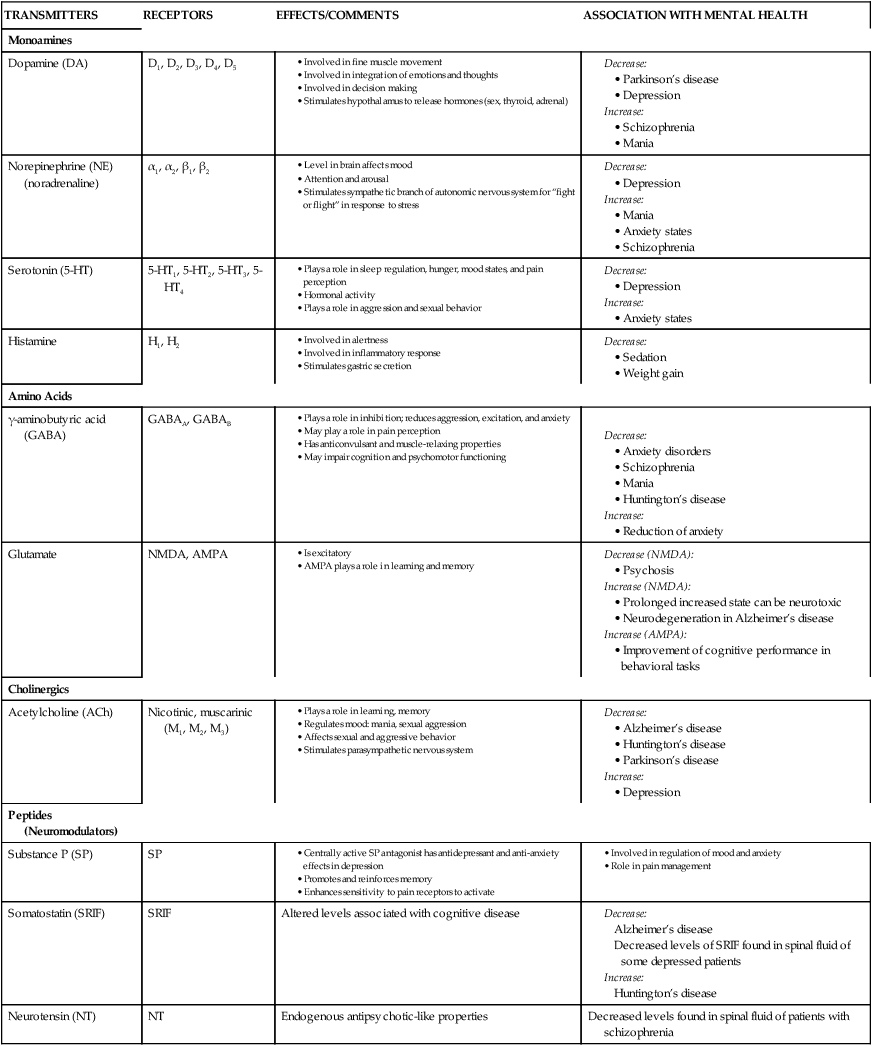

The brain is composed of approximately 100 billion neurons, nerve cells that conduct electrical impulses, as well as other types of cells that surround the neurons. Most functions of the brain, from regulation of blood pressure to the conscious sense of self, are thought to result from the actions of individual neurons and the interconnections between them. Although neurons come in a great variety of shapes and sizes, all carry out the same three types of physiological actions: (1) respond to stimuli, (2) conduct electrical impulses, and (3) release chemicals called neurotransmitters.


An essential feature of neurons is their ability to conduct an electrical impulse from one end of the cell to the other. This electrical impulse consists of a change in membrane permeability that first allows the inward flow of sodium ions and then the outward flow of potassium ions. The inward flow of sodium ions changes the polarity of the membrane from positive on the outside to positive on the inside. Movement of potassium ions out of the cell returns the positive charge to the outside of the cell. Because these electrical charges are self-propagating, a change at one end of the cell is conducted along the membrane until it reaches the other end (Figure 3-2). The functional significance of this propagation is that the electrical impulse serves as a means of communication between one part of the body and another.


Once an electrical impulse reaches the end of a neuron, a neurotransmitter is released. A neurotransmitter is a chemical substance that functions as a neuromessenger. Neurotransmitters are released from the axon terminal at the presynaptic neuron on excitation. This neurotransmitter then diffuses across a space, or synapse, to an adjacent postsynaptic neuron, where it attaches to receptors on the neuron’s surface. It is this interaction from one neuron to another, by way of a neurotransmitter and receptor, that allows the activity of one neuron to influence the activity of other neurons. Depending on the chemical structure of the neurotransmitter and the specific type of receptor to which it attaches, the postsynaptic cell will be rendered either more or less likely to initiate an electrical impulse. It is the interaction between neurotransmitter and receptor that is a major target of the drugs used to treat psychiatric disease. Table 3-1 lists important neurotransmitters and the types of receptors to which they attach. Also listed are the mental disorders associated with an increase or decrease in these neurotransmitters.


TABLE 3-1   


TRANSMITTERS AND RECEPTORS


TRANSMITTERS RECEPTORS EFFECTS/COMMENTS ASSOCIATION WITH MENTAL HEALTH
Monoamines
Dopamine (DA) D1, D2, D3, D4, D5


Norepinephrine (NE) (noradrenaline) α1, α2, β1, β2


Serotonin (5-HT) 5-HT1, 5-HT2, 5-HT3, 5-HT4


Histamine H1, H2

Amino Acids
γ-aminobutyric acid (GABA) GABAA, GABAB
 

Glutamate NMDA, AMPA


Cholinergics
Acetylcholine (ACh) Nicotinic, muscarinic (M1, M2, M3)


Peptides (Neuromodulators)
Substance P (SP) SP

Somatostatin (SRIF) SRIF Altered levels associated with cognitive disease

Neurotensin (NT) NT Endogenous antipsychotic-like properties Decreased levels found in spinal fluid of patients with schizophrenia


image


AMPA, α-Amino-3-hydroxy-5-methyl-4-isoxazolepropionic acid; NMDA, N-methyl-d-aspartate.


After attaching to a receptor and exerting its influence on the postsynaptic cell, the neurotransmitter separates from the receptor and is destroyed. The process of neurotransmitter destruction is described in Box 3-2. There are two basic mechanisms by which neurotransmitters are destroyed. Some neurotransmitters (e.g., acetylcholine) are destroyed by specific enzymes at the postsynaptic cell. The enzyme that destroys acetylcholine is called acetylcholinesterase (most enzymes start with the name of the neurotransmitter they destroy and end with the suffix –ase). Other neurotransmitters (e.g., norepinephrine) are taken back into the presynaptic cell from which they were originally released by a process called cellular reuptake and are either reused or destroyed by intracellular enzymes. In the case of the monoamine neurotransmitters (e.g., norepinephrine, dopamine, serotonin), the destructive enzyme is called monoamine oxidase (MAO).


BOX 3-2      DESTRUCTION OF NEUROTRANSMITTERS


A full explanation of the various ways in which psychotropic drugs alter neuronal activity requires a brief review of the manner in which neurotransmitters are destroyed after attaching to the receptors. To avoid continuous and prolonged action on the postsynaptic cell, the neurotransmitter is released shortly after attaching to the postsynaptic receptor. Once released, the neurotransmitter is destroyed in one of two ways.


One way is the immediate inactivation of the neurotransmitter at the postsynaptic membrane. An example of this method of destruction is the action of the enzyme acetylcholinesterase on the neurotransmitter acetylcholine. Acetylcholinesterase is present at the postsynaptic membrane and destroys acetylcholine shortly after it attaches to nicotinic or muscarinic receptors on the postsynaptic cell.


image


A second method of neurotransmitter inactivation is a little more complex. After interacting with the postsynaptic receptor, the neurotransmitter is released and taken back into the presynaptic cell, the cell from which it was released. This process, referred to as the reuptake of neurotransmitter, is a common target for drug action. Once inside the presynaptic cell, the neurotransmitter is either recycled or inactivated by an enzyme within the cell. The monoamine neurotransmitters norepinephrine, dopamine, and serotonin are all inactivated in this manner by the enzyme monoamine oxidase.


image


Looking at this second method, you might naturally ask what prevents the enzyme from destroying the neurotransmitter before its release. The answer is that before release the neurotransmitter is stored within a membrane and is protected. After release and reuptake, the neurotransmitter is either destroyed by the enzyme or reenters the membrane to be used again.


Some neurotransmitters regulate concentration at the postsynaptic receptors by exerting their own feedback inhibition of their own release. This is accomplished by the attachment of neurotransmitters to presynaptic receptors at the synapse, which act to inhibit the further release of neurotransmitters.


A neuron may release a specific neurotransmitter that stimulates or inhibits a postsynaptic membrane receptor. Negative feedback on a presynaptic receptor should maintain a normal balance of neurotransmitters; however, researchers have also found that neurons may release more than one chemical at the same time. Larger molecules, neuropeptides, may bring about long-term changes in the postsynaptic cells by joining neurotransmitters such as norepinephrine or acetylcholine. The result of these changes is an alteration of basic cell functions or genetic expression and may lead to modifications of cell shape and responsiveness to stimuli. Ultimately, this means that the action of one neuron on another affects not only the immediate response of that neuron but also its sensitivity to future influence. The long-term implications of this for neural development, normal and abnormal mental health, and the treatment of psychiatric disease are being investigated.


Communication in nerve cells goes from presynaptic to postsynaptic; however, researchers have found that the communication between neurons at a synapse does not just go in one direction. Influencing the growth, shape, and activity of presynaptic cells are protein and simple gases, such as carbon monoxide and nitrous oxide, called neurotrophic factors. These factors are thought to be particularly important during the development of the fetal brain, guiding the growing brain to form the proper neuronal connections. It is apparent that the brain retains anatomical plasticity throughout life and that internal and external influences can alter the synaptic network of the brain. Altered genetic expression or environmental trauma can change these factors and result in negative and positive consequences on mental function and psychiatric disease.


Other chemicals, such as steroid hormones, brought to the neurons by the blood can influence the development and responsiveness of neurons. Estrogen, testosterone, and cortisol can bind to neurons, where they can cause short- and long-term changes in neuronal activity. A clear example of this is evident in the psychosis that can result from the hypersecretion of cortisol in Cushing’s disease or from the use of prednisone in high doses to treat chronic inflammatory disease.


Organization of the brain


Brainstem


The central core of the brainstem regulates the internal organs and is responsible for such vital functions as the regulation of blood gases and the maintenance of blood pressure. The hypothalamus, a small area in the ventral superior portion of the brainstem, plays a vital role in such basic drives as hunger, thirst, and sex. It also serves as a crucial psychosomatic link between higher brain activities, such as thought and emotion, and the functioning of the internal organs. The brainstem also serves as an initial processing center for sensory information that is then sent on to the cerebral cortex. Through projections of the reticular activating system (RAS), the brainstem regulates the entire cycle of sleep and wakefulness and the ability of the cerebrum to carry out conscious mental activity.


Other ascending pathways, referred to as mesolimbic and mesocortical pathways, seem to play a strong role in modulating the emotional value of sensory material. These pathways project to those areas of the cerebrum collectively known as the limbic system, which plays a crucial role in emotional status and psychological function. They use norepinephrine, serotonin, and dopamine as their neurotransmitters. Much attention has been paid to the role of these pathways in normal and abnormal mental activity. For example, it is thought that the release of dopamine from the ventral tegmental pathway plays a role in psychological reward and drug addiction. The neurotransmitters released by these neurons are major targets of the drugs used to treat psychiatric disease.


Cerebellum


Located behind the brainstem, the cerebellum (Figure 3-3) is primarily involved in the regulation of skeletal muscle coordination and contraction and the maintenance of equilibrium. It plays a crucial role in coordinating contractions so that movement is accomplished in a smooth and directed manner.


Cerebrum


The human brainstem and cerebellum are similar in both structure and function to these same structures in other mammals. The development of a much larger and more elaborate cerebrum is what distinguishes human beings from the rest of the animal kingdom.


The cerebrum, situated on top of and surrounding the brainstem, is responsible for mental activities and a conscious sense of being. This is responsible for our conscious perception of the external world and our own body, emotional status, memory, and control of skeletal muscles that allow willful direction of movement. The cerebrum is also responsible for language and the ability to communicate.


The cerebrum consists of surface, the cerebral cortex, and deep areas of integrating gray matter that include the basal ganglia, amygdala, and hippocampus. Tracts of white matter link these areas with each other and the rest of the nervous system. The cerebral cortex, which forms the outer layer of the brain, is responsible for conscious sensation and the initiation of movement. Certain areas of the cortex are responsible for specific sensations. For example, the sensation of touch resides in the parietal cortex, sounds are based in the temporal cortex, and vision is housed in the occipital cortex. Likewise, a specific area of the frontal cortex controls the initiation of skeletal muscle contraction. Of course, all areas of the cortex are interconnected so that an appropriate picture of the world can be formed and, if necessary, linked to a proper response (Figure 3-4).


Both sensory and motor aspects of language reside in specialized areas of the cerebral cortex. Sensory language functions include the ability to read, understand spoken language, and know the names of objects perceived by the senses. Motor functions involve the physical ability to use muscles properly for speech and writing. In both neurological and psychological dysfunction, the use of language may become compromised or distorted. The change in language ability may be a factor in determining a diagnosis.


Underneath the cerebral cortex there are pockets of integrating gray matter deep within the cerebrum. Some of these, the basal ganglia, are involved in the regulation of movement. Others, the amygdala and hippocampus, are involved in emotions, learning, memory, and basic drives. Significantly, there is an overlap of these areas both anatomically and in the types of neurotransmitters employed. One important consequence is that drugs used to treat emotional disturbances may cause movement disorders, and drugs used to treat movement disorders may cause emotional changes.


Visualizing the brain


A variety of noninvasive imaging techniques are used to visualize brain structure, functions, and metabolic activity. Table 3-2 identifies some common brain imaging techniques and preliminary findings as they relate to psychiatry. There are two types of neuroimaging techniques: structural and functional. Structural imaging techniques (e.g., computed tomography [CT] and magnetic resonance imaging [MRI]) provide overall images of the brain and layers of the brain. Functional imaging techniques (e.g., positron emission tomography [PET] and single photon emission computed tomography [SPECT]) reveal physiological activity in the brain, as described in Table 3-2.


TABLE 3-2   


COMMON BRAIN IMAGING TECHNIQUES


TECHNIQUE DESCRIPTION USES PSYCHIATRIC RELEVANCE AND PRELIMINARY FINDINGS
Electrical: Recording Electrical Signals from the Brain
Electroencephalograph (EEG) A recording of electrical signals from the brain made by hooking up electrodes to the subject’s scalp. Can show the state a person is in —asleep, awake, anesthetized —because the characteristic patterns of current differ for each of these states. Provides support from a wide range of sources that brain abnormalities exist; may lead to further testing.
Structural: Show Gross Anatomical Details of Brain Structures
Computerized axial tomography (CT) A series of x-ray images is taken of the brain and a computer analysis produces “slices” providing a precise 3D-like reconstruction of each segment.
Get Clinical Tree app for offline access