M. Linda Workman
Assessment of the Endocrine System
Learning Outcomes
Safe and Effective Care Environment
Health Promotion and Maintenance
Psychosocial Integrity
Physiological Integrity
7 Describe the relationship between hormones and receptor sites.
8 Explain negative feedback as a control mechanism for hormone secretion.

http://evolve.elsevier.com/Iggy/
Answer Key for NCLEX Examination Challenges and Decision-Making Challenges
Audio Glossary
Key Points
Review Questions for the NCLEX® Examination
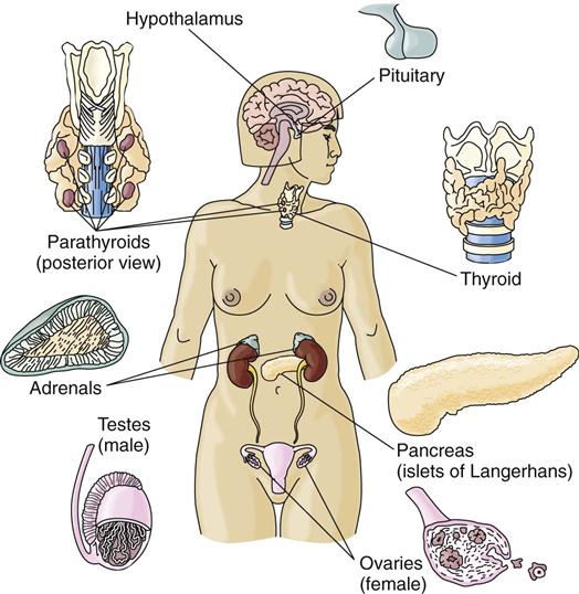
The endocrine system is made up of glands in many tissues and organs in a variety of body areas (Fig. 64-1). The purpose of all endocrine glands is to secrete hormones, which are natural chemicals that exert their effects on specific tissues known as target tissues. Target tissues are usually located some distance from the endocrine gland, with no direct physical connection between the endocrine gland and its target tissue. For this reason, endocrine glands are called “ductless” glands and must use the blood to transport secreted hormones to the target tissues (McCance et al., 2010). Endocrine glands include:
The endocrine system works with the nervous system to control overall body function, known as neuroendocrine regulation. Many interactions must occur between the endocrine system and all other body systems to ensure that each system maintains a constant normal balance (homeostasis) in response to environmental changes. For example, neuroendocrine control of body systems keeps the internal body temperature at or near 98.6° F (37° C), even when environmental temperatures vary. Other actions keep the serum sodium level between 136 and 145 mEq/L (mmol/L), regardless of whether a person eats 2 g or 12 g of sodium per day.
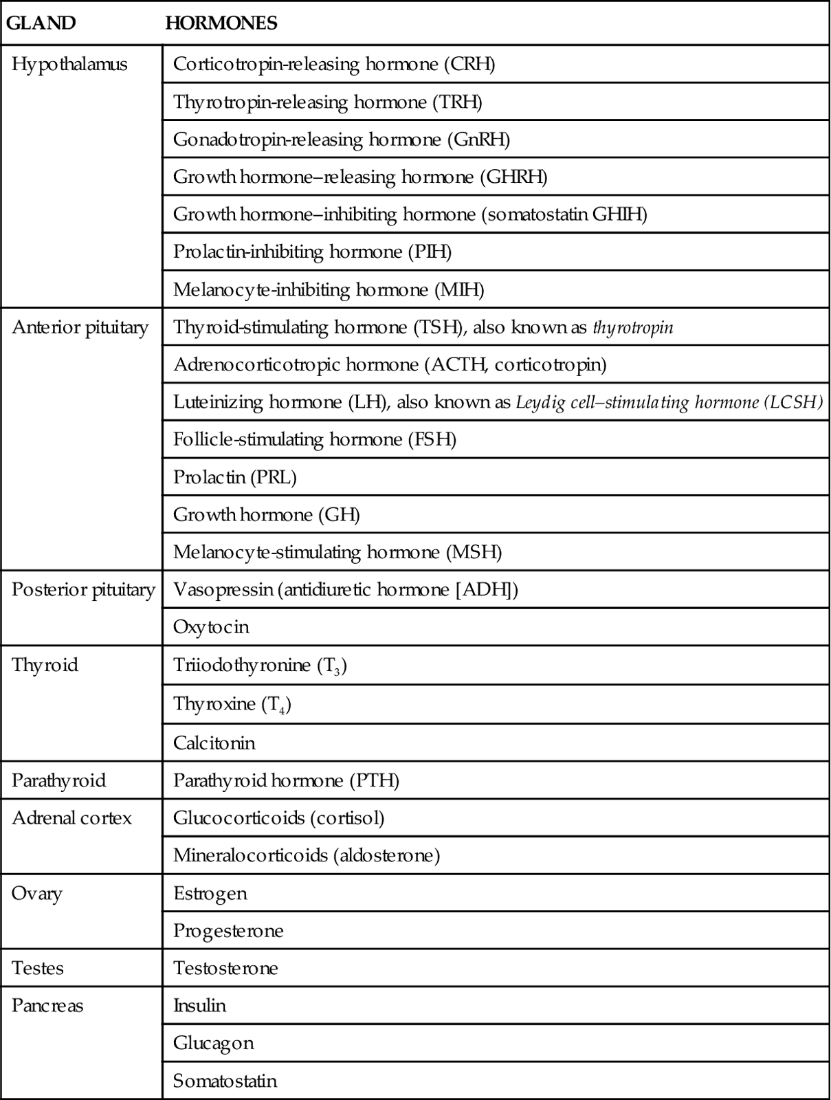
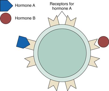
Table 64-1 lists hormones secreted by various endocrine glands. Hormones travel through the blood to all body areas but exert their actions only on target tissues. They recognize their target tissues and exert their actions by binding to receptors on or within the target tissue cells. In general, each receptor site type is specific for only one hormone. Hormone-receptor actions work in a “lock and key” manner in that only the correct hormone (key) can bind to and activate the receptor site (lock) (Fig. 64-2). Binding a hormone to its receptor causes the target tissue to change its activity, producing specific responses.
TABLE 64-1
PRINCIPAL HORMONES OF THE ENDOCRINE GLANDS
| GLAND | HORMONES |
| Hypothalamus | Corticotropin-releasing hormone (CRH) |
| Thyrotropin-releasing hormone (TRH) | |
| Gonadotropin-releasing hormone (GnRH) | |
| Growth hormone–releasing hormone (GHRH) | |
| Growth hormone–inhibiting hormone (somatostatin GHIH) | |
| Prolactin-inhibiting hormone (PIH) | |
| Melanocyte-inhibiting hormone (MIH) | |
| Anterior pituitary | Thyroid-stimulating hormone (TSH), also known as thyrotropin |
| Adrenocorticotropic hormone (ACTH, corticotropin) | |
| Luteinizing hormone (LH), also known as Leydig cell–stimulating hormone (LCSH) | |
| Follicle-stimulating hormone (FSH) | |
| Prolactin (PRL) | |
| Growth hormone (GH) | |
| Melanocyte-stimulating hormone (MSH) | |
| Posterior pituitary | Vasopressin (antidiuretic hormone [ADH]) |
| Oxytocin | |
| Thyroid | Triiodothyronine (T3) |
| Thyroxine (T4) | |
| Calcitonin | |
| Parathyroid | Parathyroid hormone (PTH) |
| Adrenal cortex | Glucocorticoids (cortisol) |
| Mineralocorticoids (aldosterone) | |
| Ovary | Estrogen |
| Progesterone | |
| Testes | Testosterone |
| Pancreas | Insulin |
| Glucagon | |
| Somatostatin |
Disorders of the endocrine system usually are related to:
Anatomy and Physiology Review
The control of cellular function by any hormone depends on a series of reactions working through negative feedback control mechanisms. Hormone secretion usually depends on the need of the body for the final action of that hormone. When a body condition starts to move away from the normal range and a specific action or response is needed to correct this change, secretion of the hormone capable of starting the correcting action or response is stimulated until the need (demand) is met and the body condition returns to the normal range. As the correction occurs, hormone secretion decreases (and may halt). This type of control for hormone synthesis is “negative feedback” because the hormone causes the opposite action of the initial condition change.
An example of a simple negative feedback hormone response is the control of insulin secretion. When blood glucose levels start to rise above normal, the hormone insulin is secreted. Insulin increases glucose uptake by the cells, causing a decrease in blood glucose levels. Thus the action of insulin (decreasing blood glucose levels) is the opposite of or negative to the condition that stimulated insulin secretion (elevated blood glucose levels).
Some hormones have more complex interactions for negative feedback. These interactions involve a series of reactions in which more than one endocrine gland, as well as the final target tissues, is stimulated. In this situation, the first hormone in the series may have another endocrine gland as its target tissue. For this type of mechanism to maintain homeostasis, this series of interactions must occur:
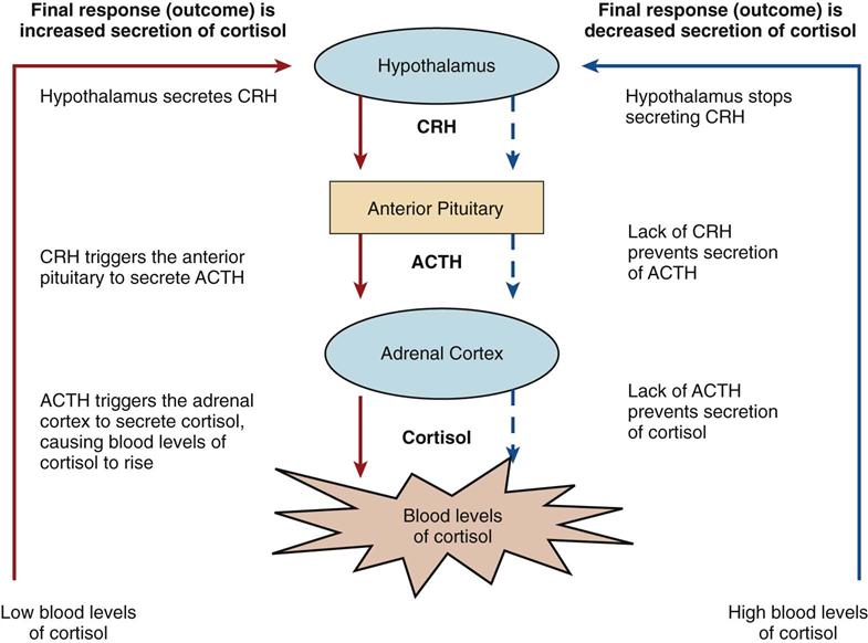
An example of complex control is the interaction of the hypothalamus and the anterior pituitary with the adrenal cortex (Fig. 64-3). Low blood levels of cortisol from the adrenal cortex stimulate the secretion of corticotropic-releasing hormone (CRH) in the hypothalamus. CRH stimulates the anterior pituitary gland to secrete adrenocorticotropic hormone (ACTH). ACTH then triggers the release of cortisol from the adrenal cortex. The rising blood levels of cortisol inhibit CRH release from the hypothalamus. Without CRH, the anterior pituitary gland stops secretion of ACTH. In response, normal blood cortisol levels are maintained.
The normal blood level range of each hormone is well defined. Excesses or deficiencies of hormone secretion can lead to pathologic conditions.
Hypothalamus and Pituitary Glands
The hypothalamus is a small area of nerve and glandular tissue located beneath the thalamus in the brain. Nerve fibers connect the hypothalamus to the rest of the central nervous system. The hypothalamus shares a small, closed circulatory system with the anterior pituitary gland, known as the hypothalamic-hypophysial portal system. This system allows hormones produced in the hypothalamus to travel directly to the anterior pituitary gland so that only very small amounts are wasted in systemic circulation.
The endocrine function of the hypothalamus is to produce regulatory hormones (see Table 64-1). Some of these hormones are released into the blood and travel to the anterior pituitary, where they either stimulate or inhibit the release of anterior pituitary hormones.
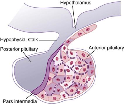
The pituitary gland is located at the base of the brain in a protective pocket of the sphenoid bone (see Fig. 64-1). The pituitary gland is about 1 cm in diameter and is divided into the anterior lobe (adenohypophysis) and the posterior lobe (neurohypophysis). Nerve fibers in the hypophysial stalk connect the hypothalamus to the posterior pituitary (Fig. 64-4).
In response to the releasing hormones of the hypothalamus, the anterior pituitary secretes tropic hormones, which are hormones that stimulate other endocrine glands. Other pituitary hormones, such as prolactin, produce their effect directly on final target tissues (Table 64-2).
TABLE 64-2
PITUITARY HORMONES: TARGET TISSUES AND SUBSEQUENT ACTIONS
| HORMONE | TARGET TISSUE | ACTIONS |
| Anterior Pituitary | ||
| TSH (thyroid-stimulating hormone or thyrotropin) | Thyroid | Stimulates synthesis and release of thyroid hormone |
| ACTH (adrenocorticotropic hormone, corticotropin) | Adrenal cortex | Stimulates synthesis and release of corticosteroids and adrenocortical growth |
| LH (luteinizing hormone [known as Leydig cell–stimulating hormone in males]) | Ovary Testis | Stimulates ovulation and progesterone secretion Stimulates testosterone secretion |
| FSH (follicle-stimulating hormone [known as interstitial cell– or Sertoli cell–stimulating hormone in males]) | Ovary Testis | Stimulates estrogen secretion and follicle maturation Stimulates spermatogenesis |
| PRL (prolactin) | Mammary glands | Stimulates breast milk production |
| GH (growth hormone) | Bone and soft tissue | Promotes growth through lipolysis, protein anabolism, and insulin antagonism |
| MSH (melanocyte-stimulating hormone) | Melanocytes | Promotes pigmentation |
| Posterior Pituitary* | ||
| Vasopressin (antidiuretic hormone [ADH]) | Kidney | Promotes water reabsorption |
| Oxytocin | Uterus and mammary glands | Stimulates uterine contractions and ejection of breast milk |
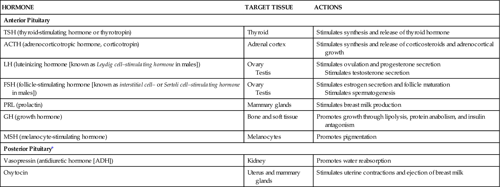
*These hormones are synthesized in the hypothalamus and are stored in the posterior pituitary gland. They are transported from the hypothalamus down the hypothalamic stalk to the posterior pituitary while bound to proteins known as neurophysins.
The hormones of the posterior pituitary, vasopressin (antidiuretic hormone [ADH]) and oxytocin, are produced in the hypothalamus and sent through the nerve tracts that connect the hypothalamus with the posterior pituitary. These hormones are stored in the nerve endings of the posterior pituitary and are released into the blood when needed.
Other factors can affect the release of hormones from the pituitary gland. Drugs, diet, lifestyle, and pathologic conditions can increase or decrease pituitary hormone secretion.
Gonads
The gonads are the male and female reproductive endocrine glands. Male gonads are the testes, and female gonads are the ovaries. Function of these glands begins at puberty.
During puberty in the male, the increased secretion of gonadotropins (luteinizing hormone [LH] and follicle-stimulating hormone [FSH]) from the anterior pituitary gland stimulates maturation of the testes, production of testosterone, and maturation of the external genitalia. During puberty in the female, increased secretion of the same gonadotropins stimulates ovarian maturation, estrogen production, ovulation, and maturation of the external genitalia. The function of the testes and ovaries is detailed in Chapter 72.
Adrenal Glands
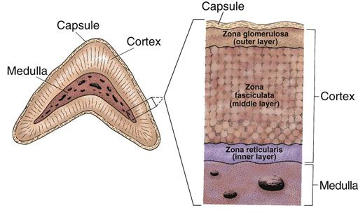
The adrenal glands are vascular, tent-shaped organs on the top of each kidney that have an outer cortex and an inner medulla (see Fig. 64-1). The adrenal hormones have effects throughout the body.
Adrenal Cortex
The adrenal cortex makes up about 90% of the adrenal gland and has cells divided into three layers (Fig. 64-5). Mineralocorticoids are the hormones produced in the zona glomerulosa and help control the body’s sodium and potassium content. Glucocorticoids, androgens, and estrogens are produced in the zona fasciculata and zona reticularis. The hormones secreted by the cortex are often called adrenal steroids or corticosteroids.
Mineralocorticoids are produced and secreted by the adrenal cortex to help control body fluids and electrolytes. Aldosterone is the major mineralocorticoid and maintains extracellular fluid volume. It promotes sodium and water reabsorption and potassium excretion in the kidney tubules. Aldosterone secretion is regulated by the renin-angiotensin system, serum potassium ion concentration, and adrenocorticotropic hormone (ACTH).
Renin is produced by specialized cells of the renal afferent arterioles. Its release is triggered by a decrease in extracellular fluid volume from blood loss, sodium loss, or posture changes. Renin converts renin substrate (formerly called angiotensinogen), a plasma protein, to angiotensin I. Angiotensin I is converted by a converting enzyme to form angiotensin II, the active form of angiotensin. In turn, angiotensin II stimulates the secretion of aldosterone. Chapter 13 (see Fig. 13-6) further explains the renin-angiotensin system. Aldosterone causes the kidney to reabsorb sodium and water to bring the plasma volume and osmolarity back to normal.
Serum potassium level also controls aldosterone secretion. The adrenal cortex secretes aldosterone when the serum potassium level increases above normal by as little as 0.1 mEq/L. Aldosterone then enhances kidney excretion of potassium to reduce the blood level back to normal.
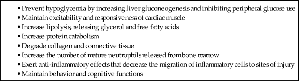
Glucocorticoids are produced by the adrenal cortex and are essential for life. The main glucocorticoid produced by the adrenal cortex is cortisol. Cortisol affects:
Cortisol also influences other important body processes. For example, it must be present for catecholamine action and maintaining the normal excitability of the heart muscle cells. Glucocorticoid functions are listed in Table 64-3.
TABLE 64-3
FUNCTIONS OF GLUCOCORTICOID HORMONES
Glucocorticoid release is regulated directly by the anterior pituitary hormone ACTH and indirectly by the hypothalamic corticotropin-releasing hormone (CRH). The release of CRH and ACTH is affected by the serum level of free cortisol, the normal sleep-wake cycle, and stress.
As described earlier and shown in Fig. 64-3, when blood cortisol levels are low, the hypothalamus secretes CRH, which triggers the pituitary to release ACTH. Then ACTH triggers the adrenal cortex to secrete cortisol. Adequate or elevated blood levels of cortisol inhibit the release of CRH and ACTH. This inhibitory effect is an example of a negative feedback system.
Glucocorticoid release peaks in the morning and reaches its lowest level 12 hours after each peak. Emotional, chemical, or physical stress increases the release of glucocorticoids.
Sex hormones (androgens and estrogens) are secreted in low levels by the adrenal cortex in both genders. Adrenal secretion of these hormones is usually not significant because the gonads (testes and ovaries) secrete much larger amounts of androgens and estrogens. In women, however, the adrenal gland is the major source of androgens. Women who have adrenal insufficiency or who have had surgical removal of the adrenals may need a small amount of testosterone replacement.
Adrenal Medulla
The adrenal medulla is a sympathetic nerve ganglion that has secretory cells. Stimulation of the sympathetic nervous system causes the release of adrenal medullary hormones, the catecholamines (which include epinephrine and norepinephrine). These hormones travel to all areas of the body through the blood and exert their effects on target cells. The adrenal medullary hormones are not essential for life because they also are secreted by other body tissues, but they do play a role in the physiologic stress response.
The adrenal medulla secretes about 15% norepinephrine (NE) and 85% epinephrine. Hormone effects vary with the specific receptor in the cell membranes of the target tissue.
These receptors are of two types: alpha adrenergic and beta adrenergic. Both types of receptors are further classified as alpha1 and alpha2 receptors and beta1, beta2, and beta3 receptors. NE acts mainly on alpha-adrenergic receptors, and epinephrine acts mainly on beta-adrenergic receptors.
Catecholamines exert their actions on many target organs (Table 64-4). Activation of the sympathetic nervous system, which then releases adrenal medullary catecholamines, is an important part of the body’s response to stress. Catecholamines are secreted in small amounts at all times to maintain homeostasis. Stress triggers increased secretion of these hormones. This sympathetic activation results in the “fight-or-flight” response, a state of heightened physical and emotional awareness.
TABLE 64-4
| ORGAN OR TISSUE | RECEPTORS | EFFECTS |
| Heart | Beta1 | Increased heart rate |
| Increased contractility | ||
| Blood vessels | Alpha | Vasoconstriction |
| Beta2 | Vasodilation | |
| Gastrointestinal tract | Alpha | Increased sphincter tone |
| Beta | Decreased motility | |
| Kidneys | Beta2 | Increased renin release |
| Bronchioles | Beta2 | Relaxation; dilation |
| Bladder | Alpha | Sphincter contractions |
| Beta2 | Relaxation of detrusor muscle | |
| Skin | Alpha | Increased sweating |
| Fat cells | Beta | Increased lipolysis |
| Liver | Alpha | Increased gluconeogenesis and glycogenolysis |
| Pancreas | Alpha | Decreased glucagon and insulin release |
| Beta | Increased glucagon and insulin release | |
| Eyes | Alpha | Dilation of pupils |
Stay updated, free articles. Join our Telegram channel

Full access? Get Clinical Tree


