M. Linda Workman
Assessment and Care of Patients with Fluid and Electrolyte Imbalances
Learning Outcomes
Safe and Effective Care Environment
Health Promotion and Maintenance
Physiological Integrity
7 Explain the relationship between weight gain or loss and fluid imbalances.
12 Prioritize interventions for patients who have dehydration or fluid overload.
13 Prioritize interventions for patients who have specific electrolyte imbalances.

http://evolve.elsevier.com/Iggy/
Answer Key for NCLEX Examination Challenges and Decision-Making Challenges
Audio Glossary
Concept Map Creator
Fluid and Electrolyte Tutorial
Key Points
Review Questions for the NCLEX® Examination
Homeostasis
Proper functioning of all body systems requires a balance of fluids and electrolytes. The body works best when these substances are kept within a narrow range of normal. For example, no body system works well if 2 liters of blood volume are gained or lost. To keep conditions as close to normal as possible (a situation called homeostasis), the body has many control actions (homeostatic mechanisms) to prevent dangerous changes.
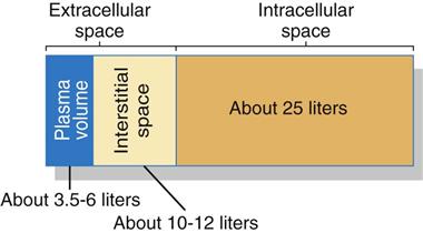
An important area for homeostasis is maintaining the body’s normal fluid volume and composition. Water is the most common substance in the body, making up about 55% to 60% of total body weight for healthy younger adults and 50% to 55% of total body weight for healthy older adults. This water (fluid) is divided into two main spaces or compartments—the fluid outside the cells, which is the extracellular fluid (ECF); and the fluid inside the cells, which is the intracellular fluid (ICF). The ECF space contains about one third (about 15 L) of the total body water. The ECF includes interstitial fluid (fluid between cells, sometimes called the “third space”); blood, lymph, bone, and connective tissue water; and the transcellular fluids. Transcellular fluids are the fluids in special body spaces and include cerebrospinal fluid, synovial fluid, peritoneal fluid, and pleural fluid. ICF contains the remaining two thirds (about 25 L) of total body water. Fig. 13-1 shows the normal distribution of total body water.
Water is needed to deliver dissolved nutrients, electrolytes, and other substances to all organs, tissues, and cells. In the healthy adult, the volume of water in the fluid compartments remains within the normal range although the water moves constantly between compartments. Changes in either the amount of water or the amount of electrolytes in body fluids can affect the functioning of all cells, tissues, and organs. For proper function, the volume of all body fluids and the types and amount of dissolved substances must be carefully balanced.
Anatomy and Physiology Review
Physiologic Influences on Fluid and Electrolyte Balance
Body fluids are composed of water and particles dissolved or suspended in water. The solvent is the water portion of fluids. Solutes are the particles dissolved or suspended in the water. Solutes vary in type and amount from one fluid space to another. When solutes express an overall electrical charge, they are known as electrolytes. Body function depends on keeping the correct balance of fluid and electrolytes within each body fluid space.
Three processes are important to control normal fluid and electrolyte balance, and they work together to maintain balance so the internal environment remains stable even when the external environment changes. These processes are filtration, diffusion, and osmosis. They determine how, when, and where fluids and particles move across cell membranes.
Filtration
Physiologic Action
Filtration is the movement of fluid through a cell or blood vessel membrane because of hydrostatic pressure differences on both sides of the membrane. Filtration occurs because of differences in water volume pressing against confining walls.
Fluid weight in a confined space is related to the amount of fluid present in that area. Water molecules in a confined space constantly press outward against the confining walls. This pressing of water molecules is hydrostatic pressure. It is a “water-pushing” pressure, because it is the force that pushes water outward from a confined space through a membrane (Fig. 13-2).
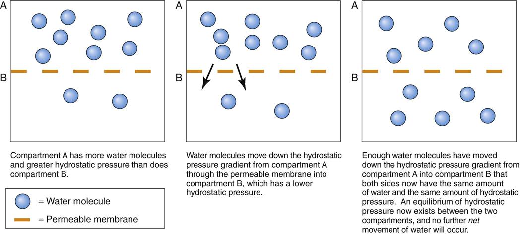
The amount of water in any body fluid space determines the hydrostatic pressure of that space. Blood, a fluid that is “thicker” than water (more viscous), is confined within the blood vessels. Blood has hydrostatic pressure because of its weight and volume. An additional factor that affects blood hydrostatic pressure in arteries is the pumping action of the heart. This action has little impact on the hydrostatic pressure in the capillaries and veins.
The hydrostatic pressures of two fluid spaces can be compared whenever a porous (permeable) membrane separates the two spaces. If the hydrostatic pressure is the same in both fluid spaces, there is no pressure difference between the two spaces and the hydrostatic pressure is now at equilibrium. If the hydrostatic pressure is not the same in both spaces, disequilibrium exists. This means that the two spaces have a graded difference (gradient) for hydrostatic pressure: one space has a higher hydrostatic pressure than the other. The human body constantly seeks equilibrium. When a gradient exists, water movement (filtration) occurs until the hydrostatic pressure is the same in both spaces (see Fig. 13-2).
When nothing stops it, water moves through the membrane (filters) from the space with higher hydrostatic pressure to the space with lower pressure. This filtration continues only as long as the hydrostatic pressure gradient exists. Equilibrium is reached when enough fluid leaves one space and enters the other space to make the hydrostatic pressure in both spaces equal. At this point, water molecules may be exchanged evenly between two spaces in equilibrium but no net further filtration of fluid occurs. Neither space gains or loses water molecules, and the hydrostatic pressure in both spaces remains the same.
Clinical Significance
Blood pressure is an example of a hydrostatic filtering force. It moves whole blood from the heart to capillaries where filtration can occur to exchange water, nutrients, and waste products between the blood and the tissues. One factor that determines whether fluid leaves the blood vessels and enters the tissue spaces (interstitial fluid) is the difference between the hydrostatic pressure of capillary blood and that of the interstitial fluid.
Capillaries are only one cell layer thick, making a thin “wall” to hold blood in the capillaries. Large spaces (pores) in the capillary membrane help water filter freely when a hydrostatic pressure gradient is present (Fig. 13-3).
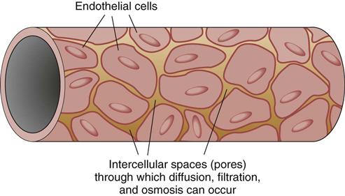
Edema (tissue swelling with fluid collection) develops with changes in normal hydrostatic pressure differences, such as in patients with right-sided heart failure. In this condition, the volume of blood in the right side of the heart increases greatly because the right ventricle is too weak to pump blood efficiently into the pulmonary blood vessels. As blood backs up into the venous system, venous hydrostatic pressure rises, which then causes capillary hydrostatic pressure to rise until it is higher than the hydrostatic pressure in the interstitial space. As a result, excess filtration of fluid from the capillaries into the interstitial tissue space occurs, forming visible edema.
Diffusion
Physiologic Action
Diffusion is the free movement of particles (solute) across a permeable membrane from an area of higher concentration to an area of lower concentration (down a concentration gradient). This action controls the movement of solute particles in solution across various body membranes.
The diffusion of particles into and out from cells and fluid spaces occurs using the energy generated by the vibration of single molecules. This motion is totally random, allowing molecules to bump into each other within a confined space. Each collision increases the speed of particle movement.
As a result of the collisions, molecules in a solution spread out evenly through the available space. They move from an area of higher amounts (concentration) of molecules to an area of lower amounts until an equal amount is present in all areas. The number of collisions is related to the amount of molecules in a confined space. Fluid spaces with many particles have more collisions and faster particle movement than spaces with fewer particles.
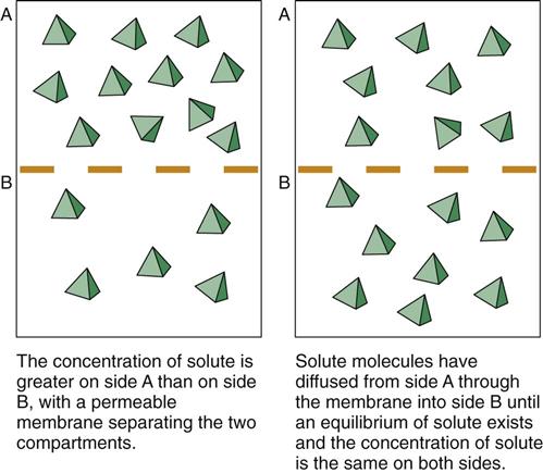
A concentration gradient exists when two fluid spaces have different amounts of the same type of particles. Particle collisions cause them to move down the concentration gradient. Any membrane that separates two spaces is struck repeatedly by particles. When the particle strikes a pore in the membrane that is large enough for it to pass through, diffusion occurs (Fig. 13-4). The chance of any single particle hitting the membrane and going through a pore is much greater on the side of the membrane with a higher solute particle concentration.
The speed of diffusion is related to the difference in amount of particles (concentration gradient) between the two sides of the membrane. The degree of difference is the steepness of the gradient: the larger the concentration difference between the two sides, the steeper the gradient. Diffusion is more rapid when the gradient is steeper (just as a ball rolls downhill more rapidly when the hill is steep than when the hill is nearly flat). Particles move from the fluid space with a higher concentration of solute particles to the fluid space with a lower concentration of solute particles.
Diffusion of particles continues as long as a concentration gradient exists between the two sides of the membrane. When the concentration of solute particles is the same on both sides of the membrane, the particles are in equilibrium and only an equal exchange of particles continues.
Clinical Significance
Diffusion is important in the transport of most electrolytes and other particles through cell membranes. Unlike capillary membranes, which permit the diffusion of most small-size particles down a gradient, cell membranes are selective in which particles can diffuse. They permit diffusion of some particles but not others. Some particles cannot move across a cell membrane, even when a steep “downhill” gradient exists, because the membrane is impermeable (not open) to that particle. For these particles, the concentration gradient is maintained across the membrane.
Impermeability and special transport systems cause differences in the amounts of specific particles from one fluid space to another. For example, usually the fluid outside the cell (the extracellular fluid [ECF]) has ten times more sodium ions than the fluid inside the cell (the intracellular fluid [ICF]). This extreme difference is caused by cell membrane impermeability to sodium and by special “sodium pumps” that move any extra sodium present inside the cell out of the cell “uphill” against its concentration gradient and back into the ECF.
In some instances, diffusion cannot occur without help, even down steep concentration gradients, because of selective membrane permeability. One example is glucose. Even though the amount of glucose may be much higher in the ECF than in the ICF (creating a steep gradient for glucose), glucose cannot cross most cell membranes without the help of insulin. When insulin is present, it binds to insulin receptors on cell membranes, which then makes the membranes much more permeable to glucose. As a result, glucose can cross the cell membrane down its concentration gradient until equilibrium of glucose concentration is achieved.
Diffusion across a cell membrane that requires the assistance of a membrane-altering system (e.g., insulin) is called facilitated diffusion or facilitated transport. This type of movement is still a form of diffusion.
Osmosis
Physiologic Action
Osmosis is the movement of water only through a selectively permeable (semipermeable) membrane. For osmosis to occur, a membrane must separate two fluid spaces and one space must have particles that cannot move through the membrane. (The membrane is impermeable to this particle.) Some of the reasons a membrane may be impermeable to a particle are that the particle is too large or that it expresses an electrical charge. A concentration gradient of this particle must also exist. Because the membrane is impermeable to these particles, they cannot cross the membrane but water molecules can. (Usually water can always move through a cell membrane.)
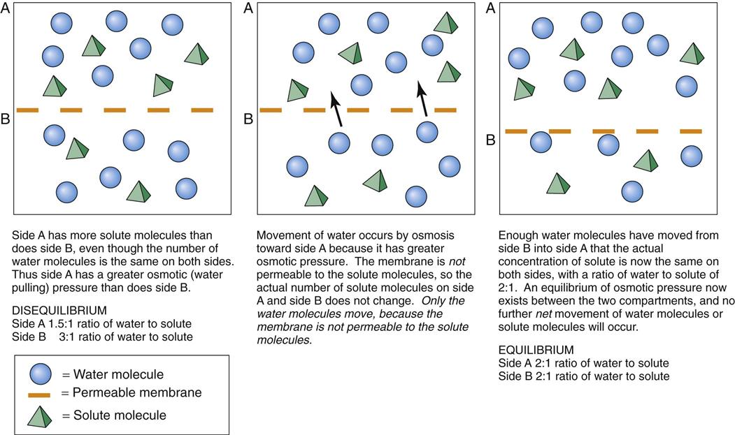
For the fluid spaces to have equal concentrations of the particle, the water molecules move down their concentration gradient from the side with the higher concentration of water molecules (and thus a lower concentration of particles along with a greater hydrostatic pressure) to the side with the lower concentration of water molecules (and a higher concentration of particles along with a lower hydrostatic pressure). This movement continues until both spaces contain the same proportions of particles to water. Dilute (less concentrated) fluid has fewer particles and more water molecules than the more concentrated fluid. Thus water moves by osmosis down its hydrostatic pressure gradient from the dilute fluid to the more concentrated fluid until a concentration equilibrium occurs (Fig. 13-5).
At this point, the concentrations of particles in the fluid spaces on both sides of the membrane are equal even though the total amounts of particles and volumes of water are different. The concentration equilibrium occurs by the movement of water molecules rather than the movement of solute particles.
Particle concentration in body fluids is the major factor that determines whether and how fast osmosis and diffusion occur. This concentration is expressed in milliequivalents per liter (mEq/L), millimoles per liter (mmol/L), and milliosmoles per liter (mOsm/L). Osmolarity is the number of milliosmoles in a liter of solution; osmolality is the number of milliosmoles in a kilogram of solution. The normal osmolarity value for plasma and other body fluids ranges from 270 to about 300 mOsm/L. The body functions best when the osmolarity of the fluids in all body fluid spaces is close to 300 mOsm/L. When all body fluids have this particle concentration, the body fluids are isosmotic to each other. Another term with the same meaning is isotonic (also called normotonic).
Fluids with osmolarities greater than 300 mOsm/L are hyperosmotic, or hypertonic, compared with isosmotic fluids. These fluids have a greater osmotic pressure than do isosmotic fluids and tend to pull water from the isosmotic fluid space into the hyperosmotic fluid space until an osmotic balance occurs. If a hyperosmotic IV solution (e.g., 2% saline) were infused into a patient with normal ECF osmolarity, the infusing fluid would make the person’s blood hyperosmotic. To balance this situation, the interstitial fluid would be pulled into the circulation in an attempt to dilute the blood osmolarity back to normal. As a result, the interstitial volume would shrink and the plasma volume would expand.
Fluids with osmolarities of less than 270 mOsm/L are hypo-osmotic, or hypotonic, compared with isosmotic fluids. Hypo-osmolar fluids have a lower osmotic pressure than isosmotic fluids, and water is pulled from the hypo-osmotic fluid space into the isosmotic fluid space.
Solubility is how well a particle type dissolves in water. Fluids that have particles with greater solubility have higher osmotic pressures than fluids with insoluble particles.
Amount of membrane available for osmosis affects the speed of osmosis. More membrane increases the chances that water molecules will hit the membrane at a point where penetration is possible and thus increases the speed of osmosis.
Clinical Significance
Osmosis and filtration act together at the capillary membrane to maintain both extracellular fluid (ECF) and intracellular fluid (ICF) volumes within their normal ranges. The thirst mechanism is an example of how osmosis helps maintain homeostasis. The feeling of thirst is caused by the activation of cells in the brain that respond to changes in ECF osmolarity. These cells are so sensitive to changes in ECF osmolarity that they are called osmoreceptors. When a person loses body water but most of the particles remain, such as through excessive sweating, ECF volume is decreased and osmolarity is increased (a hypertonic condition exists). The cells in the thirst center shrink as water moves from the cells into the hypertonic ECF. The shrinking of these cells triggers a person’s awareness of thirst and increases the urge to drink. Drinking replaces the amount of water lost through sweating and dilutes the ECF osmolarity, restoring it to normal. It is important to remember that the thirst mechanism is less sensitive in older adults, making them more at risk for dehydration.
Fluid Balance
Body Fluids
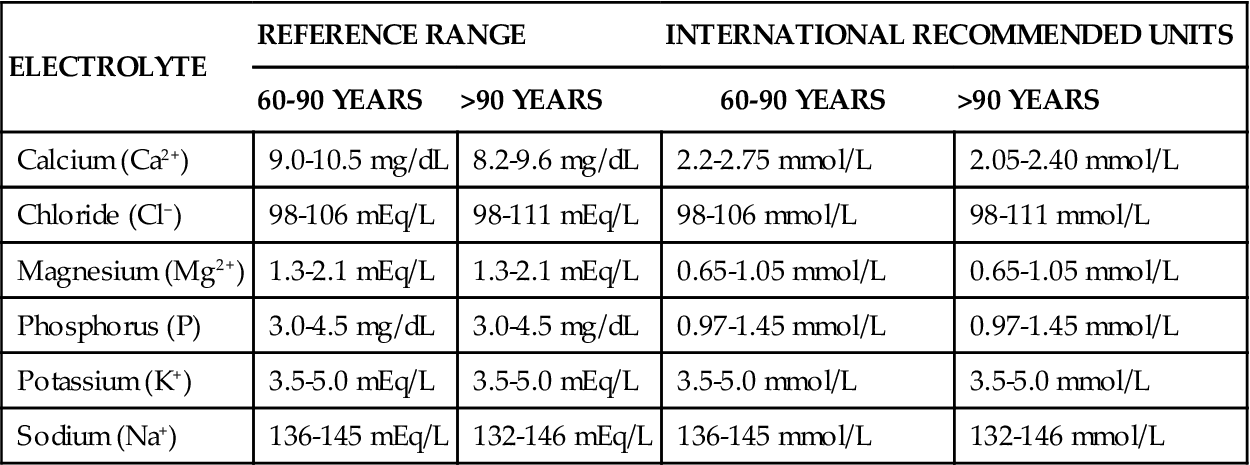
Fluid balance is closely linked to and affected by electrolyte concentrations. Table 13-1 lists the normal ranges of the major serum electrolytes. Chart 13-1 lists the normal electrolyte values for people older than 60 years.
TABLE 13-1
MAJOR SERUM ELECTROLYTE CONCENTRATIONS AND SIGNIFICANCE OF ABNORMAL VALUES

Data from Pagana, K., & Pagana, T. (2010). Mosby’s manual of diagnostic and laboratory tests (4th ed.). St. Louis: Mosby.
A person’s age, gender, and amount of fat affect the amount and distribution of body fluids. An older adult has less total body water than a younger adult. Chart 13-2 discusses age-related changes in fluid balance. An obese person has less total water than a lean person of the same weight because fat cells contain almost no water.
Body fluids are constantly filtered and replaced as fluid balance is maintained through intake and output. The total amount of water within each fluid space is stable, but individual water molecules move continually among all spaces. As a result, water in all spaces is exchanged continually while maintaining constant fluid volume.
Fluid intake is regulated through the thirst drive. Fluids enter the body mainly as liquids (Table 13-2). Solid foods also contain up to 85% water, allowing some fluid to enter the body with ingested solid foods.
TABLE 13-2
ROUTES OF FLUID INGESTION AND EXCRETION
| INTAKE | OUTPUT |
| Measurable | |
| Oral fluids | Urine |
| Parenteral fluids | Emesis |
| Enemas* | Feces |
| Irrigation fluids* | Drainage from body cavities |
| Not Measurable | |
| Solid foods | Perspiration |
| Metabolism | Vaporization through the lungs |
*Measured by subtracting the amount returned from the amount instilled.
A rising blood osmolarity or a decreasing blood volume triggers the sensation of thirst. Sensations such as mouth dryness or the thought that a person has not had a drink recently also triggers the thirst drive. An adult drinks an average of 1500 mL of fluid per day and ingests an additional 800 mL of fluid from food.
Fluid loss occurs through several routes (see Table 13-2). Of all the water loss pathways, the kidney is the most important and the most sensitive. Water loss by the kidney is regulated and is adjustable. The volume of urine excreted daily varies depending on the amount of fluid taken in and the body’s need to conserve fluids.
The minimum amount of urine per day needed to excrete toxic waste products is 400 to 600 mL. This minimum volume is called the obligatory urine output. If the 24-hour urine output falls below the obligatory output amount, wastes are retained and can cause lethal electrolyte imbalances, acidosis, and a toxic buildup of nitrogen.
The ability of the kidneys to make either concentrated or very dilute urine helps maintain fluid balance. The kidney works with various hormones to maintain fluid balance when extracellular fluid concentrations, volumes, or pressures change.
Other normal water loss occurs through the skin, the lungs, and the intestinal tract. Water losses also can result from salivation, drainage from fistulas and drains, and GI suction.
Water loss from the skin, lungs, and stool is called insensible water loss, because the body has no mechanisms to control this loss. The amount lost can be significant. In a healthy adult, insensible water loss is about 500 to 1000 mL/day. This loss increases greatly during thyroid crisis, trauma, burns, states of extreme stress, and fever. (For every Celsius-degree increase in body temperature, insensible water loss increases by 10% to 25%.) Insensible water loss also increases when the environment is hot and dry. Patients at risk for increased insensible water loss include those being mechanically ventilated and those with rapid respirations (tachypnea). Insensible water loss, if not balanced by intake, can lead to severe dehydration and electrolyte imbalances.
Loss by sweating is variable and can reach a maximum rate of about 2 L/hr. Sweating is influenced by the autonomic nervous system, body temperature, and blood flow in the skin.
Water loss through stool is normally minimal. However, this loss can increase greatly with severe diarrhea or excessive fistula drainage.
Hormonal Regulation of Fluid Balance
The endocrine system helps control fluid and electrolyte balance. Three hormones that help control these critical balances are aldosterone, antidiuretic hormone (ADH), and natriuretic peptide (NP).
Aldosterone is a hormone secreted by the adrenal cortex whenever sodium levels in the extracellular fluid (ECF) are decreased. Aldosterone prevents both water and sodium loss. When aldosterone is secreted, it acts on the kidney nephrons, triggering them to reabsorb sodium and water from the urine back into the blood. This action increases blood osmolarity and blood volume. Aldosterone prevents excessive kidney excretion of sodium. It also helps prevent blood potassium levels from becoming too high.
Antidiuretic hormone (ADH), or vasopressin, is produced in the brain and stored in the posterior pituitary gland. ADH release from the posterior pituitary gland is controlled by the hypothalamus in response to changes in blood osmolarity. The hypothalamus contains specialized cells (osmoreceptors) that are sensitive to changes in blood osmolarity. Increased blood osmolarity, especially an increase in the level of plasma sodium, results in a slight shrinkage of these cells and triggers ADH release from the posterior pituitary gland.
ADH acts directly on kidney tubules and collecting ducts, making them more permeable to water. As a result, more water is reabsorbed by these tubules and returned to the blood, decreasing blood osmolarity by making it more dilute. When blood osmolarity decreases, especially when the plasma sodium level is below normal, the osmoreceptors swell slightly and inhibit ADH release. Less water is then reabsorbed, and more is lost from the body in the urine. As a result, the amount of water in the extracellular fluid (ECF) decreases, bringing osmolarity up to normal.
Natriuretic peptides (NPs) are hormones secreted by special cells that line the atria of the heart (atrial natriuretic peptide [ANP]) and the ventricles of the heart. (The peptide secreted by the heart ventricular cells is known as brain natriuretic peptide [BNP] because it was first discovered in the brain.) These peptides are secreted in response to increased blood volume and blood pressure, which stretch the heart tissue. NP binds to receptors in the nephrons, creating effects that are opposite of aldosterone. Kidney reabsorption of sodium is inhibited at the same time that glomerular filtration is increased, causing increased urine output. The outcome is decreased circulating blood volume and decreased blood osmolarity.
Significance of Fluid Balance
The Renin-Angiotensin II Pathway
The human body requires a balance of body fluids, electrolytes, and acids and bases for best function. The most important fluids to keep in balance are the blood volume (plasma volume) and the fluid inside the cells (intracellular fluid). Of these two, the most critical fluid balance to prevent death is maintaining blood volume at a sufficient level for blood pressure to remain high enough to ensure adequate perfusion and oxygenation of all organs and tissues. Balance of both water and electrolytes is needed for this very vital function.
Although having an excess of blood volume can be a problem, more conditions have the potential to lower blood volume and blood pressure to the point that tissues are not perfused well and do not receive sufficient oxygen to prevent cell death. Because low blood volume and low blood pressure can rapidly lead to death, the body has many specific actions (mechanisms) that guard against excessive fluid loss from the plasma volume. These actions involve specific hormone levels, kidney function, and blood vessel responses to change how water and sodium are handled to maintain blood pressure.
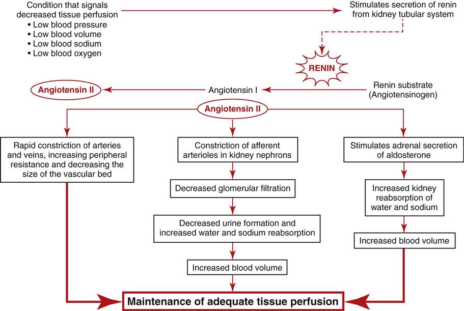
Because the kidney is a major regulator of water and sodium balance to maintain blood pressure and perfusion to all tissues and organs, the kidneys monitor blood pressure, blood volume, blood oxygen levels, and blood osmolarity (related to sodium concentration). When the kidneys sense that any one of these parameters is getting low, they begin to secrete a substance called renin that sets into motion a group of hormonal and blood vessel responses to ensure that blood pressure is raised back up to normal. Fig. 13-6 summarizes these responses.
So, the triggering event is any change in the blood that indicates to the kidney that tissue and organ perfusion are at risk. Low blood pressure is a triggering event because when it gets too low, blood cannot flow through vessels into tissues and organs. Low blood volume is a triggering event because it is directly related to blood pressure. Anything that reduces blood volume (e.g., dehydration, hemorrhage) below a critical level always lowers blood pressure. Low blood oxygen levels also are triggering events because with too little oxygen in the blood, even if the blood reaches the tissues and organs, it cannot supply the needed oxygen and the tissues and organs could die. A low blood sodium level also is a triggering event because sodium and water are closely linked. Where sodium goes, water follows. So, anything that causes the blood to have too little sodium prevents water from staying in the blood. The result is low blood volume with low blood pressure and poor tissue perfusion.
Once the kidneys sense that tissue and organ perfusion are at risk, special cells in the kidney tubule (in an area of the nephron known as the juxtaglomerular complex) begin to secrete renin into the bloodstream. Renin is an enzyme that activates some blood proteins, one of which is angiotensinogen. Activated angiotensinogen is angiotensin I, which is relatively weak and has little action. It is then acted on by another enzyme known as angiotensin-converting enzyme or ACE, which converts angiotensin I into its most active form, angiotensin II.
Angiotensin II starts several different activities that all work to increase blood volume and blood pressure. First, because angiotensin II is a powerful vasoconstrictor, it causes constriction of small arteries and veins throughout the body. This action increases peripheral resistance and reduces the size of the vascular bed, which raises blood pressure without adding more blood volume. At the same time, a second action of angiotensin II is that it constricts the size of the arterioles that feed the kidney nephrons. This action results in a lower glomerular filtration rate and a huge reduction of urine output. Decreasing urine output prevents further loss of water so that more is retained in the blood to help raise blood pressure. The last and slightly slower action of angiotensinogen II is to cause the adrenal glands to secrete the hormone aldosterone. Aldosterone is nicknamed the “water- and sodium-saving hormone” because it causes the kidneys to reabsorb water and sodium, preventing them from being excreted into the urine. This response allows more water and sodium to be returned to the blood, increasing blood pressure and blood volume. All of these actions help maintain perfusion to vital organs.
Clinical Application
The renin-angiotensin II pathway is greatly stimulated whenever the patient is in shock or is stressed to the point that the stress response is stimulated. This is why urine output is used as an indicator of perfusion adequacy after surgery or any time the patient has undergone an invasive procedure.
An additional application of this pathway is related to management of hypertension (high blood pressure). Patients who have hypertension are often asked to limit their intake of sodium. The reason for this is that a high sodium intake raises the blood level of sodium, causing more water to be retained in the blood volume and raising blood pressure. Drug therapy for hypertension management may include diuretic drugs that increase the excretion of sodium so that less is present in the blood, resulting in a lower blood volume. Another class of drugs often used to manage blood pressure is the “ACE inhibitors.” These drugs disrupt the renin-angiotensin II pathway by reducing the amount of angiotensin-converting enzyme (ACE) made so that less angiotensin II is present. With less angiotensin II, there is less vasoconstriction and reduced peripheral resistance, less aldosterone production, and greater excretion of water and sodium in the urine. All of these responses lead to decreased blood volume and blood pressure. One more class of drugs used to manage hypertension is the angiotensin receptor blockers (ARBs). These drugs disrupt the renin-angiotensin II pathway by blocking the receptors that bind with angiotensin II so that the tissues cannot respond to it and blood pressure is lowered.
Fluid Imbalances
All patients are at risk for some degree of fluid imbalance because many health problems can disrupt fluid intake or output. Fluid imbalances can occur in any setting.
Dehydration
Pathophysiology
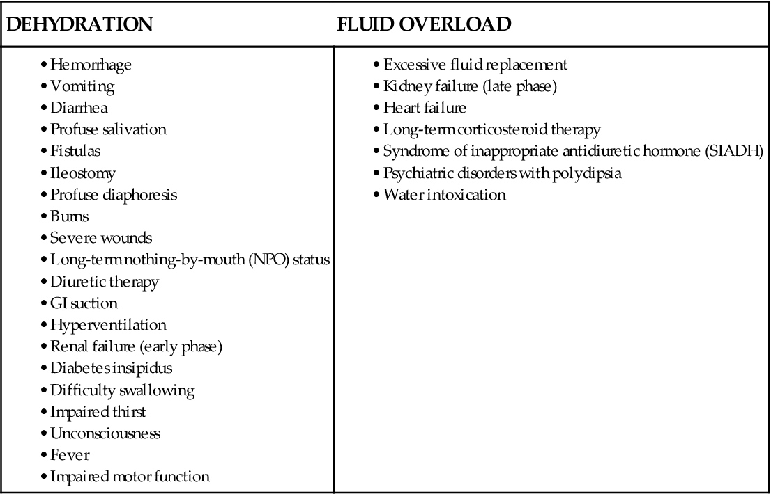
In dehydration, fluid intake or fluid retention is less than what is needed to meet the body’s fluid needs, resulting in a fluid volume deficit, especially a plasma volume deficit. It is a condition rather than a disease and can be caused by many factors (Table 13-3). Dehydration may be an actual decrease in total body water caused by either too little intake of fluid or too great a loss of fluid. It also can occur without an actual loss of total body water, such as when water shifts from the plasma into the interstitial space. This condition is called relative dehydration.
TABLE 13-3
COMMON CAUSES OF FLUID IMBALANCES
| DEHYDRATION | FLUID OVERLOAD |
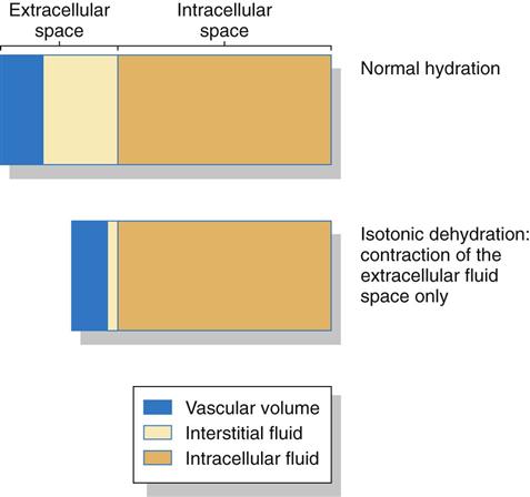
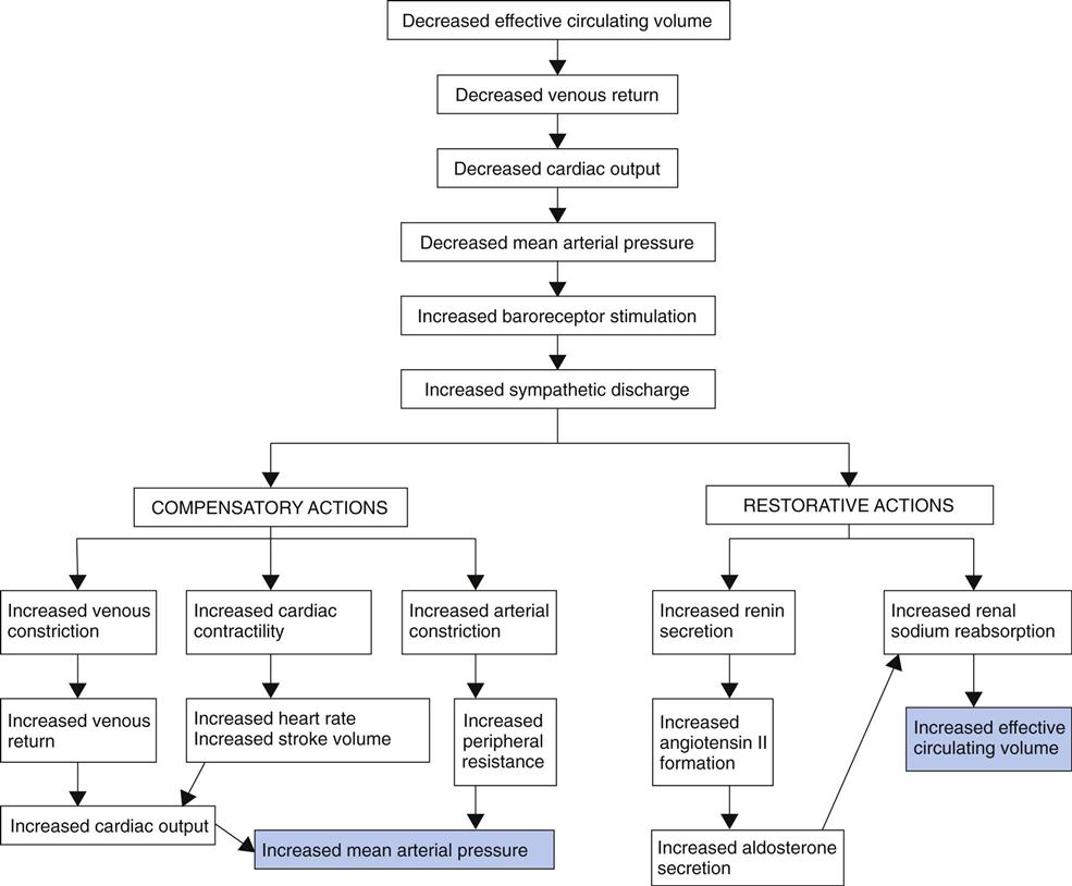
Dehydration may occur with just water loss or with water and electrolyte loss (isotonic dehydration). Isotonic dehydration is the most common type of fluid loss problem. Fluid is lost only from the extracellular fluid (ECF) space, including both the plasma and the interstitial spaces. There is no shift of fluids between spaces, so the intracellular fluid (ICF) volume remains normal (Fig. 13-7). Circulating blood volume is decreased (hypovolemia) and leads to inadequate tissue perfusion. The body’s defenses adapt (compensate) during dehydration to maintain adequate blood flow to vital organs in spite of hypovolemia (Fig. 13-8).
Health Promotion and Maintenance
Mild dehydration is very common among healthy adults and is corrected or prevented easily by matching fluid intake with fluid output. Problems occur when people perform heavy exercise, especially in warm environments, without taking the time to replace excessive fluid losses. Dry climates and higher altitudes also increase fluid loss through the respiratory tract. Teach all patients to drink more fluids, especially water, whenever they engage in heavy physical activity or live in dry climates or at higher altitudes. Beverages with caffeine can increase fluid loss, as can drinks containing alcohol; thus these beverages should not be used to prevent or treat dehydration.
Moderate to severe dehydration is more likely to occur in people who are unable to obtain fluids without help. Older adults living in long-term care facilities and those adults with cognitive or motor problems depend on others for hydration. Dehydration in this population can be prevented with careful hydration programs. These programs include routinely offering residents a choice of fluids every hour or two during the day and at any other logical opportunity, such as when administering medications.
Patient-Centered Collaborative Care
Assessment
History
One way of organizing history data to assess the patient’s fluid status is to use Gordon’s Functional Health Patterns (Gordon, 2010). The patterns that most affect fluid status are the Nutritional-Metabolic Pattern and the Elimination Pattern (Chart 13-3).
The nutritional history can reveal problems that affect fluid balance. Ask specific questions about food and liquid intake, and guide the patient in reporting accurately the amount of fluid ingested because he or she may not understand the connection between dietary intake and the onset of fluid imbalances. Also assess the types of fluids and foods ingested to determine amount and osmolarity. Many patients do not know that solid foods contain liquid. Solid foods such as ice cream, gelatin, and ices are liquids at body temperature, and these must be included when calculating fluid intake.
Collect specific information about output because exact intake and output volumes are important, as are serial daily weight measurements. If possible, weigh the patient directly rather than ask his or her weight because weight loss is an indication of dehydration. Because 1 L of water weighs 2.2 pounds (1 kg), changes in daily weights are the best indicators of fluid losses or gains. A weight change of 1 pound corresponds to a fluid volume change of about 500 mL.
Output includes losses not only as urine but also as sweat, diarrhea, and insensible loss during fevers. Ask specific questions about prescribed and over-the-counter drugs, and check the dosage, the length of time taken, and the patient’s adherence with the drug regimen.
Other important areas of the patient history include a sense of thirst or excessive drinking, exposure to hot environments, living at higher altitudes, and the presence of kidney or endocrine diseases. Assess the patient’s level of consciousness and mental status, because changes in mental status occur with fluid imbalance. In such cases, you may need to check the accuracy of information with family members. Ask the patient about changes in ring or shoe tightness. A sudden decrease in tightness may indicate dehydration.
Older adults often use diuretics and laxatives, which can disturb fluid balance. Misuse and overuse of these drugs can lead to dehydration and electrolyte imbalances. An important issue for many older adults is that they may depend on other people to provide assistance in meeting fluid needs.
Physical Assessment/Clinical Manifestations
Nearly all body systems are affected by dehydration to some degree. The most obvious changes occur in the cardiovascular and integumentary systems.
Cardiovascular changes are good indicators of hydration status because of the relationship between plasma fluid volume and blood pressure. Heart rate increases in an attempt to maintain blood pressure with less blood volume. Peripheral pulses are weak, difficult to find, and easily blocked with light pressure. The blood pressure also decreases, as does the pulse pressure, with a greater decrease in the systolic blood pressure. Hypotension is more severe with the patient in the standing position than in the sitting or lying position (orthostatic or postural hypotension). Because the blood pressure with the patient standing may be much lower than in other positions, first measure blood pressure with the patient lying down, then sitting, and finally standing. (These measures are also called “ortho checks” or “ortho changes.”) As the blood pressure decreases when changing position, the person may not have sufficient blood flow to the brain, causing the sensations of light-headedness and dizziness. This problem increases the risk for falling, especially among older adults.
Neck veins are normally distended when a patient is in the supine position, and hand veins are distended when lower than the level of the heart. Neck veins normally flatten when the patient moves to a sitting position. With dehydration, neck and hand veins are flat, even when the neck and hands are not raised above the level of the heart.
Respiratory changes include an increased rate because the decreased blood volume reduces perfusion and oxygenation. The increased respiratory rate is an attempt to maintain oxygen delivery when perfusion is decreased.
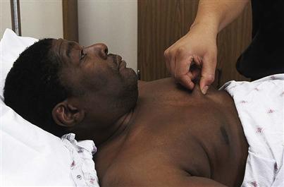
Skin changes can indicate dehydration. Assess the skin and mucous membranes for color, moisture, and turgor. In older patients, this information is less reliable because of poor skin turgor resulting from the loss of elastic tissue and increased skin dryness from the loss of tissue fluids with aging. Assess skin turgor by checking:
In generalized dehydration, skin turgor is poor, with the tent remaining for minutes after pinching the skin, and no skin depressions occur with gentle pressure. The skin is dry and scaly.
In dehydration, oral mucous membranes are not moist. They may be covered with a thick, sticky, pastelike coating and may have cracks and fissures. The surface of the tongue may have deep furrows. This manifestation may not be accurate for assessing dehydration in patients taking drugs that have the side effect of dry mouth.
Neurologic changes with dehydration include alterations of mental status and body temperature status because blood flow in the brain is reduced. Mental status changes, especially confusion, are more common among older adults and may be the first indication of a fluid balance problem. Check to determine whether the patient is alert and oriented. Chapter 43 provides more information about assessment of mental status.
The patient with dehydration often has a low-grade fever, and fever can also cause dehydration. A patient with a temperature higher than 102° F (39° C) for longer than 6 hours is especially at risk because the increased body temperature increases the rate at which fluids are lost. For every degree (Celsius) increase in body temperature above normal, a minimum of an additional 500 mL of body fluid is lost. The older adult begins to lose more body water at lower levels of fever.
Kidney changes in dehydration affect urine volume and composition. Monitor urine output, comparing total output with total fluid intake and daily weights. The urine may be concentrated, with a specific gravity greater than 1.030. The color is dark amber and has a strong odor. Urine output below 500 mL/day for any patient without kidney disease is cause for concern. Weigh the patient each day at the same time and on the same scale. When possible, have the patient wear the same amount and type of clothing for each weigh-in. Any weight loss over a half pound per day is fluid loss.
Laboratory Assessment
No single laboratory test result confirms or rules out dehydration. Instead, dehydration is determined by laboratory findings along with clinical manifestations (see Table 13-1). Usually, laboratory findings with dehydration show elevated levels of hemoglobin, hematocrit, serum osmolarity, glucose, protein, blood urea nitrogen, and various electrolytes because more water is lost and other substances remain, increasing the osmolarity or concentration of the blood (hemoconcentration). Hemoconcentration is not present when dehydration is caused by hemorrhage, because loss of all blood and plasma products occurs together.
Interventions
The focus of management for the patient with dehydration is to prevent injury, prevent further fluid losses, and increase fluid compartment volumes to normal ranges. Nursing priorities include patient safety, fluid replacement, and drug therapy.
Patient safety issues and strategies are priorities of care before and during other therapies for dehydration. Monitor vital signs, especially heart rate and blood pressure. The patient with dehydration is at risk for falls because of the accompanying orthostatic hypotension, dysrhythmia, muscle weakness, and possible confusion. Assess his or her muscle strength, gait stability, and level of alertness. Instruct the patient to get up slowly from a lying or sitting position and to immediately sit down if he or she feels light-headed. Implement the falls precautions listed in Chart 13-4.
