Antiviral agents II: drugs for HIV infection and related opportunistic infections

CHAPTER 94


Antiviral agents II: drugs for HIV infection and related opportunistic infections


In this chapter we discuss drug therapy of infection with the human immunodeficiency virus (HIV), the microbe that causes acquired immunodeficiency syndrome (AIDS). HIV promotes immunodeficiency by killing CD4 T lymphocytes (CD4 T cells), which are key components of the immune system (see Chapter 67). As a result of HIV-induced immunodeficiency, patients are at risk of opportunistic infections and certain neoplasms.


It is important to appreciate that HIV infection is not synonymous with AIDS, which develops years after HIV infection is acquired. The definition of AIDS, established by the Centers for Disease Control and Prevention (CDC) in 1993, is a syndrome in which the individual is HIV positive and has either (1) CD4 T-cell counts below 200 cells/mL or (2) an AIDS-defining illness. Included in the CDC’s long list of AIDS-defining illnesses are Pneumocystis pneumonia, cytomegalovirus retinitis, disseminated histoplasmosis, tuberculosis, and Kaposi’s sarcoma.


Since being identified as a new disease in 1981, AIDS has become a global epidemic. In the United States, nearly 1.2 million people are now infected, about 50,000 more become infected each year, and nearly 600,000 have died since the epidemic began. Worldwide, an estimated 33 million people are now infected, and about 25 million have died, including 270,000 children in 2007 alone. However, there is good news: According to a United Nations report, released in 2011, the number of new HIV infections and HIV-related deaths is on the decline, due in large part to more widespread use of HIV drugs.


Therapy of HIV infection has made dramatic advances. Today, standard antiretroviral therapy (ART) consists of three or four drugs. These combinations, often referred to as HAART (for highly active antiretroviral therapy), can decrease plasma HIV to levels that are undetectable with current technology, and can thereby delay or reverse loss of immune function, decrease certain AIDS-related complications, preserve health, prolong life, and decrease HIV transmission. In the United States, ART has reduced AIDS-related deaths by 72%—from a peak of 50,000 in 1995 to 16,000 in 2008. However, these benefits have not come without a price: ART is expensive, poses a risk of long-term side effects and serious drug interactions, and must continue lifelong. Accordingly, if treatment is to succeed, patients must be highly motivated and well informed about all aspects of the treatment program. A strong support network is extremely valuable too.


ART cannot cure HIV infection. Although treatment can greatly reduce HIV levels—often rendering the virus undetectable—discontinuation has consistently been followed by a rebound in HIV replication. Because ART does not eliminate HIV, patients continue to be infectious and must be warned to avoid behaviors that can transmit the virus to others.


Understanding this chapter requires a basic understanding of the immune system. Accordingly, you may find it helpful to read Chapter 67 before proceeding.


Pathophysiology


Characteristics of HIV


HIV is a retrovirus. Like all other viruses, retroviruses lack the machinery needed for self-replication, and hence are obligate intracellular parasites. However, in contrast to other viruses, retroviruses have positive-sense, single-stranded RNA as their genetic material. Accordingly, in order to replicate, retroviruses must first transcribe their RNA into DNA. The enzyme employed for this process is viral RNA-dependent DNA polymerase, commonly known as reverse transcriptase. (The enzyme is called reverse transcriptase to distinguish it from DNA-dependent RNA polymerase, the host enzyme that transcribes DNA into RNA, which is the usual [“forward”] transcription process.) The name retrovirus is derived from the first two letters of reverse and transcriptase.


There are two types of HIV, referred to as HIV-1 and HIV-2. HIV-1 is found worldwide, whereas HIV-2 is found mainly in West Africa. Although HIV-1 and HIV-2 differ with respect to genetic makeup and antigenicity, they both cause similar disease syndromes. Not all drugs that are effective against HIV-1 are also effective against HIV-2.


Target cells

The principal cells attacked by HIV are CD4 T cells (helper T lymphocytes). As discussed in Chapter 67, these cells are essential components of the immune system. They are required for production of antibodies by B lymphocytes and for activation of cytolytic T lymphocytes. Accordingly, as HIV kills CD4 T cells, the immune system undergoes progressive decline. As a result, infected individuals become increasingly vulnerable to opportunistic infections, a major cause of death among people with AIDS. HIV targets CD4 T cells because the CD4 proteins on the surface of these cells provide points of attachment for HIV (see below). Without such a receptor, HIV would be unable to connect with and penetrate these cells. Once HIV has infected a CD4 T cell, the cell dies in about 1.25 days. It is important to appreciate that only a few percent of CD4 T cells circulate in the blood; the vast majority reside in lymph nodes and other lymphoid tissues.


In addition to infecting CD4 T cells, HIV infects macrophages and microglial cells (the central nervous system [CNS] counterparts of macrophages), both of which carry CD4 proteins. Since macrophages and microglial cells are resistant to destruction by HIV, they can survive despite being infected. As a result, they serve as a reservoir of HIV during chronic infection.


Structure of HIV

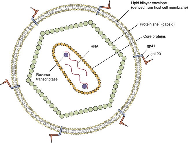
The structure of HIV is very simple. As shown in Figure 94–1, the HIV virion (ie, the entire virus particle) consists of nucleic acid (RNA) surrounded by core proteins, which in turn are surrounded by a capsid (protein shell), which in turn is surrounded by a lipid bilayer envelope (derived from the membrane of the host cell).


The central core contains two separate but identical single strands of RNA, each with its own molecule of reverse transcriptase attached. The RNA serves as the template for DNA synthesis.


The outer envelope of HIV contains glycoproteins that are needed for attachment to host cells. Each glycoprotein (gp) consists of two subunits, known as gp41 and gp120. The smaller protein (gp41) is embedded in the lipid bilayer of the viral envelope; the larger protein (gp120) is connected firmly to gp41. (The numbers 41 and 120 simply indicate the mass of these glycoproteins in thousands of daltons.)


Replication cycle of HIV

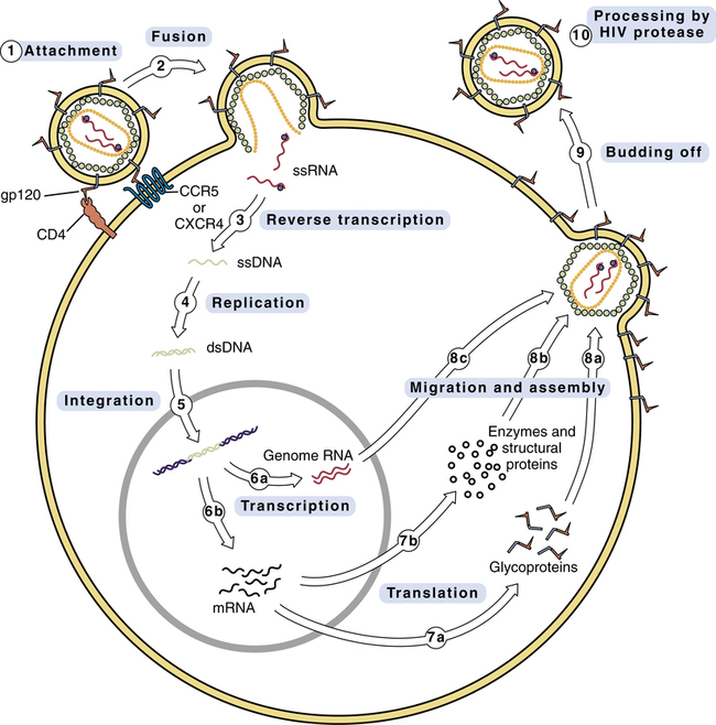
The replication cycle of HIV is depicted in Figure 94–2. The numbered steps below correspond to the numbers in the figure.


• Step 1—The cycle begins with attachment of HIV to the host cell. The primary connection takes place between gp120 on the HIV envelope and a CD4 protein on the host cell membrane. Other host proteins, known as co-receptors, act in concert with CD4 to tighten the bond with HIV. Two of these co-receptors—known as CCR5 and CXCR4—are of particular importance. One drug—maraviroc—blocks HIV entry by binding CCR5.


• Step 2—The lipid bilayer envelope of HIV fuses with the lipid bilayer of the host cell membrane. Fusion is followed by release of HIV RNA into the host cell. One drug—–enfuvirtide—–works by blocking the fusion process.


• Step 3—HIV RNA is transcribed into single-stranded DNA by HIV reverse transcriptase. Twelve antiretroviral drugs inhibit this enzyme.


• Step 4—Reverse transcriptase converts the single strand of HIV DNA into double-stranded HIV DNA.


• Step 5—Double-stranded HIV DNA becomes integrated into the host’s DNA, under the direction of a viral enzyme known (aptly) as integrase. One drug—raltegravir—inhibits this enzyme.


• Step 6—HIV DNA undergoes transcription into RNA. Some of the resulting RNA becomes the genome for daughter HIV virions (step 6a). The rest of the RNA is messenger RNA that codes for HIV proteins (step 6b).


• Step 7—Messenger RNA is translated into HIV glycoproteins (step 7a) and HIV enzymes and structural proteins (step 7b).


• Step 8—The components of HIV migrate to the cell surface and assemble into a new virus. Prior to assembly, HIV glycoproteins become incorporated into the host cell membrane (step 8a). In steps 8b and 8c, the other components of the virion migrate to the cell surface, where they undergo assembly into the new virus.


• Step 9—The newly formed virus buds off from the host cell. As indicated, the outer envelope of the virion is derived from the cell membrane of the host.


• Step 10—In this step, which occurs either during or immediately after budding off, HIV undergoes final maturation under the influence of protease, an enzyme that cleaves certain large polyproteins into their smaller, functional forms. If protease fails to cleave these proteins, HIV will remain immature and noninfectious. HIV protease is the target of several important drugs.


Replication rate

HIV replicates rapidly during all stages of the infection. During the initial phase of infection, replication is massive. Why? Because (1) the population of CD4 cells is still large, thereby providing a large viral breeding ground; and (2) the host has not yet mounted an immune response against HIV, hence replication can proceed unopposed. As a result of massive replication, plasma levels of HIV can exceed 10 million virions/mL. During this stage of high viral load, patients often experience an acute retroviral syndrome (see below).


Over the next few months, as the immune system begins to attack HIV, plasma levels of HIV undergo a sharp decline and then level off. A typical steady-state level is between 1000 and 100,000 virions/mL. Please note, however, that steady-state numbers can be deceptive. The plasma half-life of HIV is only 6 hours; that is, every 6 hours, half of the HIV virions in plasma are lost. Accordingly, in order to maintain the steady-state levels typically seen during chronic HIV infection, the actual rate of replication is between 1 and 10 billion virions/day. Despite this high rate of ongoing replication, infected persons typically remain asymptomatic for about 10 years, after which symptoms of advanced HIV disease appear.


Mutation and drug resistance

HIV mutates rapidly. Why? Because HIV reverse transcriptase is an error-prone enzyme. Hence, whenever it transcribes HIV RNA into single-stranded DNA and then into double-stranded DNA, there is a high probability of introducing base-pair errors. In fact, according to one estimate, up to 10 incorrect bases may be incorporated into HIV DNA during each round of replication. Because of these errors, HIV can rapidly mutate from a drug-sensitive form into a drug-resistant form. The probability of developing resistance in the individual patient is directly related to the total viral load. Hence, the more virions the patient harbors, the greater the likelihood that at least one will become resistant. To minimize the emergence of resistance, patients must be treated with a combination of antiretroviral drugs. This is the same strategy we employ to prevent emergence of resistance when treating tuberculosis (see Chapter 90).


Transmission of HIV


HIV is transmitted sexually and by other means. The virus is present in all body fluids of infected individuals. Transmission can be via intimate contact with semen, vaginal secretions, and blood. The disease can be transmitted by sexual contact, transfusion, sharing IV needles, and accidental needle sticks. In addition, it can be transmitted to the fetus by an infected mother, usually during the perinatal period. Initially, HIV infection was limited largely to homosexual males, injection-drug users, and hemophiliacs. However, the disease can now be found routinely in the population at large. The risk of acquiring HIV sexually can be reduced by male circumcision, limiting sexual partners, and use of condoms—as well as by complete sexual abstinence. In addition, acquisition can be prevented with drugs, as discussed below under Preventing HIV Infection with Drugs.


Clinical course of HIV infection


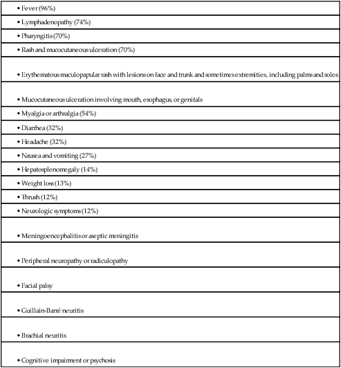
HIV infection follows a triphasic clinical course. During the initial phase, HIV undergoes massive replication, causing blood levels of HIV to rise very high. As a result, between 50% and 90% of patients experience a flu-like acute retroviral syndrome. Signs and symptoms include fever, lymphadenopathy, pharyngitis, rash, myalgia, and headache (Table 94–1). Soon, however, the immune system mounts a counterattack, causing HIV levels to fall. As a result, symptoms of the acute syndrome fade. Very often, the acute retroviral syndrome is perceived as influenza, and hence goes unrecognized for what it really is.


The middle phase of HIV infection is characterized by prolonged clinical latency. Blood levels of HIV remain relatively low, and most patients are asymptomatic. However, as noted above, HIV continues to replicate despite apparent dormancy. Because of persistent HIV replication, CD4 T cells undergo progressive decline. The average duration of clinical latency is 10 years.


During the late phase of HIV infection, CD4 T cells drop below a critical level (200 cells/mL), rendering the patient highly vulnerable to opportunistic infections and certain neoplasms (eg, Kaposi’s sarcoma). The late phase is when AIDS occurs.


Many patients with HIV infection experience neurologic complications. Both the peripheral and central nervous systems may be involved. Peripheral neuropathies affect 20% to 40% of patients and may develop at any time over the course of HIV infection. In contrast, CNS complications usually occur late in the disease. Symptoms of CNS injury include decreased cognition, reduced concentration, memory loss, mental slowness, and motor complaints (eg, ataxia, tremors). Neuronal injury may be the direct result of HIV infection, or may develop secondary to an opportunistic infection in the CNS.


Classification of antiretroviral drugs


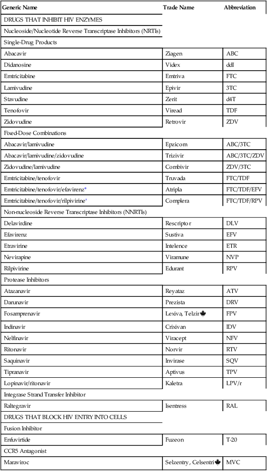
At this time, we have five types of antiretroviral drugs. Three types—reverse transcriptase inhibitors, integrase strand transfer inhibitors (INSTIs), and protease inhibitors (PIs)—inhibit enzymes required for HIV replication. The other two types—fusion inhibitors and chemokine receptor 5 (CCR5) antagonists—block viral entry into cells. As discussed below, the reverse transcriptase inhibitors are subdivided into two groups: nucleoside/nucleotide reverse transcriptase inhibitors (NRTIs), which are structural analogs of nucleosides or nucleotides, and (2) non-nucleoside reverse transcriptase inhibitors (NNRTIs). Drugs that belong to these groups are listed in Table 94–2. All NRTIs, NNRTIs, PIs, INSTIs, and CCR5 antagonists are administered orally, and one NRTI—zidovudine—may also be given IV. The one fusion inhibitor available—enfuvirtide [Fuzeon]—is administered subQ.


Nucleoside/nucleotide reverse transcriptase inhibitors


The nucleoside/nucleotide reverse transcriptase inhibitors (NRTIs) were the first drugs used against HIV infection, and remain mainstays of therapy today. In fact, these drugs constitute the backbone of all treatment regimens. As their name suggests, the NRTIs are chemical relatives of naturally occurring nucleosides or nucleotides, the building blocks of DNA. Antiretroviral effects derive from suppressing synthesis of viral DNA by reverse transcriptase. To be effective, all of the NRTIs must first undergo intracellular conversion to their active (triphosphate) forms. The NRTIs have few drug interactions, and most can be taken without regard to meals. Rarely, these agents cause a potentially fatal syndrome characterized by lactic acidosis and hepatomegaly with steatosis; pregnant women taking two NRTIs may be at increased risk. At this time, seven NRTIs are available. Major properties are summarized in Table 94–3.


TABLE 94–3 


Properties of Nucleoside/Nucleotide Reverse Transcriptase Inhibitors


  Abacavir (ABC) Didanosine (ddI) Emtricitabine (FTC) Lamivudine (3TC) Stavudine (d4T) Tenofovir (TDF) Zidovudine (ZDV)
Trade Name Ziagen Videx, Videx EC Emtriva Epivir Zerit Viread Retrovir
Formulations Tablets: 300 mg
PO soln: 20 mg/mL
EC capsules: 125, 200, 250, 400 mg
Buffered powder for PO soln: 100, 167, 250 mg
Capsules: 200 mg
PO soln: 10 mg/mL
Tablets: 150, 300 mg
PO soln: 10 mg/mL
Capsules: 15, 20, 30, 40 mg
PO soln: 1 mg/mL
Tablets: 300 mg Capsules: 100 mg
Tablets: 300 mg
PO soln: 10 mg/mL
IV soln: 10 mg/mL
Dosage 300 mg 2 times/day or 600 mg once/day EC capsules:
≥60 kg: 400 mg once/day (or 250 mg once/day with tenofovir)
<60 kg: 250 mg once/day (or 200 mg once/day with tenofovir)
PO solution:
≥60 kg: 200 mg 2 times/day
<60 kg: 125 mg 2 times/day
Capsules: 200 mg once/day
PO soln: 240 mg once/day
Adults: 150 mg 2 times/day or 300 mg once/day
Children: 4 mg/kg 2 times/day (max 150 mg 2 times/day)
>60 kg: 40 mg 2 times/day
<60 kg: 30 mg 2 times/day
Note: WHO recommends 30 mg 2 times/day regardless of weight
300 mg once/day 200 mg 3 times/day or 300 mg 2 times/day
Impact of Food Take without regard to meals—but alcohol increases levels by 41% Take 30 min before meals or 2 hr after Take without regard to meals Take without regard to meals Take without regard to meals Take without regard to meals Take without regard to meals
Bioavailability 83% 30%–40% 93% 86% 86% 39% (with food) 60%
Serum Half-life 1.5 hr 1.5 hr 10 hr 5–7 hr 1 hr 17 hr 1.1 hr
Intracellular Half-life 12–26 hr More than 20 hr More than 20 hr 18–22 hr 7.5 hr More than 60 hr 7 hr
Elimination Metabolized by alcohol dehydrogenase, then excreted in the urine Partial metabolism followed by renal excretion Renal excretion Renal excretion (unchanged) Partial metabolism followed by renal excretion Renal excretion Hepatic metabolism followed by renal excretion
Adverse Effects
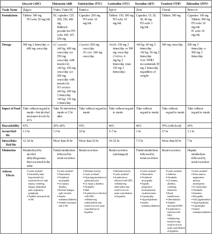


image


BMD = bone mineral density, EC = enteric coated, HBV = hepatitis B virus, MI = myocardial infarction, WHO = World Health Organization.


*Lactic acidosis with hepatic steatosis is a rare but potentially fatal toxicity associated with all NRTIs.


Adapted from Guidelines for the Use of Antiretroviral Agents in HIV-1–Infected Adults and Adolescents, prepared by the Panel on Clinical Practices for Treatment of HIV Infection, convened by the DHHS, as updated on October 14, 2011.


Zidovudine


Zidovudine [Retrovir] was the first NRTI available and will serve as our prototype for the group. The drug is an analog of thymidine, a naturally occurring nucleoside. When employed in combination with other antiretroviral drugs, zidovudine can decrease viral load, increase CD4 T-cell counts, delay onset of disease symptoms, and reduce symptom severity. The drug’s principal dose-limiting toxicities are severe anemia and neutropenia. Abbreviations for this agent are ZDV (for zidovudine) and AZT (for azidothymidine, its original name).


Therapeutic use

Zidovudine is used to treat infection with HIV-1. Because monotherapy with any antiretroviral drug can rapidly lead to resistance, zidovudine should always be combined with other antiretroviral agents. Zidovudine penetrates to the CNS better than most other antiretroviral drugs, and hence can be especially valuable for relieving cognitive symptoms. Zidovudine is also the drug of choice for preventing mother-to-infant HIV transmission during labor and delivery. The role of zidovudine and other agents in the management of HIV infection is discussed at length later in the chapter.


Adverse effects


Anemia and neutropenia from bone marrow suppression.

Severe anemia and neutropenia are the principal toxic effects. Multiple transfusions may be required. The risk of hematologic toxicity is increased by high-dose therapy, advanced HIV infection, deficiencies in vitamin B12 and folic acid, and concurrent use of drugs that are myelosuppressive, nephrotoxic, or directly toxic to circulating blood cells. Anemia and neutropenia generally resolve following zidovudine withdrawal.


Hematologic status (hemoglobin concentration and neutrophil counts) should be determined before treatment and at least every 4 weeks thereafter. Hemoglobin levels may fall significantly within 2 to 4 weeks; neutrophil counts may not fall until after week 6. For patients who develop severe anemia (hemoglobin below 5 gm/dL or down 25% from baseline) or severe neutropenia (neutrophil count below 750 cells/mL or down 50% from baseline), zidovudine should be interrupted until there is evidence of bone marrow recovery. If neutropenia and anemia are less severe, a reduction in dosage may be sufficient. Transfusions may permit some patients to continue drug use.


Granulocyte colony-stimulating factors may be given to reverse zidovudine-induced neutropenia. Also, if erythropoietin levels are not already elevated, epoetin alfa (recombinant erythropoietin) can be given to reduce transfusion requirements in patients with anemia. Granulocyte colony-stimulating factors and epoetin alfa are discussed in Chapter 56.


Lactic acidosis with hepatic steatosis.

Rarely, zidovudine causes a syndrome of lactic acidosis with severe hepatomegaly (liver enlargement) and hepatic steatosis (fatty degeneration of the liver). Symptoms include nausea, vomiting, abdominal pain, malaise, fatigue, anorexia, and hyperventilation (blowing off carbon dioxide can reduce acidosis). Left untreated, the syndrome can be fatal. Diagnosis is based on lactic acid measurement in arterial blood. If clinically significant lactic acidosis is present, zidovudine should be discontinued. Lactic acidosis is caused by toxicity to mitochondria.


Combining NRTIs during pregnancy may increase the risk of lactic acidosis. Fatalities have occurred in pregnant women who were taking the NRTIs didanosine and stavudine. Because lactic acidosis and hepatitic steatosis are potential side effects of all NRTIs, it may be prudent to avoid combining any of these drugs during pregnancy.


Drug interactions

Drugs that are myelosuppressive, nephrotoxic, or directly toxic to circulating blood cells can increase the risk of zidovudine-induced hematologic toxicity. Notable among these is ganciclovir, an antiviral agent used to treat cytomegalovirus retinitis, a common infection in patients with AIDS. Other drugs of concern include dapsone, pentamidine, pyrimethamine, trimethoprim/sulfamethoxazole, amphotericin B, flucytosine, vincristine, vinblastine, and doxorubicin.


Preparations, dosage, and administration


Intravenous therapy: adults with pneumocystis pneumonia.


Intravenous zidovudine is indicated for adults with AIDS who have a history of cytologically confirmed Pneumocystis pneumonia or a CD4 T-cell count below 200 cells/mL. The IV dosage is 1 to 2 mg/kg (infused over 1 hour) every 4 hours around-the-clock. Rapid infusion and bolus injection must be avoided. Intravenous therapy should be stopped as soon as oral therapy is appropriate.


Intravenous solutions are prepared by withdrawing the calculated dose from the stock vial and diluting it to 4 mg/mL (or less) in 5% dextrose for injection. The solution should not be mixed with biologic or colloidal fluids (eg, blood products, protein solutions) and should be administered within 8 hours (if held at room temperature) or within 24 hours (if held under refrigeration).


Other NRTIs


Didanosine


Actions and uses.


Didanosine [Videx, Videx EC], also known as dideoxyinosine (ddI), is an analog of inosine, a naturally occurring nucleoside. The drug is taken up by host cells, where it undergoes conversion to its active form, dideoxyadenosine triphosphate (ddATP). Like the active form of zidovudine, ddATP suppresses viral replication primarily by causing premature termination of the growing DNA strand. In addition, ddATP competes with natural nucleoside triphosphates for binding to the active center of reverse transcriptase, and thereby further suppresses DNA synthesis. In clinical trials, didanosine increased CD4 T-cell counts, decreased viremia, and reduced symptoms in patients with AIDS.


Didanosine is approved only for HIV-1 infection. Because monotherapy with any antiretroviral drug can rapidly lead to resistance, the regimen should always include other antiretroviral drugs.


Adverse effects. 


Drug interactions.


For treatment of HIV infection, didanosine can be used in various regimens (see Table 94–8). Buffered didanosine formulations can interfere with the absorption of drugs that require gastric acidity, including delavirdine and indinavir. Ribavirin and allopurinol can increase levels of didanosine, and may thereby pose a risk of toxicity. Accordingly, these combinations should be avoided.


Preparations, dosage, and administration.


Didanosine is available in two formulations: enteric-coated capsules [Videx EC], and a buffered powder for oral solution [Videx].


Stavudine


Adverse effects. 


Lamivudine


Actions and uses.


Lamivudine [Epivir], also known as dideoxy-3′-thiacytidine (3TC), is an analog of cytidine, a naturally occurring nucleoside. Following uptake by cells, the drug is converted to its active form, lamivudine triphosphate, which then suppresses HIV replication by (1) causing premature termination of the growing DNA strand and (2) competing with natural nucleoside triphosphates for binding to reverse transcriptase.


Lamivudine is approved for treating infection with HIV-1 and hepatitis B virus (HBV). The formulation used for HIV-1 is marketed as Epivir, and the formulation for HBV is marketed as Epivir HBV (see Chapter 93). Like all other drugs used against HIV, lamivudine should be combined with at least one other antiretroviral agent, so as to decrease the risk of resistance.


Stay updated, free articles. Join our Telegram channel

Jul 24, 2016 | Posted by in NURSING | Comments Off on Antiviral agents II: drugs for HIV infection and related opportunistic infections

Full access? Get Clinical Tree

Get Clinical Tree app for offline access