Fetal Development
Objectives
2. Define chromosome and state the number in each human body cell.
3. Compare a gene and a chromosome.
4. Explain how the sex of an individual is determined.
5. Describe human fertilization and implantation.
7. Explain the development and function of the placenta.
8. Review the functions of amniotic fluid and the umbilical cord.
9. Diagram fetal circulation to circulation after birth.
10. Discuss multifetal pregnancy, and compare two types of twins.
Key Terms
age of viability (p. 37)
amnion (ĂM-nē-ŏn, p. 32)
blastocyst (BLĂS-tō-sĭst, p. 31)
chorion (KŌ-rē-ŏn, p. 33)
chorionic villi (KŌ-rē-ŏn-ĭk VĭL-ī, p. 33)
chromosomes (KRŌ-mō-sōmz, p. 29)
conception (p. 31)
decidua (d-sidū- p. 31)
deoxyribonucleic acid (DNA) (dē-ŎK-sē-rī-bō-nū-KLĀ-ĭk, p. 29)
dizygotic (p. 37)
ductus arteriosus (DŬK-tŭs ăhr-TĒR-ē-Ō-sŭs, p. 35)
ductus venosus (vĕn-Ō-sŭs, p. 35)
embryonic period (ĕm-brē-ŎN-ĭk, p. 37)
fertilization (p. 30)
foramen ovale (fŏrā-mĕn ō-VĂL, p. 35)
gametes (GĂM-ētz, p. 30)
gametogenesis (găm-ĕ-tō-JĔN-ĕ-sĭs, p. 30)
genes (jēnz, p. 29)
hydramnios (hī-DRĂM-nē-ŏs, p. 33)
implantation (p. 31)
meiosis (mī-Ō-sĭs, p. 30)
mitosis (mī-TŌ-sĭs, p. 30)
monozygotic (p. 37)
morula (MŌR-ū-lă, p. 31)
multifetal pregnancy (p. 37)
oligohydramnios (ol-ĭgō-hī-DRĂM-nē-ŏs, p. 33)
placenta (plăSĔN-tă, p. 33)
teratogenic agents (p. 31)
trophoblasts (TRŌF-ō-blăsts, p. 31)
Wharton’s jelly (p. 35)
zygote (ZĪ-gōt, p. 31)
 http://evolve.elsevier.com/Leifer/maternity
Genetics
At birth, the human body consists of many millions of cells, but life begins with a single cell. The first cell of a new human being is formed by the fusion of a sperm from the male and an ovum from the female. Genetic codes are programmed into the new individual’s cells by deoxyribonucleic acid (DNA).
DNA is viewed as spiral-shaped strands found in the nucleus of all human cells. Figure 3-1 shows the relationships of the cell, nucleus, chromosome, genes, and DNA. DNA is the master protein that controls the development and functioning of all cells.
Genes are programmed to use DNA through specific codes of instructions from four molecules: adenine (A), thymine (T), cytosine (C), and guanine (G). A single mistake in a code or variation in the genetic sequence means a cell may make the wrong amount or type of protein, which can disrupt the cell’s function and lead to changes that may have serious effects on the developing organism. These mistakes can lead to mutations or disease. Defects within the cell’s DNA code are the root of many inherited disorders. The master blueprint for a person’s characteristics is determined by genes in chromosomes, which are located within the nucleus of each body cell.
Chromosomes
Chromosomes are threadlike, spiral structures found within the nucleus of each cell. Chromosomes are made up of long chains or building blocks of DNA that control heredity. Chromosomes occur in pairs; one member of the pair is supplied by the mother and the other by the father. Each cell in the human body contains 46 chromosomes, or 22 arranged pairs, known as autosomes (body chromosomes), and one pair of gametes, or “sex cells,” which determine the sex of the fetus. Each chromosome is composed of genes, which are defined as segments of DNA that control heredity. A gene can be described as a single bead, and a chromosome is like a string of beads.
Cell Division and Gametogenesis
The division of a cell begins in its nucleus, which contains the gene-bearing chromosomes. The two types of cell division are mitosis and meiosis.
Mitosis is a continuous process by which the body grows and develops and dead body cells are replaced. In this type of cell division, each daughter cell contains the same number of chromosomes as the parent cell. The 46 chromosomes in a body cell are called the diploid number of chromosomes. The process of mitosis in the sperm is called spermatogenesis, and in the ovum is called oogenesis (the development of an oocyte into an ovum).
Meiosis is a different type of cell division in which the reproductive cells undergo two sequential divisions. During meiosis, the number of chromosomes in each cell is reduced to half, or 23 chromosomes, each with one sex chromosome only. This is called the haploid number of chromosomes. This process is completed in the sperm before it travels toward the fallopian tube and in the ovum after ovulation if fertilized. At the moment of fertilization (when the sperm and ovum unite), the new cell contains 23 chromosomes from the sperm and 23 chromosomes from the ovum, thus returning to the diploid number of chromosomes (46); traits are therefore inherited from both the mother and the father. The formation of gametes by this type of cell division is called gametogenesis.
As soon as fertilization occurs, a chemical change in the membrane around the fertilized ovum prevents penetration by another sperm.
Genes
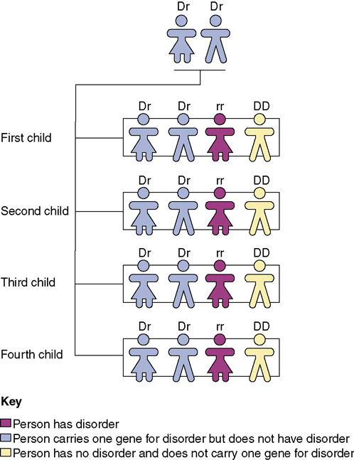
Each gene (a segment of the DNA chain) is coded for inheritance. The coded information carried by the DNA in the gene is responsible for individual traits, such as eye and hair color, facial features, and body shape. Genes carry instructions for dominant and recessive traits. Dominant traits usually overpower recessive traits and are passed on to the offspring. If only one parent carries a dominant trait, 50% of the offspring will have that dominant trait. If each parent carries a recessive trait, there is a chance that one of the offspring will display that trait (Figure 3-2).
 Sex Determination
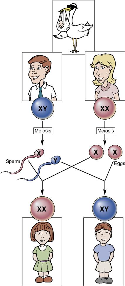
The sex of an individual is determined at the time of fertilization. It depends on the type of spermatozoon that penetrates the ovum (egg). The spermatozoon (sperm cell) has an X or Y chromosome, whereas the female ovum always contains the X chromosome. When the spermatozoon with an X factor unites with the ovum, a female (XX) will result. When an ovum is fertilized with a spermatozoon that contains the Y factor, a male (XY) will result. Because the father may contribute either a Y- or X-bearing sperm, it is the male sperm that is responsible for determining the sex of the embryo (Figure 3-3).
Beginning of Embryonic Development
Fertilization
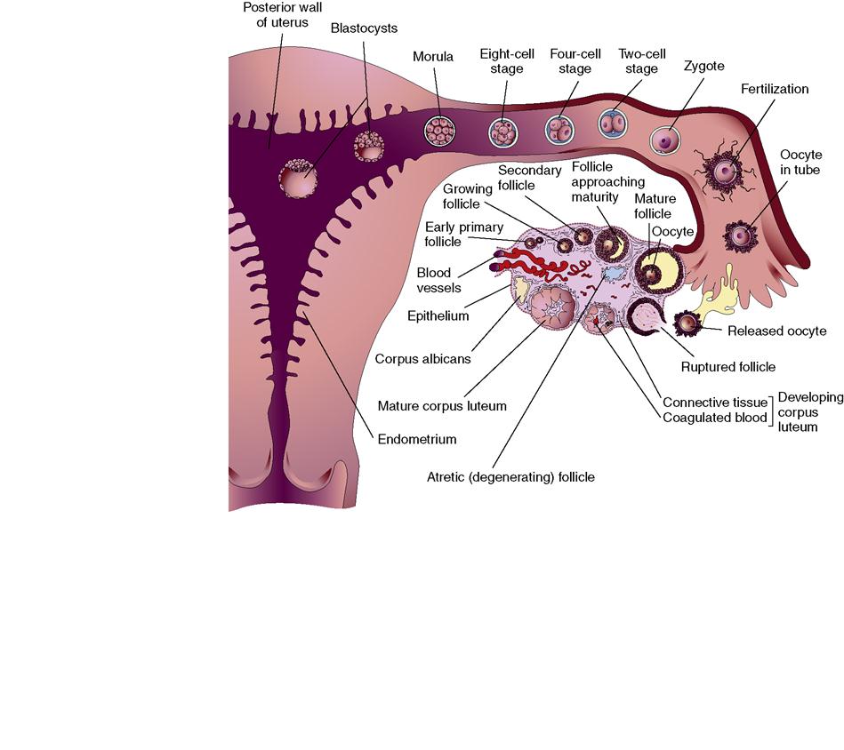
Conception is the union of the egg and sperm, also known as fertilization. Fertilization normally takes  place in the outer third of the uterine (fallopian) tube. Approximately 300 million spermatozoa are deposited into the vagina at ejaculation. Most of the sperm remain in the vagina and within the cervical mucus in the endometrium. After a single sperm enters the ovum, a membrane forms that prevents the entry of more sperm. The ovum can be fertilized for approximately 6 to 24 hours after its release from the ovary, whereas sperm are viable for up to 5 days after ejaculation and their entrance into the female genital tract. At fertilization, the nucleus of the sperm and the nucleus of the ovum unite to form a zygote.
Implantation
The zygote begins a rapid series of cell divisions while traveling down the fallopian tube toward the uterus. Within approximately 3 days, cell division results in a solid mass of 16 cells, called a morula. The morula cells continue to multiply as the morula floats free in the uterine cavity for another 3 or 4 days. Large cells tend to mass at the periphery of the morula ball, leaving a fluid space surrounding an inner cell mass. At this stage, the formed structure is termed a blastocyst. The cells in the outer ring are known as trophoblasts. At this time, the blastocyst contains various components: (1) a fluid-filled cavity; (2) the trophoblast (outer wall), which later will become the placenta; and (3) an inner cell mass, which will form the embryo. The trophoblast cells secrete enzymes that permit them to invade the thickened uterine lining, or endometrium (decidua). During implantation (the cell mass typically takes root in the upper segment of the uterus), trophoblastic cells provide nutrition for the embryo from nutrients carried in the maternal blood. The trophoblastic layer (which later forms the chorion) of blastocyst cells begin to mature rapidly (Figure 3-4). As early as the twelfth day, miniature villi, or rootlike projections, reach out from the layer of cells into the uterine endometrium. These projections (chorionic villi) extend from the chorion into the endometrium.
During the stages of development that immediately follow implantation, teratogenic agents such as drugs, viruses, or radiation may exert profound and damaging effects on the developing embryo. The embryonic period of development is from the second to the eighth week after conception, when most of the basic organ structures are formed. From the eighth week until birth, this developing human being is called a fetus (Box 3-1).
Embryonic Cell Differentiation
During the second and third weeks after conception, three cell layers form: the ectoderm, mesoderm, and endoderm. These cell layers become all of the tissues and organs of the embryo. Each germ layer has specific characteristics (Box 3-2). The differentiated cells stay attached by a body stalk, which later forms the umbilical cord by day 14. Small spaces soon form the amniotic cavity. This hollow space will eventually surround the developing embryo and fetus like a protective, transparent sac.
Fetal Membranes and Amniotic Fluid
The two membranes, called the chorion and the amnion, begin to form at the time of implantation. The amnion, the inner membrane, protects the developing embryo. It forms a cavity in which the embryo or fetus floats, suspended in amniotic fluid. The amnion expands to accommodate the growing fetus. The chorion is the outer membrane that encloses the growing amnion. As the fetus grows, the amnion expands until the chorion and the amnion fuse together to become the amniotic sac, or “bag of waters.” The amniotic sac contains a clear, slightly straw-colored liquid, called the amniotic fluid. This fluid consists of 98% water and contains traces of protein, glucose, fetal lanugo (hair), fetal urine, and vernix caseosa (a cheesy material that covers the fetal skin). Examination of the amniotic fluid for diagnostic purposes (amniocentesis) is discussed in Chapter 5. Most amniotic fluid appears to be derived from the maternal blood. Later, the fetus also adds to the fluid by excreting urine into it. Some amniotic fluid is swallowed by the fetus and is subsequently absorbed by its gastrointestinal tract. The amount of amniotic fluid present at term varies from 800 to 1000 mL. The presence of more than 2 L is called hydramnios (excessive fluid). An excess of amniotic fluid is associated with malformations of the fetal central nervous system and the gastrointestinal tract. An amount of amniotic fluid that is less than 300 mL is oligohydramnios, and it is associated with renal abnormalities. The functions of the amniotic fluid are listed in Box 3-3.


