Transient Ischemic Attacks and Acute Ischemic Stroke


Transient Ischemic Attacks and Acute Ischemic Stroke


Sarah L. Livesay

Joanne V. Hickey


Stroke is the fourth leading cause of death in the United States, surpassed only by heart disease, cancer, and chronic lower respiratory disease.1 It represents an enormous public health and economic burden, estimated at $53.9 billion for direct and indirect costs in 2010.1 The 2012 update on stroke published by the American Heart Association (AHA) reports the following.1


  • Each year about 7,95,000 people experience a new or recurrent stroke (6,10,000 first attack and 1,85,000 recurrent); by gender about 55,000 more women than men have a stroke.


  • On the average, every 40 seconds, someone in the United States has a stroke.


  • From 1998 to 2008, the stroke death rate fell 34.8%, and the actual number of stroke deaths declined 19.4%. However, there continues to be increased numbers of strokes annually related to population growth and increased numbers of older Americans.


  • First-time stroke for African Americans is almost double that for Caucasians.


  • Stroke age-adjusted rates by state report a death rate per 1,00,000 of 51.5 to 58.1 for the so called “stroke belt” states which includes the Carolinas, Georgia, Tennessee, Alabama, Mississippi, Louisiana, Arkansas, and Oklahoma. The stroke belt has the highest incidence of stroke compared to other parts of the country.


  • Projections for 2030 suggest an additional 4 million people will have a stroke, a 24.9% increase in prevalence from 2010.2

In the past several years, management of stroke has undergone a fundamental transformation as a result of research and technological advances, including improved pathophysiologic models of stroke to understand changes in the biochemical and cellular levels; superior neuroimaging using magnetic resonance imaging (MRI), magnetic resonance angiography (MRA), magnetic resonance with diffusion-weighted imaging (DWI) and perfusion-weighted imaging (PWI), and improved computed tomography (CT) scanning techniques; the introduction of new treatment technologies including pharmacologic agents and targeted temperature management (TTM); the definitive role of thrombolytic agents in early treatment; advances in radiologic interventional procedures, such as angioplasty, cerebrovascular stenting, and embolic protection; the preventive benefit of carotid endarterectomy (CEA) in patients with symptomatic high-grade stenosis; neurotransplantation; and other studies and investigative tools that continue to shape and refine patient management.

An appreciation has developed of the stroke timeline associated with the development of neurological deficits and the window of opportunity that exists for reversal of neurological deficits with new interventions. Cardiac resuscitation training programs in basic life support (BLS) and advanced cardiac life support (ACLS) have been revised and now include identification of stroke symptoms and rapid response to save brain tissue as well as cardiac muscle. Improved emergency medical services’ (EMS) recognition of stroke symptoms and triage and the creation of dedicated stroke centers at selected hospitals have significantly enhanced rapid stroke interventions.

A repository of guidelines related to stroke is available at the AHA website. Examples of evidence-based guidelines include primary prevention of stroke, spontaneous intracerebral hemorrhage,
comprehensive overview of nursing and interdisciplinary rehabilitation care, and other guidelines that address the management of stroke patients along a continuum of care through rehabilitation. They are available at the AHA website and are updated periodically to reflect the latest scientific information to assist health care providers in providing best practices in managing patients.3, 4, 5 Other respected groups such as the Veterans Administration have also published evidence-based guidelines that are available at a variety of websites.6

Interdisciplinary clinical pathways for stroke management are the norm in practice. The emphasis is on providing coordinated interdisciplinary care focused on stabilization through acute care and treatment with early rehabilitation of patients for optimal recovery of function and prevention of recurrent stroke. The processes of care are driven by achievement of identified outcomes that are indicators of quality.


DISEASE-SPECIFIC CERTIFICATION: PRIMARY AND COMPREHENSIVE STROKE CENTERS

In December 2003, The Joint Commission (TJC) launched the Primary Stroke Center (PSC) certification program which was developed in collaboration with the American Heart Association/American Stroke Association (AHA/ASA).7 The stroke certification is part of other disease-specific certifications offered by TJC. As of 2011, there are more than 800 certified PSCs in 49 states. The PSC certification program is a voluntary program focused on quality and safety within the framework of standards, guidelines, and outcomes. Certified PSCs


  • use a standardized method for delivering care based on the Brain Attack Coalition recommendations for the establishment of PSCs.


  • support a patient’s self-management activities.


  • tailor treatment and interventions to individual needs.


  • promote the flow of patient information across settings and providers, while protecting patient rights, security, and privacy.


  • analyze and use standardized performance measure data to continually improve treatment plans.


  • demonstrate application and compliance with the clinical practice guidelines published by the AHA/ASA or equivalent evidence-based guidelines.

In addition, performance measures must be collected and reported quarterly on eight National Inpatient Hospital Quality Measures for stroke. The measures include venous thromboembolism (VTE) prophylaxis; discharge on antithrombotic therapy; anticoagulation therapy for atrial fibrillation/flutter; thrombolytic therapy; antithrombotic therapy by the end of hospital day 2; discharge on statins; stroke education; and assessment for rehabilitation. Additional information about application, eligibility, and review process are available at TJC website. For those facilities that meet the established criteria, PSC certification is awarded for a period of 2 years.

A Disease-Specific Care Advanced Certification Program for Comprehensive Stroke Centers (CSCs) is the newest addition to stroke certification options developed by TJC in collaboration with AHA/ASA. Described as rigorous, the CSC requires additional technology and resources when compared to PSC. In addition to meeting all of the requirements for a PSC, the CSC requires the following.7, 8


  • Data supporting minimum volume of cases including evidence of subarachnoid patients, craniotomies for aneurysm clippings, endovascular coiling procedures, and administration of intravenous (IV) thrombolytics.


  • Capacity of advanced imaging capabilities such as carotid ultrasound, catheter angiography, CT angiography, MRA, MRI, transcranial Doppler (TCD), transesophageal echocardiography (TEE), and others.


  • Posthospital care coordination of patients.


  • Dedicated neurological intensive care unit beds for complex stroke patients.


  • Peer review process.


  • Participation in stroke research.


  • Collection of additional performance measures as identified for CSC.

The latest stroke care guidelines published by the AHA/ASA9 encourages certification as a stroke center by an outside facility such as TJC. Experts also encourage the development of more certified CSCs nationwide. Regardless of certification, all hospitals should develop a multidisciplinary performance improvement initiative reviewing stroke care. When a facility is unable to provide the expertise of neuroradiology or neurology, the provision of these services through teleradiology or telestroke programs is advisable.9 Development of Acute Stroke Ready Facilities (ASRFs) may be beneficial in some areas of the country to stabilize the stroke patient and provide acute treatment and then rapidly transfer the patient to a higher level of care. ASRF, PSCs, and CSCs should work together to provide care using the Stroke Systems of Care Model (SSCM).9


PUBLIC AND PROFESSIONAL EDUCATION


  • Stroke is a preventable health problem; it is a treatable condition, in most cases, if treatment is prompt and evidence based. A well-developed public education program is critical to have an informed public who can recognize the signs and symptoms of a stroke and know how to respond. In addition, the health care system must be organized to provide evidence-based care provided by stroke-competent health care providers. The following recommendations are made by the AHA.5, 10 Activation of the 911 system by patients and others is strongly supported because it speeds treatment of stroke.


  • Approved resources are available. The National Stroke Association’s Stroke Rapid Response is a new innovative program that teaches Emergency Medical System and prehospital providers basic and advanced stroke information, including field assessment and hospital treatment, and provides reference tools that can be used when caring for stroke patients; it is available online and includes other tools to assist rapid delivery of appropriate care.


  • The American Association of Neuroscience Nurses (AANN) now offers a stroke certification program for nurses to demonstrate expertise and competency in the delivery of stroke care.9


  • Public education programs to increase public awareness of stroke are supported to increase the number of patients who can be seen and treated in the first few hours after stroke.


  • Education of all health care providers and EMS personnel will increase the number of patients promptly and properly treated.


  • Since EMS personnel are often the first responders, education in brief assessment according to an established protocol will facilitate communication of information for decisions about transport to
    the appropriate health care facility and needed care that alerts health providers.


  • EMS personnel should begin the initial management of stroke in the field according to approved protocols.


  • The use of a stroke identification algorithm such as the Los Angeles Prehospital Stroke Scale (LAPSS) or the Cincinnati Prehospital Stroke Scale (CPSS) screens is encouraged.


  • Patients should be transported for evaluation and treatment to the closest facility that provides emergency stroke care, even if it means bypassing other health care facilities not prepared to provide emergency stroke care.


  • Many states have enacted legislation guiding the transportation and treatment of acute stroke patients based on the ASA Stroke Systems of Care work in the 2000s.


  • The latest ASA guidelines support the triage and transport of stroke patients to the nearest PSC or CSC.9


Risk Factors for Acute Ischemic Stroke

The major risk factors for acute ischemic stroke are hypertension, atrial fibrillation, diabetes mellitus, cigarette smoking, and hyperlipidemia.11 In special situations, systemic disease associated with hypercoagulable state and the use of birth control pills are also risk factors for ischemic stroke. In addition, approximately 40% of patients with ischemic stroke caused by atherothrombosis will have transient ischemic attacks (TIAs), making the history of TIAs a significant risk factor for acute ischemic stroke.


TRANSIENT ISCHEMIC ATTACKS

TIAs are temporary focal brain or retinal ischemia, caused by vascular disease, which fit a known vascular territory and clear completely in less than 24 hours. Most TIAs are much shorter, usually reversing completely within 1 hour. The transient nature of the disease leads to underreporting and exact epidemiologic numbers of people affected are difficult to find. Most recent estimates suggest an incidence of somewhere between 2,00,000 to 5,00,000 cases per year with a population prevalence of 2.3%.12 TIA exists on the same continuum of ischemia as ischemic stroke. Not surprisingly, the risk factors for TIA mirror the risk factors for ischemic stroke. TIAs are more common with advancing age and more common in African Americans and Mexican Americans compared to Caucasians.12 A TIA is a significant warning sign for impending stroke. Between 10% and 15% of patients who experience a TIA will develop an ischemic stroke within 3 months, with half occurring within 48 hours.12 As many as 40% of patients experiencing ischemic stroke have a history of TIA suggesting the transient attack represents an opportune time to employ secondary prevention strategies to prevent permanent ischemia.

The traditional definition of TIA based on duration of symptoms has recently been questioned. Traditionally, a TIA was defined as symptoms of focal ischemia that resolved within 24 hours and most often within 1 hour. However, this definition was challenged as advanced imaging revealed that many patients with improving or resolving symptoms actually had permanent ischemic changes.12 Imaging studies reveal that a third of patients whose symptoms resolve in 1 hour have permanent ischemic changes on perfusion and diffusion imaging. Further, patients with treatable ischemia may have symptoms that wax and wane during the acute event. The assumption that the symptoms are improving and therefore, the patient should not be treated may be contributing to undertreatment of ischemic strokes. Based on the most recent evidence, the clinical definition of TIA was revised. A patient should be diagnosed with TIA only when advanced imaging such as MRI shows no permanent ischemic changes.


TABLE 22-1 COMPARISON OF SIGNS AND SYMPTOMS OF CAROTID AND VERTEBROBASILAR TRANSIENT ISCHEMIC ATTACKS


CAROTID TERRITORY


VERTEBROBASILAR TERRITORY


Related to Ophthalmic Artery


Amaurosis fugax (temporary monocular blindness)


Transient graying, fogging, or blurred vision


A “shade” descending over line of vision


Related to Middle Cerebral Artery


Hemiparesis (more arm than leg weakness)


Hemianesthesia


Contralateral motor or sensory deficits to face or limbs


Related to Anterior Cerebral Artery


Hemiparesis (more leg than arm weakness)


Related to Posterior Cerebral Artery


Dysarthria


Dysphagia


Diplopia


Bilateral blindness


Unilateral or bilateral motor and sensory weakness


Quadriparesis


Related to Cerebellar Arteries


Ataxia


Vertigo


Dizziness


TIAs are classified into TIAs arising from the carotid and TIAs associated with vertebrobasilar vascular territories. One of the most important warning signs of a stroke is a TIA.

TIAs of the carotid (anterior circulation) cause lateralizing signs. When the carotid territory is involved, the symptoms reflect ischemia to the ipsilateral eye or cerebral hemisphere. The first major small branch of the internal carotid artery (ICA) after branching from the common carotid artery is the ophthalmic artery, which supplies the eye. Transient ischemia involving this vessel results in transitory monocular blindness in one eye, also called amaurosis fugax. Hemispherical ischemia usually causes weakness or numbness of the contralateral face or limb; language deficits and cognitive and behavioral changes may also occur.

TIAs of the vertebrobasilar (posterior) circulation cause diffuse signs. When the vertebrobasilar territory is involved, the symptoms often include dysarthria, vertigo, dizziness, ataxia, abnormalities of eye movement resulting in diplopia, and unilateral or bilateral motor and sensory deficits (Table 22-1).13

The ABCD and subsequent ABCD1 score evolved over the past several years as a means to risk stratify TIA patients and better understand the patients at high risk for ischemic stroke in the hours and days following the TIA. Patients receive points for advanced age, elevated blood pressure (BP), unilateral symptoms, speech symptoms, duration greater than 60 minutes, and a history of diabetes (Table 22-2). Risk of stroke in the following 48 hours is 0% with an ABCD1 score of 0 or 1, 1.3% for a score of 2 or 3, 4.1% for a score of 4 or 5, and 8.1% for a score of 6 or 7 (Table 22-3).12 Some programs use the risk stratification to guide hospitalization and workup. An ABCD1 score of 3 or greater is considered a high risk for stroke and requires treatment. The diagnostic workup and secondary stroke prevention for TIA mimics the workup and treatment of ischemic stroke.


DEFINITION AND CLASSIFICATION OF STROKE

Stroke is a heterogeneous, neurological syndrome characterized by gradual or rapid, nonconvulsive onset of neurological deficits that
fit a known vascular territory and that last for 24 hours or more. Stroke occurs when oxygen supply to a localized area in the brain is interrupted, resulting in a series of intricate processes that lead to the destruction of neural tissue and consequent brain damage.


TABLE 22-2 ABCD SCORE


SYMPTOM


SCORE


Age >60


+1


BP >140/90 mm Hg at initial evaluation


+1


Unilateral weakness


+2


Speech disturbance without weakness


+1


Other symptoms


+0


Duration of symptoms <10 mins


+0


Duration of symptoms 10-59 mins


+1


Duration of Symptoms >60 mins


+2


From: Heidenreich, P. A., Trogdon, J. G., Khavjou, O. A., Butler, J., Dracup, K., Ezekowitz, M. D., … Woo, Y. J.; American Heart Association Advocacy Coordinating Committee; Stroke Council; Council on Cardiovascular Radiology and Intervention; Council on Clinical Cardiology; Council on Epidemiology and Prevention; Council on Arteriosclerosis; Thrombosis and Vascular Biology; Council on Cardiopulmonary; Critical Care; Perioperative and Resuscitation; Council on Cardiovascular Nursing; Council on the Kidney in Cardiovascular Disease; Council on Cardiovascular Surgery and Anesthesia, and Interdisciplinary Council on Quality of Care and Outcomes Research. (2011). Forecasting the future of cardiovascular disease in the United States: a policy statement from the American Heart Association. Circulation, 123(8), 933-944. doi:10.1161/CIR.0b013e31820a55f5. Available at: http://circ.ahajournals.org.


There are two major categories of brain injury in stroke: (1) ischemia, which is a lack of blood flow depriving the brain tissue of needed fuel and oxygen; and (2) hemorrhage, which is the release of blood into the brain and into extravascular spaces within the cranium.14 Ischemic and hemorrhagic strokes are further subdivided, as outlined in Figure 22-1. Both ischemic stroke and hemorrhagic stroke will be discussed separately with acute ischemic stroke in this chapter and intracerebral hemorrhagic stroke addressed in Chapter 23.


Acute Ischemic Stroke

Acute ischemic stroke is caused by cerebral ischemia, a reduction of blood flow to the brain that can last from several seconds to minutes. Focal cerebral ischemia differs fundamentally from global ischemia. In focal cerebral ischemia there is usually some degree of circulation from collateral blood vessels that contribute to providing a varying degree of oxygenated blood and glucose. In global ischemia, there is no cerebral blood flow (CBF) to the entire brain as is the case of cardiac arrest. With no blood flow to the brain there is irreversible destruction of neurons that occur within 4 to 8 hours at normal body temperature.11


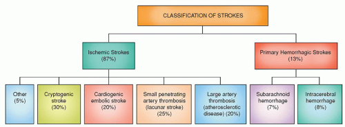
Figure 22-1 ▪ Classification of stroke types.


TABLE 22-3 ABCD SCORE AND RISK OF NEW ISCHEMIC STROKE WITHIN 48 HOURS


TOTAL SCORE


RISK OF STROKE (%)


0-1


0


2-3


1.3


4-5


4.1


6-7


8.1


From: Heidenreich, P. A., Trogdon, J. G., Khavjou, O. A., Butler, J., Dracup, K., Ezekowitz, M. D., … Woo, Y. J.; American Heart Association Advocacy Coordinating Committee; Stroke Council; Council on Cardiovascular Radiology and Intervention; Council on Clinical Cardiology; Council on Epidemiology and Prevention; Council on Arteriosclerosis; Thrombosis and Vascular Biology; Council on Cardiopulmonary; Critical Care; Perioperative and Resuscitation; Council on Cardiovascular Nursing; Council on the Kidney in Cardiovascular Disease; Council on Cardiovascular Surgery and Anesthesia, and Interdisciplinary Council on Quality of Care and Outcomes Research. (2011). Forecasting the future of cardiovascular disease in the United States: a policy statement from the American Heart Association. Circulation, 123(8), 933-944. doi:10.1161/CIR.0b013e31820a55f5. Available at: http://circ.ahajournals.org.


Ischemia can be further subdivided into three different mechanisms: thrombosis, embolism, and decreased systemic perfusion. A plumbing analogy may be useful to understand the differences among the mechanisms of ischemia. A pipe can be plugged partially or completely due to a local occlusion process. In a blood vessel the local obstruction is due to a thrombus. A pipe may also be blocked from a substance that originated elsewhere in the system such as the water tank. The substance becomes dislodged from its place of origin and plugs a pipe upstream. This is an example of an embolism. The third mechanism is that of pressure that is intermittently low thus resulting in the low flow of water to the sinks. The analogy to blood vessels is that of systematic hypotension and hypoperfusion.14


Thrombosis is an obstruction of blood flow due to a localized occlusive process within one or more blood vessels. The lumen of the vessel is narrowed or occluded due to a localized occlusive process within one or more blood vessels that is caused by an alteration in the vessel wall or by a superimposed clot formation. The most common type of vascular pathology is atherosclerosis, in which fibrous and muscular tissues overgrow in the subintima, and fatty materials form plaques that can narrow the lumen. Platelets can adhere to the plaque and clump together thus serving as a site for the deposition of fibrin, thrombin, and clot formation. Atherosclerosis mainly affects the larger extracranial (EC) and intracranial (IC) arteries.

An embolism is formed elsewhere within the systemic vascular system; it lodges in a cerebral artery and blocks blood flow. Blockage can be transient or may persist for hours or days before moving distally in the blood vessel. As compared to a thrombus, an embolism lumina blockage is not caused by a localized process originating within the blocked artery. The material arises away from the involved cerebral vessel, most commonly from the heart, from major arteries such as the aorta, carotid, and vertebral arteries (VAs), or from systemic veins. Cardiac sources of embolism include the heart valves and clots or tumors within the atrial or ventricular cavities. Artery-to-artery emboli are composed of clots, platelet clumps, or fragments of plaques that break off from the primary cerebral source. Clots originating in systemic veins travel to the brain through cardiac defects such as an atrial septal defect or a patent foramen ovale (PFO).

Systemic hypoperfusion occurs as a result of a significant reduction in systemic BP due to any cause. Susceptibility to ischemia varies among neurons with gray matter particularly vulnerable. Systemic hypotension from any cause can result in global cerebral ischemia. Global ischemia, which may be caused by cardiac arrest, causes the greatest damage to areas between the territories of the major cerebral and cerebellar arteries known as the “watershed area.” A watershed or border zone infarction is an infarcted area that occurs between the terminal distributions of two adjacent cerebral arteries, such as the anterior cerebral and middle cerebral arteries. Because the terminal distributions are at the end of the pipeline, watershed areas are subject to low arterial pressure under normal circumstances (Fig. 22-2). They are also the first to fail when adequate systemic BP drop. If systemic hypotension occurs, there is failure to maintain adequate cerebral perfusion.


ISCHEMIC STROKE SUBTYPES

Ischemic stroke accounts for 87% of all strokes and is subdivided into thrombotic atherosclerotic large vessel disease (20%); small vessel (penetrating) artery disease, or “lacunae” (25%); cardiogenic embolic (20%); cryptogenic (30%); and other (5%). Note that these percentages are approximate for each category with variations noted depending on resource consulted. Atherosclerosis of large and small cerebral arteries that results in thrombosis is the most common cause of ischemic stroke in North America and Europe and accounts for 45% of strokes in the United States.


Large Artery Atherosclerotic Stroke

The large EC and IC arteries are subject to atherosclerosis with an associated atheroma plaque that narrows the lumen of the vessel. Plaque enlargement occurs slowly over decades, and the person is asymptomatic until the plaque intrudes on a substantial percentage of the arterial lumen diameter. Typically, luminal thrombi are associated with luminal surface disruption or ulceration of the endothelial lining, leading a surface ripe for the development of a thrombus that results in arterial obstruction. Thrombosis of a vessel can result in artery-to-artery embolism. The atheroma can also be the site for thrombus formation. Both conditions can lead to hypoperfusion, ischemia, and ischemic stroke.


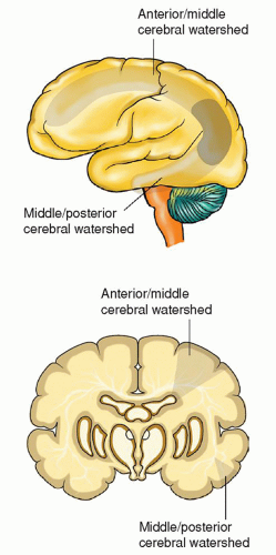
Figure 22-2 ▪ Location of watershed areas.

If a major artery is involved, large areas of both gray and white matter become ischemic, infarcted, and necrotic. Neuronal ischemia causes changes in the cell membrane, resulting in intracellular edema and compression of the capillaries, further compromising adequate blood supply. Cerebral edema peaks approximately 2 to 4 days after the stroke. Symptoms of ischemic stroke often develop in a stepwise progression relating to cerebral edema and infarction, reaching a peak in 1 to 3 days before stabilizing.


Small Artery Stroke (Lacunar Stroke)

The term lacuna describes the small cavity remaining in the brain tissue that develops after the necrotic tissue of a small, deep infarct has been removed. A lacunar stroke is a type of ischemic stroke caused by microatheroma and thrombosis of a small penetrating artery, resulting in a small, softened area in the deep white matter structures of the brain. As the softened tissue sloughs away, small cavities or lakes remain, which are called lacuna (diameter of 0.5 mm or less). Occlusion occurs in the presence of lipohyalinosis,
a condition characterized by pathologic thickening of these small vessels, and leads to a specific clinical stroke syndrome. Hypertension is the principal risk factor for lacunar strokes. Diabetes mellitus is also associated with the development of lacunar strokes. Lacunar strokes are seen predominately in the basal ganglia, especially the putamen, the thalamus, and the white matter of the internal capsule and pons; they occur occasionally in the white matter of the cerebral gyri. They are rare in the gray matter of the cerebral surface, the corpus callosum, visual radiations, or medulla (Fig. 22-3). Most lacunae occur in the lenticulostriate branches of the anterior cerebral artery (ACA) and middle cerebral artery (MCA), the thalamoperforate branches of the posterior cerebral arteries, and the paramedian branches of the basilar artery (BA).15


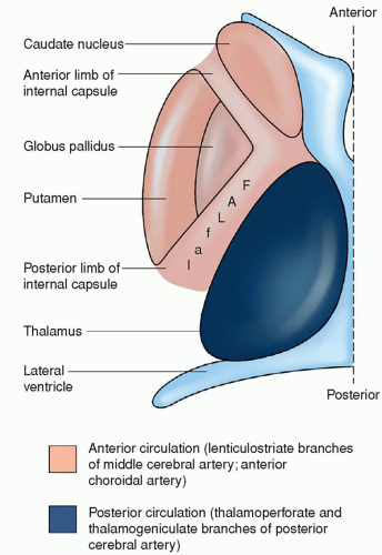
Figure 22-3 ▪ Primary location of lacunar strokes.

There are distinct signs and symptoms associated with several recognized lacunar syndromes, including pure motor hemiplegia, pure sensory stroke, homolateral ataxia and leg paresis, dysarthria/clumsy hand syndrome, sensorimotor stroke, and basilar branch syndromes. Even though a lacunar stroke is small, it can cause considerable deficits if a critical area, such as the internal capsule, is involved. Patients may have several lacunae, as evidenced on CT or MRI, and have diffuse white matter changes associated with dementia.


Cardiogenic Embolic Stroke

About 20% of ischemic strokes result from cardiogenic embolism from atrial fibrillation (the most common), PFO, valvular disease, ventricular thrombi, myocardial infarction, congestive heart failure, atrial septal aneurysm, and other cardiac problems. Atherosclerosis and atherogenic plaques of the proximal aorta are another source of cardiac emboli detectable with the use of TEE. The atherogenic plaques commonly found in coronary vessels, in the heart, and at the bifurcation of the aorta are precursors for hypertension and atrial fibrillation. Unstable plaques can break off and become microemboli to the brain, causing stroke. Microemboli from the heart are mobilized and enter the cerebral system most often through the carotid arteries, flowing until the vessel is too narrow to allow further passage of the embolus and the vessel becomes occluded. The left MCA is affected most often because it is a relatively straight vessel and provides the path of least resistance for the embolus. Cardiogenic strokes associated with PFO occur in approximately 20% to 25% of persons older than 30 years of age and usually occur when the patient is awake and active.16 The development of ischemia is very rapid with maximal deficit present within minutes.


Cryptogenic Stroke

About 30% of ischemic strokes are cryptogenic in origin, which means that no cause of the stroke could be found after diagnostic evaluation.


Stroke from Other Causes

About 5% of ischemic strokes result from nonatherosclerotic vasculopathies, hypercoagulable states, hematologic disorders, arteritis, migraine/vasospasm, and cocaine use.17


Arterial Dissection

Arterial dissection is an unusual cause of stroke and accounts for 1% to 5% of all strokes, occurring commonly in younger persons (aged between 25 and 45 years), usually in the absence of atherosclerosis. Arterial dissection is typically caused by trauma to a vessel wall (as in the case of iatrogenic trauma associated with catheter passage during an angiographic procedure), vessel abnormality, migraine, and fibromuscular dysplasia. After the vessel wall is injured, a hematoma forms in the medial layer and breaks through the intimal wall. Injury to the intimal wall allows blood to dissect through the medial layer, resulting in luminal narrowing; a pseudoaneurysm may result in some cases.

The patient with an arterial dissection is at risk for ischemic stroke due to resulting thrombosis, embolization, or subarachnoid hemorrhage due to vessel rupture. The most common locations for dissection are the cervical carotid, IC carotid (usually in the middle cerebral or supraclinoid ICA), and VA. Vertebral dissections are often associated with trauma.

The signs and symptoms of an arterial dissection may evolve over hours to days. The patient commonly experiences a severe unilateral headache, scalp throbbing, and/or neck pain. Other presenting signs and symptoms include transient monocular blindness, oculosympathetic paralysis, pulsatile tinnitus, TIAs, or stroke in evolution.

Emergency MRA or cerebral angiography is indicated if an arterial dissection is suspected. After the diagnosis has been established, cautious heparinization is initiated; other measures such as stenting, angioplasty, grafting, and bypass may be undertaken as appropriate. Chronic oral anticoagulation may be necessary in unresolved cases.


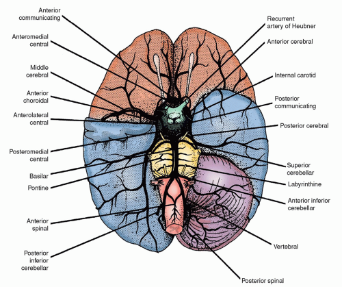
Figure 22-4 ▪ Arteries that supply the brain, as seen from the ventral surface. The right cerebral hemisphere and the tip of the right temporal lobe have been removed.


ANATOMY, ATHEROGENESIS, AND PATHOPHYSIOLOGY RELATED TO ACUTE ISCHEMIC STROKE


Anatomical and Physiological Basis for Ischemic Stroke

There are four major cerebral arteries that supply the brain: two ICAs, constituting the anterior circulation, and two VAs, which constitute the posterior circulation (Fig. 22-4). See Chapter 5 for a detailed discussion of cerebral circulation. Normally, CBF is approximately 50 to 60 mL/100 g/min with some variation in different parts of the brain. The overall CBF to the brain is about 650 to 700 mL/min.

The rate of CBF to the entire brain is relatively constant, and it does not change in response to alterations in mean systemic BP over a range of 50 to 150 mm Hg. This phenomenon, known as autoregulation, protects the brain from possible hypotension (i.e., hypoperfusion) or hypertension (i.e., cerebrovascular hemorrhage caused by excessive intravascular pressure). In response to ischemia, the cerebral autoregulatory mechanisms compensate for the reduced CBF by local vasodilation, opening the collaterals, and increasing the extraction of oxygen and glucose from the blood. However, when the CBF falls below 20 mL/100 g/min, electrical silence occurs, and the activity at the synapses is greatly diminished in an attempt to preserve energy stores. When the CBF falls below 10 mL/100 g/min, irreversible neuronal injury occurs.18, 19, 20 Occlusion seldom completely abolishes the delivery of oxygen and glucose to the affected vascular territory because CBF to the affected vascular territory is usually partly maintained by collateral circulation. Collateral circulation to an occluded vessel is possible because of anastomosis between the vessels. However, anomalies of cerebral vessels are common, making it difficult to predict if viable collateral circulation is available to an occluded area.


Pathophysiology of Acute Ischemic Stroke

The pathophysiology that leads to an acute ischemic stroke is a dynamic process that evolves over time. The progression and extent of ischemic injury is influenced by a number of ischemic modifying factors which include rate of onset and duration of ischemia; state of collateral circulation; adequate functionality of the systemic circulation; hematological factors; elevated temperature; and hyper-or hypoglycemia states (Table 22-4). The pathophysiology of ischemic stroke due to thrombosis and embolism is the same. At the cellular level, the mechanisms of neuronal injury are driven by hypoxia or anoxia regardless of cause. Focal cerebral ischemia that
can lead to infarction follows two distinct pathways. The first is a necrotic pathway in which cellular cytoskeletal breakdown is rapid, due primarily to energy failure of the cell. The second is an apoptotic pathway in which cells become programmed to die.21


TABLE 22-4 FACTORS CONTRIBUTING TO POOR OUTCOME FOLLOWING ACUTE ISCHEMIA


ISCHEMIC MODIFYING FACTOR


DESCRIPTION


TREATMENT IMPLICATIONS


Rate of onset and duration of ischemia


Ischemia of slower onset and shorter duration is better tolerated by the brain than fast onset with a longer duration


Blood pressure targets may be impacted by lack of collateral circulation. The patient’s neurologic status may guide ideal blood pressure range following new ischemia


Collateral circulation


The effect of ischemic injury is greatly influenced by the state of collateral circulation to the affected area of the brain; good collaterals provide a compensatory mechanism to supply blood to affected areas


Patients may resolve their symptoms or experience less significant symptoms than expected if collateral circulation is adequate


Adequate systemic circulation


Cerebral perfusion pressure is dependent on adequate systemic blood pressure; systemic hypotension can result in global cerebral ischemia


Permissive hypertension is the standard care of a new ischemic stroke patient. Induced hypertension is rarely warranted due to complications of therapy


Hematological factors


Hypercoagulable states increase the progression and extent of microscopic thrombi thus exacerbating vascular occlusion


Patients without typical stroke risk factors should be evaluated for hypercoagulable states and antiplatelet and anticoagulation therapy should be planned accordingly


Elevated temperature


Elevated body temperature is related to increased cerebral ischemic injury and in poorer outcomes


Normothermia is desired; elevations in temperature should be treated aggressively


Hypothermia is a proven neuroprotective treatment in patients with cardiac arrest that leads to improve patient outcomes


Hyper- or hypoglycemia states


Adversely influence the size of an infarct.


Control of glycemia within a range of <150 mg/dL is recommended


Complex and concurrent biochemical and molecular cascades result from ischemic damage to neurons in response to deprivation of oxygen and cessation of oxidative metabolism at the cell membrane level. Without oxygen, adenosine triphosphate (ATP), necessary for energy-dependent mitochondrial function (e.g., the cellular respiratory chain, lipid metabolism, and maintenance of the transmembrane ion channels) rapidly ceases. Impairment of the respiratory chain results in anaerobic glycolysis of remaining available glucose. Anaerobic glycolysis proceeds only to pyruvate, which reduces to lactate. Lactic acid and free fatty acid accumulation causes intracellular acidosis, further inhibiting mitochondrial function. Without ATP, the membrane ion pump ceases function and neurons depolarize allowing an intracellular influx of sodium and calcium.11

Concurrently, other cell destruction processes occur that include excitotoxicity, increased intracellular calcium, and generation of free radicals. The hypoxic-ischemic cascade triggers a state of “excitotoxicity” caused by depletion of cellular energy stores from failure of the cellular membrane ion pump. The excitotoxicity is due to a hyperreaction of certain neurotransmitters, primarily glutamate and aspartate. Glutamate is normally stored inside the synaptic terminals. These neurotransmitters are released by ischemic cells. Excess extracellular glutamate produce neurotoxicity by activating postsynaptic glutamate receptors that increase neuronal sodium, chloride, and calcium channels resulting in an intracellular influx of calcium as well as sodium and chloride ions with water into the cell, causing acute cellular swelling. The voltage-dependent calcium channels allow influx of calcium into the intracellular space and cause an exit of potassium. (Intracellular calcium is normally maintained at a low level by active transport mechanisms.) The high intracellular calcium activates calcium-dependent degradative enzymes (proteases, phospholipases, and endonucleases) that attack the cell membranes and DNA and further inhibit mitochondrial function.

Oxygen-free radicals are produced by membrane lipid degradation and mitochondrial dysfunction which cause catalytic destruction of membranes and likely damage other vital functions of cells. Free radicals with resultant lipid peroxidation occur in inadequately perfused areas and during reperfusion of previously ischemic areas. Oxygen-free radicals, superoxide peroxide, and hydroxyl ions destroy fatty acids and disrupt calcium homeostasis, further contributing to cellular demise. Approximately 8 to 12 hours after the ischemic insult, the neuron becomes smaller and more angular. The cytoplasm and nucleus shrink, followed by complete dissolution of the cell and cell death.22


Infarction and Ischemic Penumbra

Ischemia severe enough to cause neuronal death of cerebral cells is called ischemic necrosis or cerebral infarction. With infarction, a core of necrotic tissue exists from a lack of adequate oxygen supply and nutrients (e.g., glucose) resulting in rapid depletion of energy stores. Cerebral infarcted areas vary in their composition. The infarcted tissue is softened with a varying amount of congestion and hemorrhage. Some infarctions are pale because there is an absence of blood (pale infarction) while others include mild congestion from dilated blood vessels and escaped red blood cells, especially at the margins. Still others have extensive extravasation of blood from many small vessels within the infarction (hemorrhagic infarction). In addition, many infarctions are a mixture of both types.11

Within an hour of a hypoxic-ischemic event, a core of infarction surrounded by an oligemic region called the ischemic penumbra occurs (Fig. 22-5 and Table 22-5). Although the neuronal cells of the penumbra do not function normally due to a decreased blood supply and loss of autoregulation, they remain viable for a limited
period of time. The critical time period during which the penumbra is at risk is referred to as the “window of opportunity” because the neurological deficits created by the ischemia can be partly or completely reversed if reperfusion of the ischemic area occurs within a critical time frame of 2 to 4 hours.24 Lesser degrees of ischemia, seen within the ischemic penumbra, result in apoptotic cellular death causing cells to die days to weeks later if a timely reversal of ischemia did not occur.21


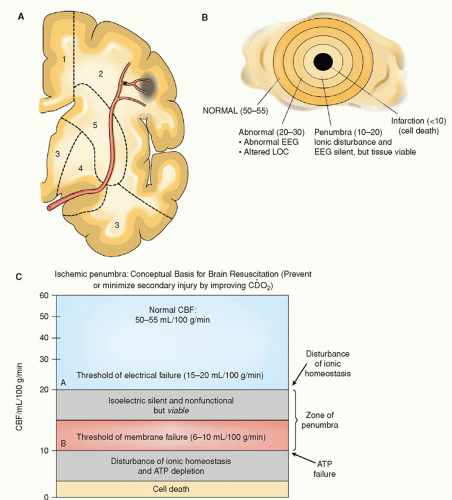
Figure 22-5 ▪ Cerebral blood supply during ischemic events. A: See distribution of middle cerebral artery and occlusion of a branch. Note area of infarction and surrounding penumbra with viable but nonfunctional cells. These cells will either become infarcted or recover, depending on treatment. B: Ischemic penumbra: normal CBF = 50-55 mL/100 g/min. Variations in CBF are noted. Penumbra is the critical area that may be salvageable with appropriate treatment, or cell death will occur if adequate CBF is not restored. C: Ischemic penumbra: conceptual basis for brain resuscitation. ATP, adenosine triphosphate; CBF, cerebral blood flow; EEG, electroencephalogram; LOC, level of consciousness.

The penumbra is the target for pharmacologic interventions to re-establish adequate perfusion, thus salvaging neuronal cells from infarction. Neuroprotective agents, including the use of mild to moderate brain cooling, are being tested in clinical trials to assess their safety and efficacy in protecting the cells from the secondary injury associated with ischemia. Thus far, there are no drugs tested that provide neuroprotection against ischemia.


Neuronal Death

Apoptosis, also known as programmed cell death, can occur as a result of ischemia. First, nuclear damage occurs although the integrity of
the plasma and mitochondrial membrane is intact until later in the process when mitochondrial damage occurs. Ischemia activates proteins within the nuclei, triggers an autolytic process which results in cell death.19


TABLE 22-5 THRESHOLDS OF CEREBRAL ISCHEMIA


Normal range


40-50 mL/100 g/min


Oligemia


30-40 mL/100 g/min


Mild ischemia


30-30 mL/100 g/min


Moderate ischemia (penumbra)


Electrical function is affected 10-12 mL/100 g/min Reversible cellular damage


Severe ischemia (lesion core)


0-10 mL/100 g/min Irreversible cellular damage


From: Hock, N. H. (1999). Brain attack. The stroke continuum. Nursing Clinics of North America, 34(3), 697.


SIGNS AND SYMPTOMS OF STROKE SYNDROMES ACCORDING TO THE INVOLVED VESSEL

The type and severity of neurological deficits from stroke encompass a wide range and gradation of signs and symptoms. The presenting signs and symptoms of stroke depend on the extent of CBF compromise, the particular cerebral vessel involved, and size of the infarction. When a cerebral artery is occluded by a thrombus or embolus, classic syndromes are said to develop. The clinical features of stroke are commonly classified as carotid artery (anterior circulation) syndromes and vertebrobasilar (posterior circulation) syndromes. Table 22-6 summarizes the signs and symptoms associated with stroke according to the cerebral vessel involved.


Comparison of Left-Sided and Right-Sided Stroke

Some generalizations can be made about the deficits incurred with left-sided and right-sided stroke (Table 22-7). A stroke is a form of cerebral injury. The injury to the brain results from ischemia that develops over time or suddenly, as may be the case in thrombotic or embolic strokes, or from a ruptured blood vessel, in the case of hemorrhagic stroke. In all cases of stroke, areas of the brain are deprived of an adequate oxygen supply. The particular type and degree of neurological deficits incurred depends on the particular area of the brain involved, because the brain is composed of the most highly specialized tissue in the body. If the blood supply is cut off for an extended period, the involved cerebral tissue may become necrotic, resulting in permanent neurological deficits. In instances of ischemia, temporary neurological impairment may result.


Diagnostics for Transient Ischemic Attack and Acute Ischemic Stroke

A number of diagnostic studies are useful for the investigation of TIA and stroke patients. Diagnostic testing proceeds in a stepwise fashion. When the most common tests fail to uncover the cause of the stroke, other less common tests are ordered. For example, stroke in a young person without the usual stroke risk factors suggests other causes such as PFO or antiphospholipid abnormalities. Table 22-8 describes the commonly recommended diagnostic procedures and laboratory tests.

Initially, the following tests are recommended for all patients: noncontrast brain CT or brain MRI, blood glucose, electrolytes, renal function tests (blood urea nitrogen, creatinine), complete blood count (CBC), platelets, prothrombin time/international normalized ratio (INR), activated partial thromboplastin time, and markers of cardiac ischemia. Oxygen saturation is also monitored. Selected patients may require hepatic function tests, toxicology screen, blood alcohol level, pregnancy test, arterial blood gases, chest x-ray (only if lung disease is suspected), and lumbar puncture (only if subarachnoid hemorrhage is suspected and a CT scan is negative for blood). Diagnostic testing required prior to administration of tissue plasminogen activator (tPA) has been debated over the years and waiting for test results is thought to contribute to delays in patient treatment. Limited hematologica, coagulation, and chemistry testing are recommended during the first few minutes of treatment and clinicians should not wait for testing to result to start tPA if indicated. A baseline assessment of cardiac injury such as troponin should be obtained. However, waiting for the results of testing prior to the initiation of tPA is not warranted. If an x-ray of the chest or electrocardiogram (ECG) is indicated, they should not delay the initiation of acute stroke treatment.9

CT without contrast is considered the gold standard imaging test for acute ischemic stroke. However, new onset ischemia is not visible in CT imaging until hours or days after the event. The purpose of a noncontrast head CT is to rule out the presence of blood as the cause of focal neurologic deficits. Therefore, noncontrast CT imaging may appear normal in the first few hours after ischemia. Advanced imaging such as CT angiography and CT perfusion are gaining popularity based on recent research. However, the addition of angiography and/or perfusion may delay the time to treatment with thrombolytic agents in some circumstances. Balancing the need for advanced imaging information with timely treatment is key.26

Other tests may be ordered in the course of treatment for special reasons. Besides neuroimaging tests (CT or MRI), blood flow studies (TCD) may be ordered. If a vascular anomaly is suspected, a cerebral angiography or MRA may be ordered. Recent reports recommend replacement of CT with MRI as the primary neuroimaging technique for evaluation of acute stroke.27, 28 The multimodal MRI provides more information about brain ischemic pathophysiology, can localize perfusion deficits and ischemic injury including the penumbra within minutes after onset of ischemia, and are useful to guide treatment decisions. DWI and PWI are distinctly different techniques; they are interrelated physiologic parameters, and both are usually performed during the same MRI examination.29 However, the standard initial diagnostic imaging procedure is an emergency CT scan without contrast medium to differentiate ischemic stroke from hemorrhagic stroke. Advanced CT imaging with angiography and perfusion or a rapid MR image may be warranted during the acute phase to determine which patients may be eligible for neurointerventional radiology to reperfuse the ischemic territory.

Otherwise, they should not be considered a routine part of the work-up of acute ischemic stroke.30


TABLE 22-6 NAME, DESCRIPTION, AND SIGNS/SYMPTOMS RELATED TO STROKE BY INVOLVED VESSEL


NAME


DESCRIPTION


SIGNS AND SYMPTOMS


Internal carotid artery (ICA) syndrome


  • Paralysis of the contralateral face, arm, and leg


  • Sensory deficits of the contralateral face, arm, and leg


  • Aphasia, if the dominant hemisphere is involved


  • Apraxia, agnosia, and unilateral neglect, if the nondominant hemisphere is involved


  • Homonymous hemianopsia


Middle cerebral artery (MCA) syndrome


  • MCA is the most common of all cerebral occlusions


  • If the main stem of MCA is occluded, a massive infarction of most of the hemisphere results


  • Initially, there may be vomiting and a rapid onset of coma, which may last a few weeks


  • Cerebral edema is extensive


  • Hemiplegia (involving face and arm on the contralateral side; the leg is spared or has fewer deficits than the arm)


  • Sensory deficits (same area as hemiplegia)


  • Aphasia (global aphasia if the dominant hemisphere is involved)


  • Homonymous hemianopsia


Anterior cerebral artery (ACA) syndrome


  • ACA is least often occluded


  • If occlusion occurs proximal to a patent anterior communicating artery (ACom), the blood supply may be compromised


  • If occlusion is distal or if the ACom artery is inadequate, there will be infarction of the medial aspect of one frontal lobe


  • Bilateral medial frontal lobe infarction occurs if one ACA is occluded and the other artery is small and dependent on blood flow


  • Paralysis of contralateral foot and leg (foot drop is a consistent finding)


  • Impaired gait


  • Sensory loss over toes, foot, and leg


  • Abulia (slowness and prolonged delays to perform acts voluntarily or to respond)


  • Flat affect, lack of spontaneity, slowness, distractibility, and lack of interest in surroundings


  • Cognitive impairment, such as perseveration and amnesia


  • Urinary incontinence


  • Note that aphasia and hemianopsia are not part of the syndrome


Vertebral artery syndrome


  • Occlusion of vessels within the vertebrobasilar system produces unique syndromes


  • Vertebral and basilar arteries and their branches supply the brainstem and cerebellum


  • Posterior cerebral arteries are the terminal branches of the basilar artery and supply the medial temporal and occipital lobes, as well as part of the corpus callosum


  • Wallenberg’s syndrome (lateral medullary syndrome)


  • Dizziness


  • Nystagmus


  • Dysphagia and dysarthria


  • Pain in face, nose, or eye


  • Ipsilateral numbness and weakness of face


  • Staggering gait and ataxia


  • Clumsiness


Basilar artery (BA) syndrome


  • Quadriplegia


  • Possibly the “locked-in” syndrome


  • Weakness of facial, lingual, and pharyngeal muscles


Anterior inferior cerebellar artery (AICA) syndrome


Occlusion of the AICA is also known as the lateral inferior pontine syndrome


  • Vertigo


  • Nausea and vomiting


  • Tinnitus


  • Nystagmus


Ipsilateral side


  • Paresis of lateral conjugate gaze


  • Horner’s syndrome


  • Cerebellar signs (ataxia, nystagmus)


Contralateral side


  • Impaired pain and temperature sensation in trunk and limbs (may also involve face)


Posterior inferior cerebellar artery (PICA) syndrome (also called Wallenberg’s syndrome)


PICA involves the lateral portion of the medulla as a result of the occlusion of the posterior inferior cerebellar artery


  • Nausea and vomiting


  • Dysphagia and dysarthria


  • Horizontal nystagmus


  • Ipsilateral Horner’s syndrome


  • Cerebellar signs (ataxia and vertigo)


  • Loss of pain and temperature sensation on contralateral side of trunk and limbs


Posterior cerebral artery (PCA) syndrome


  • If superficial occlusion (peripheral areas) of a PCA is involved, contralateral homonymous hemianopsia is seen


  • If penetrating branches (central areas) are occluded, the cerebral peduncle, thalamus, and upper brainstem are involved


  • There is wide variation in the manifestations of the syndrome


Peripheral area


  • Homonymous hemianopsia


  • Memory deficits


  • Perseveration


  • Several visual deficits (cortical blindness, lack of depth perception, failure to see objects not centrally located, visual hallucinations)


Central area


  • If the thalamus is involved, sensory loss of all modalities, spontaneous pain, intentional tremors, and mild hemiparesis


  • If the cerebral peduncle is involved, Weber’s syndrome (oculomotor nerve palsy with contralateral hemiplegia)


  • If the brainstem is involved, deficits involving conjugate gaze, nystagmus, and pupillary abnormalities, with other possible symptoms of ataxia and postural tremors


TABLE 22-7 COMPARISON OF SIGNS AND SYMPTOMS ASSOCIATED WITH RIGHT-SIDED AND LEFT-SIDED HEMIPLEGIA


STROKE SYNDROME ON LEFT SIDE OF BRAIN (RIGHT-SIDED HEMIPLEGIA)


STROKE SYNDROME ON RIGHT SIDE OF BRAIN (LEFT-SIDED HEMIPLEGIA)


  • Expressive aphasia


  • Receptive aphasia


  • Global aphasia


  • Intellectual impairment


  • Slow and cautious behavior


  • Defects in right visual fields


  • Spatial-perceptual deficits


  • Denial and the deficits of the affected side require special safety considerations


  • Tendency for distractibility


  • Impulsive behavior; apparently unaware of deficits


  • Poor judgment


  • Defects in left visual fields


TABLE 22-8 DIAGNOSTIC PROCEDURES FOR TRANSIENT ISCHEMIC ATTACKS (TIAs) AND STROKE


DIAGNOSTIC PROCEDURE


INFORMATION PROVIDED


Computed tomography (CT) scan without contrast


Important immediate diagnostic to differentiate between ischemic and hemorrhagic stroke; if hemorrhagic, antiplatelets or anticoagulants are not given because of the increased risk of more bleeding; important for treatment decisions


CT scan with contrast


Useful to rule out lesions that many mimic a TIA, especially when symptoms are related to hemispheric deficits; hypodense areas on CT scan suggest infarction


Magnetic resonance imaging (MRI)


Offers excellent soft-tissue contrast discrimination with superior demarcation of mass lesion from surrounding structures including areas of ischemia and infarction; good visualization of vascular structures when questioning a vascular lesion; useful for diagnosis of stroke in first 72 hrs; a diffusion-weighted MRI can show ischemia in first few hours


Magnetic resonance angiography (MRA)


Less available and higher cost; noninvasive imaging of the carotid, vertebral, basilar, and major intracranial and extracranial arteries to determine occlusion; useful for clot visualization


Carotid ultrasonography


Noninvasive imaging; widely used initial diagnostic in patients with carotid territory symptoms for whom carotid endarterectomy (CEA) is considered; cervical carotid artery imaging often required to exclude high-grade stenosis, which is an exclusion for CEA; less sensitive in assessing mild to moderate stenosis


Transcranial Doppler (TCD)


TCDs are now part of standard work-up for stroke, especially when CEA is considered; useful to detect severe intracranial stenosis, evaluate the carotid and vertebrobasilar vessels, assess patterns and extent of collateral circulation in patients with known arterial stenosis or occlusion, and detect microemboli


Cerebral angiography


Ordered for patients considered candidates for CEA to define precisely the percentage of occlusion and in patients with unusual presentation with aneurysm, vasculitis, and high-grade stenosis


Transthoracic echocardiography (TTE)


Helpful in search for cardio embolic sources; TTE is particularly helpful for diagnosing left ventricular thrombi, left atrial myxomas, and thrombi that protrude into the atrial cavity; they are less reliable for small tumors, laminated thrombi, and thrombi limited to the left or right atrium


Transesophageal echocardiography (TEE)


Benefit of TEE is in greater sensitivity for source of cardioemboli (except ventricular disease); TEE provides better visualization of cardiac structures, especially those at greater depth from chest wall and lesions of the atria (atrial appendage thrombi associated with atrial fibrillation), interarterial septum defects (patent foramen ovale, atrial septal defects), mitral valvular vegetation, and atherosclerotic disease of ascending aortic arch


Electrocardiogram (ECG); 12-lead is recommended initially


12-lead ECG recommended immediately because of the high incidence of heart disease in patients with stroke; ECG also useful when cardiogenic embolic stroke or concurrent coronary artery disease is suspected


Ambulatory ECG monitoring


Reserved for patients who have suspicious palpitations, arrhythmias, or enlarged left atrium


Prothrombotic states


Protein C, protein S, antithrombin III, thrombin time, hemoglobin, electrophoresis, anticardiolipin antibody, lupus anticoagulant, and syphilis serology


From: Adams, H. P, Jr., del Zoppo, G., Alberts, M. J., Bhatt, D. L., Brass, L., Furlan, A., … Wijdicks, E. F.; American Heart Association; American Stroke Association Stroke Council; Clinical Cardiology Council; Cardiovascular Radiology and Intervention Council; Atherosclerotic Peripheral Vascular Disease and Quality of Care Outcomes in Research Interdisciplinary Working Groups. (2007). Guidelines for the early management of adults with ischemic stroke. Stroke, 38(5), 1655—1711.

Only gold members can continue reading. Log In or Register to continue

Stay updated, free articles. Join our Telegram channel

Jul 14, 2016 | Posted by in NURSING | Comments Off on Transient Ischemic Attacks and Acute Ischemic Stroke

Full access? Get Clinical Tree

Get Clinical Tree app for offline access