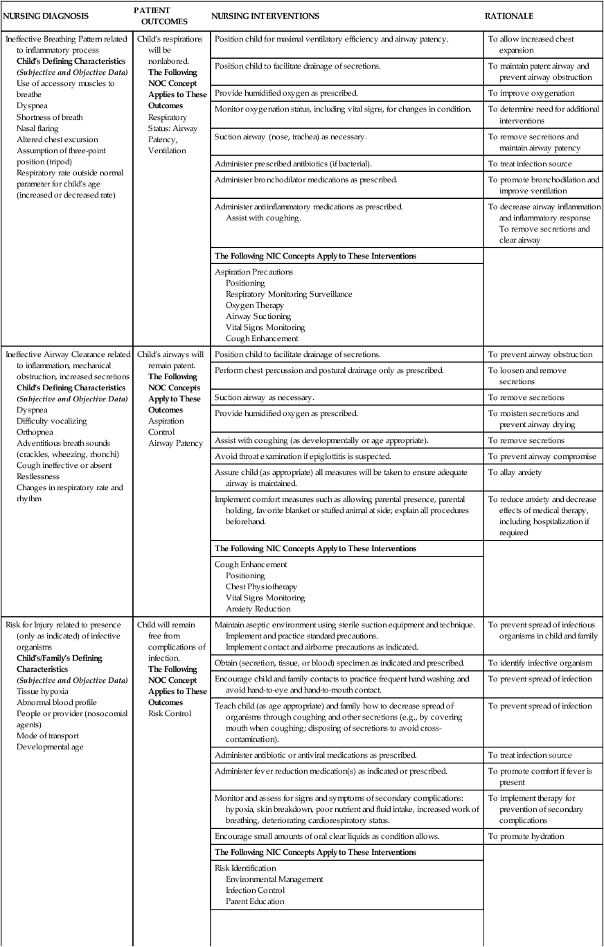
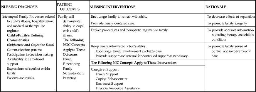
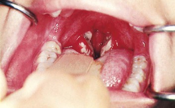
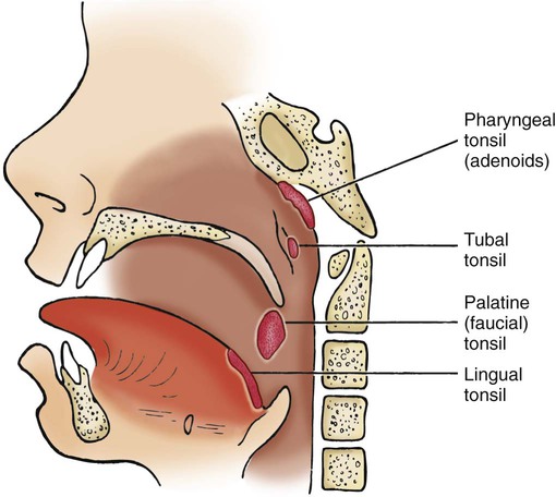
On completion of this chapter the reader will be able to: • Identify the factors leading to respiratory tract infection in infants and young children. • Contrast the effects of various respiratory infections observed in infants and children. • Describe the postoperative nursing care of a child with a tonsillectomy. • Outline a nursing care plan for a child with croup. • Describe priorities of nursing care for a child with acute otitis media. • Identify priorities of nursing care for an infant with respiratory syncytial virus bronchiolitis. • Describe the various therapeutic measures to relieve the symptoms of asthma. • Outline a plan for teaching home care management of a child with asthma. • Describe the physiologic effects of cystic fibrosis on the gastrointestinal and pulmonary systems. • Outline a care plan for a child with cystic fibrosis. • List the major signs of respiratory distress in infants and children. • Describe emergent procedures for the relief of foreign body obstruction in an infant or child. http://evolve.elsevier.com/wong/essentials Animations—Asthma; Bag Ventilation; Bronchi and Bronchioles; Intubation; Intubation in Infant; Intubation, Incorrect Placement; Lung Sounds; Pediatric CPR; Pneumonia; Respiratory Failure, Infant Case Studies—Acute Epiglottitis; Asthma; Bronchiolitis; Cystic Fibrosis; Mononucleosis; Tonsillitis Nursing Care Plans—The Child with Acute Respiratory Infection; The Child with Asthma; The Child with Bronchiolitis and Respiratory Syncytial Virus (RSV) Infection; The Child with Cystic Fibrosis; The Child with Respiratory Failure; The Child with Tonsillectomy Dehydration is a potential complication when children have respiratory tract infections and are febrile or anorectic, especially when vomiting or diarrhea is present. Infants are especially prone to fluid and electrolyte deficits when they have a respiratory illness because a rapid respiratory rate that accompanies such illnesses precludes adequate oral fluid intake. In addition, the presence of fever increases the total body fluid turnover in infants. If the infant has nasal secretions, this further prevents adequate respiratory effort by blocking the narrow nasal passages when the infant reclines to bottle feed or breastfeed and ceases the compensatory mouth breathing effort, thus causing the child to limit intake of fluids. Adequate fluid intake is encouraged by offering small amounts of favorite fluids (clear liquids if vomiting) at frequent intervals. Oral rehydration solutions, such as Infalyte or Pedialyte, should be considered for infants, and water or a low-carbohydrate (≤5 g per 8 oz) flavored drink should be considered for older children. Fluids with caffeine (tea, coffee) are avoided because these may act as diuretics and promote fluid loss. Sports drinks and energy drinks are not recommended for oral rehydration (American Academy of Pediatrics [AAP], 2011). Breastfeeding infants should continue to be breastfed because human milk confers some degree of protection from infection (see Chapter 8). Fluids should not be forced, and children should not be awakened to take fluids. Forcing fluids creates the same problem as urging unwanted food. Gentle persuasion with preferred beverages or sugar-free popsicles is usually more successful. Younger children may like to drink smaller amounts from a plastic medicine cup. To assess their child’s level of hydration (see Chapters 9 and 24), parents are advised to observe the frequency of voiding and to notify the nurse or practitioner if there is insufficient voiding. Counting the number of wet diapers in a 24-hour period is a satisfactory method to assess output in infants and toddlers. In the hospital, diapers are weighed to assess output, which should be at least 1 ml/kg/hr up to 30 kg in weight. Then it should be at least 30 ml per hour in patients weighing more than 30 kg. The practitioner should be notified if the urine output is low. Loss of appetite is characteristic of children with acute infections. In most cases, children can be permitted to determine their own need for food. Many children show no decrease in appetite, and others respond well to foods such as gelatin, popsicles, and soup (see Feeding the Sick Child, Chapter 22). Urging solid foods for children who are sick may precipitate nausea and vomiting and cause an aversion to feeding that may extend into the convalescent period and beyond. Young children with respiratory tract infections are irritable and difficult to comfort; therefore, the family needs support, encouragement, and practical suggestions concerning comfort measures and administration of medication. In addition to antipyretics and nose drops, the child may require antibiotic therapy. Parents of children receiving oral antibiotics must understand the importance of regular administration and of continuing the drug for the prescribed length of time regardless of whether the child appears ill. Parents are cautioned against giving their children any medications that are not approved by the health practitioner and are cautioned to avoid giving antibiotics left over from a previous illness or prescribed for another child. Administering unprescribed antibiotics can produce serious side effects and adverse reactions (see Chapter 22 for administration of medications and teaching parents). See Nursing Care Plan. Acute nasopharyngitis, or the equivalent of the “common cold,” is caused by the rhinovirus, RSV, adenoviruses, enteroviruses, influenza virus, and parainfluenza virus. Symptoms are more severe in infants and children than in adults. Fever is common in young children, and older children have low-grade fevers, which appear early in the course of the illness. Other clinical manifestations are listed in Box 23-3. Symptoms may last up to 10 days. Children with nasopharyngitis are managed at home. There is no specific treatment, and effective vaccines are not available. Antipyretics may be indicated for mild fever and discomfort (see Chapter 22 for management of fever). Rest is recommended. The provision of a humidified environment and increasing oral fluids may be beneficial to some children with a cold. Decongestants may be prescribed for children and infants older than 12 months of age to shrink swollen nasal passages (they should be used with caution in infants younger than 1 year of age). Cough suppressants containing dextromethorphan should be used with caution (cough is a protective way of clearing secretions) but may be prescribed for a dry, hacking cough, especially at night. However, some preparations contain 22% alcohol and can cause adverse effects such as confusion, hyperexcitability, dizziness, nausea, and sedation. Parents should monitor the child carefully for potential adverse effects. Recent concerns regarding serious side effects of cough and cold preparations in young children, particularly infants, and lack of convincing evidence that such medications are effective in reducing symptoms have prompted recommendations by health experts to carefully evaluate the benefits and risks of recommending such preparations for children younger than 6 years of age (Ryan, Brewer, and Small, 2008). Over-the-counter cold preparation such as pseudoephedrine and some antihistamines are not appropriate for the treatment of the common cold in infants and toddlers; these may cause serious side effects in such children and have been associated with death in infants (Rimsza and Newberry, 2008; Ryan, Brewer, and Small, 2008). Support and reassurance are important elements of care for families of young children with recurrent upper respiratory infections (URIs). Because URIs are frequent in children younger than 3 years of age, families may feel they are on an endless roller coaster of illness. They need reassurance that frequent colds are a normal part of childhood and that by 5 years of age, their children will have developed immunity to many viruses. When children spend time in daycare centers, their infection rate is higher than if they are cared for in the home because of increased exposure. Parents should know the signs of respiratory complications and should notify a health professional if complications occur or the child does not improve within 2 or 3 days (Box 23-4). Children who experience GABHS infection of the upper airway (strep throat) are at risk for rheumatic fever (RF), an inflammatory disease of the heart, joints, and central nervous system (CNS) (see Chapter 25), and acute glomerulonephritis (AGN), an acute kidney infection (see Chapter 27). Permanent damage can result from these sequelae, especially RF. GABHS may also cause skin manifestations, including impetigo and pyoderma. Group A β-hemolytic streptococci infection is generally a relatively brief illness that varies in severity from subclinical (no symptoms) to severe toxicity. The onset is often abrupt and characterized by pharyngitis, headache, fever, and abdominal pain. The tonsils and pharynx may be inflamed and covered with exudate (Fig. 23-1), which usually appear by the second day of illness. However, streptococcal infections should be suspected in children older than 2 years of age who have pharyngitis without exudate or nasal symptoms. The tongue may appear edematous and red (strawberry tongue), and the child may have a fine sandpaper rash on the trunk, axillae, elbows, and groin seen in scarlet fever (caused by a strain of group A streptococcus). The uvula is edematous and red. Anterior cervical lymphadenopathy (in ≈30%–50% of cases) usually occurs early, and the nodes are often tender. Pain can be relatively mild to severe enough to make swallowing difficult. Clinical manifestations usually subside in 3 to 5 days unless complicated by sinusitis or parapharyngeal, peritonsillar, or retropharyngeal abscess. Nonsuppurative complications may appear after the onset of GABHS—AGN in about 10 days and RF in an average of 18 days. Children who are GABHS carriers may have a positive throat culture but often experience a coincidental viral illness. Although antibiotic administration is not indicated for most GABHS carriers, some conditions require antibiotic therapy; these are published in the AAP’s Red Book (AAP, Committee on Infectious Diseases and Pickering, 2009). Rapid identification of GABHS with diagnostic test kits (rapid antigen detection test) is possible in the office or clinic setting. Because of the high specificity of these rapid tests, a positive test result generally does not require throat culture confirmation. However, the sensitivities of these kits vary considerably, and a confirmatory throat culture is recommended in patients who have a negative test result (AAP, Committee on Infectious Diseases and Pickering, 2009). Intramuscular (IM) benzathine penicillin G is an appropriate therapy, but it is painful and is not the first choice for children. Oral erythromycin is indicated for children who are allergic to penicillin. Other antibiotics used to treat GABHS are azithromycin, clarithromycin, oral cephalosporins, amoxicillin, and amoxicillin with clavulanic acid (AAP, Committee on Infectious Diseases and Pickering, 2009). Special emphasis is placed on correct administration of oral medication and completion of the course of antibiotic therapy (see Administration of Medication, and Compliance, Chapter 22). If an injection is required, it must be administered deep into a large muscle mass (e.g., vastus lateralis or ventrogluteal muscle). To prevent pain, application of a topical anesthetic cream such as EMLA (an eutectic mixture of lidocaine and prilocaine) over the injection site Several pairs of tonsils are part of a mass of lymphoid tissue encircling the nasal and oral pharynx, known as the Waldeyer tonsillar ring (Fig. 23-2). The palatine, or faucial, tonsils are located on either side of the oropharynx behind and below the pillars of the fauces (opening from the mouth). A surface of the palatine tonsils is usually visible during oral examination. The palatine tonsils are those removed during tonsillectomy. The pharyngeal tonsils, also known as the adenoids, are located above the palatine tonsils on the posterior wall of the nasopharynx. Their proximity to the nares and eustachian tubes causes difficulties in instances of inflammation. The lingual tonsils are located at the base of the tongue. The tubal tonsils, found near the posterior nasopharyngeal opening of the eustachian tubes, are not part of the Waldeyer tonsillar ring. Tonsillectomy is the surgical removal of the palatine tonsils. Absolute indications for a tonsillectomy are recurrent peritonsillar abscess, airway obstruction, tonsillitis resulting in febrile convulsions, and tonsils requiring tissue pathology (American Academy of Otolaryngology—Head and Neck Surgery, 2011). Relative indications include three or more tonsil infections per year, persistent foul taste or breath caused by chronic tonsillitis, unilateral tonsil hypertrophy presumed to be malignant, and chronic tonsillitis in a streptococcus carrier who fails to respond to antibiotics (American Academy of Otolaryngology—Head and Neck Surgery, 2011). Adenoidectomy (the surgical removal of the adenoids) is recommended for children who have hypertrophied adenoids that obstruct nasal breathing; additional indications for adenoidectomy include recurrent adenoiditis and sinusitis, chronic otitis media (OM) with effusion (especially if associated with hearing loss), airway obstruction and subsequent sleep-disordered breathing, persistent mouth-breathing, nasal speech, and recurrent nasopharyngitis (Benninger and Walner, 2007a). For some children, the effectiveness of tonsillectomy or adenoidectomy is modest and may not justify the risk of surgery. In practice, many physicians rely on individualized decision making and do not subscribe to an absolute set of eligibility criteria for these surgical procedures. Contraindications to either tonsillectomy or adenoidectomy are (1) cleft palate because the tonsils help minimize escape of air during speech, (2) acute infections at the time of surgery because locally inflamed tissues increase the risk of bleeding, (3) uncontrolled systemic diseases or blood dyscrasias, and (4) poor anesthetic risk. If surgery is required, the child requires the same psychologic preparation and physical care as for any other surgical procedure (see Chapters 21 and 22). Most tonsillectomy and adenoidectomy (T&A) surgeries now take place in outpatient settings; however, the priorities of preoperative and postoperative care remain the same. The following discussion focuses on postoperative nursing care for T&A, although both procedures may not be performed. The throat is sore after surgery. An ice collar may provide relief, but many children find it bothersome and refuse to use it. Most children experience moderate pain after a T&A and need pain medication regularly for at least the first few days. Analgesics may be given rectally or intravenously to avoid the oral route. Because the pain is continuous, analgesics should be administered at regular intervals even at night (see Pain Management, Chapter 7). An antiemetic such as ondansetron (Zofran) may be administered postoperatively if nausea or vomiting is present. H1N1 (swine flu) is a subtype of influenza type A. In 2009, a pandemic of H1N1 caused significant morbidity and mortality, particularly in Mexico and the United States; it was declared at an end in August 2010. A pandemic is defined by the World Health Organization (2011) as the spread of a new disease to which the population has little or no immunity and that spreads rapidly from human to human. The signs and symptoms of H1N1 flu are the same as those mentioned below for influenza. H1N1 vaccine was combined with the seasonal influenza vaccine in the 2011 to 2012 season.
The Child with Respiratory Dysfunction
Respiratory Infection
Nursing Care Management
![]() Assessment of the respiratory system follows the guidelines described in Chapter 6 (for assessment of the ears, nose, mouth and throat, chest, and lungs). The assessment should include respiratory rate, depth and rhythm, heart rate, oxygenation, hydration status, body temperature, activity level, and level of comfort. Special attention should also be given to the components and observations listed in Box 23-2. A noninvasive pulse oximeter (oxygen saturation) measurement should be performed on all children as part of the routine physical assessment. The nursing process in the care of the child with acute respiratory tract infection is outlined in the Nursing Process box.
Assessment of the respiratory system follows the guidelines described in Chapter 6 (for assessment of the ears, nose, mouth and throat, chest, and lungs). The assessment should include respiratory rate, depth and rhythm, heart rate, oxygenation, hydration status, body temperature, activity level, and level of comfort. Special attention should also be given to the components and observations listed in Box 23-2. A noninvasive pulse oximeter (oxygen saturation) measurement should be performed on all children as part of the routine physical assessment. The nursing process in the care of the child with acute respiratory tract infection is outlined in the Nursing Process box.
![]() Animations—Bronchi and Bronchioles; Lung Sounds
Animations—Bronchi and Bronchioles; Lung Sounds
![]() Nursing Care Plan—The Child with Acute Respiratory Infection
Nursing Care Plan—The Child with Acute Respiratory Infection
Promote Hydration
Provide Nutrition
Provide Family Support and Home Care
Upper Respiratory Tract Infections
Acute Viral Nasopharyngitis
Therapeutic Management
Nursing Care Management
Family Support
Acute Streptococcal Pharyngitis
Clinical Manifestations
Diagnostic Evaluation
Therapeutic Management
Nursing Care Management
 hours before the injection or LMX4 (4% lidocaine) over the site 30 minutes before the injection is helpful (see Administration of Medication: Intramuscular Administration, Chapter 22). The injection site may be tender for 1 to 2 days.
Tonsillitis
![]() Nursing Care Plan—The Child with Tonsillectomy
Nursing Care Plan—The Child with Tonsillectomy
Pathophysiology
Therapeutic Management
Nursing Care Management
Influenza
![]()
Stay updated, free articles. Join our Telegram channel

Full access? Get Clinical Tree


The Child with Respiratory Dysfunction
Get Clinical Tree app for offline access


