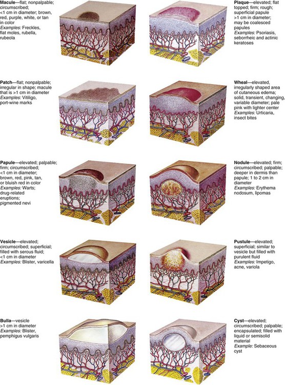
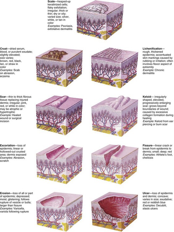
The Child with Integumentary Dysfunction


The Child with Integumentary Dysfunction


Marilyn J. Hockenberry, Rose U. Baker and Mary A. Mondozzi


image


http://evolve.elsevier.com/wong/essentials


Integumentary Dysfunction


Skin Lesions


Lesions of the skin result from a variety of etiologic factors. Skin lesions originate from (1) contact with injurious agents (infective organisms, toxic chemicals, and physical trauma), (2) hereditary factors, (3) external factors (e.g., allergens), or (4) systemic diseases (e.g., measles, lupus erythematosus, nutritional deficiency diseases). Responses to these agents or factors are highly individualized. An agent that is harmless to one individual may be damaging to another, and a single agent may produce varying degrees of response


An important factor in the etiology of skin manifestations is the child’s age. Infants are subject to “birthmark” malformations and atopic dermatitis (AD) that appear early in life, school-age children are susceptible to ringworm of the scalp, and acne is a characteristic skin disorder of puberty. Contact dermatitis, such as poison ivy, is seen only when the noxious agent is found in the environment. Tension and anxiety may produce, modify, or prolong skin conditions.


Pathophysiology of Dermatitis

More than half of the dermatologic problems in children are forms of dermatitis. This implies a sequence of inflammatory changes in the skin that are grossly and microscopically similar but diverse in course and causation. Acute responses produce intercellular and intracellular edema, the formation of intradermal vesicles, and an initial infiltration of inflammatory cells into the epidermis. In the dermis, there is edema, vascular dilation, and early perivascular cellular infiltration. The location and manner of these reactions produce the lesions characteristic of each disorder. The changes are usually reversible, and the skin ordinarily recovers without blemish unless complicating factors such as ulceration from the primary irritant, scratching, and infection are introduced or underlying vascular disease develops. In chronic conditions, permanent effects are seen that vary according to the disorder, the general condition of the affected individual, and the available therapy.


Diagnostic Evaluation

Although the history and subjective symptoms of skin lesions are explored first, the obvious objective characteristics of the lesions are often noted simultaneously. Many skin lesions are easily diagnosed after careful inspection.


History and Subjective Symptoms

Many cutaneous lesions are associated with local symptoms. The most common local symptom is itching (pruritus), which varies in intensity. Pain or tenderness often accompanies some skin lesions. Other skin sensations such as burning, prickling, stinging, or crawling are also described. Alterations in local feeling include absence of sensation (anesthesia); excessive sensitivity (hyperesthesia); diminished sensation (hypesthesia or hypoesthesia); or abnormal sensation, such as burning or prickling (paresthesia). These symptoms may remain localized or migrate; may be constant or intermittent; and may be aggravated by a specific activity, such as exposure to sunlight.


It is important to determine whether the child has an allergic condition such as asthma or hay fever or history of a previous skin disease. AD, often associated with allergies, frequently begins in infancy. Important questions for the parent include when the lesion or symptom first appeared; whether it occurred with ingestion of a food or other substance, including any medication; and whether the condition was related to activity such as contact with plants, insects, or chemicals.


Types of Lesions

Skin lesions assume distinct characteristics that are related to the pathologic process. Nurses should become familiar with the common terms that are applied to skin lesions because these terms are used in the processes of record keeping and communication. These terms include:


Erythema—A reddened area caused by increased amounts of oxygenated blood in the dermal vasculature


Ecchymoses (bruises)—Localized red or purple discolorations caused by extravasation of blood into dermis and subcutaneous tissues


Petechiae—Pinpoint, tiny, and sharp circumscribed spots in the superficial layers of the epidermis


Primary lesions—Skin changes produced by a causative factor; common primary lesions in pediatric skin disorders are macules, papules, and vesicles (Fig. 30-1)


Secondary lesions—Changes that result from alteration in the primary lesions, such as those caused by rubbing, scratching, medication, or involution and healing (Fig. 30-2)


Distribution pattern—The pattern in which lesions are distributed over the body, whether local or generalized, and the specific areas associated with the lesions


Configuration and arrangement—The size, shape, and arrangement of a lesion or groups of lesions (e.g., discrete, clustered, diffuse, or confluent)


Wounds


Wounds are structural or physiologic disruptions of the skin that activate normal or abnormal tissue repair responses. Wounds are classified as acute or chronic. Acute wounds are those that heal uneventfully within 2 to 3 weeks. Chronic wounds are those that do not heal in the expected time frame or are associated with complications. Cofactors that disrupt or delay wound healing include compromised perfusion, malnutrition, and infection. In children, most wounds are acute and can be prevented from becoming chronic wounds through appropriate nursing care. Wounds are also classified as surgical and nonsurgical and then further classified in the same manner as burns: superficial, partial thickness, or full thickness (complex wounds that include muscle or bone).


Process of Wound Healing

When the skin is injured, its normal protective barrier function is broken. In a healthy immunocompetent individual, acute traumatic abrasions, lacerations, and superficial skin and soft tissue injuries heal spontaneously without complications. The process of tissue healing involves complex cellular interactions and biochemical reactions. The healing process is segregated into four phases that are characterized by the particular cells involved and the chemicals produced. The four stages of wound healing are hemostasis, inflammation, proliferation, and remodeling (Krasner, Rodeheaver, and Sibbald, 2007). Some authorities combine the first two phases.


In the hemostasis phase, platelets act to seal off the damaged blood vessels and to form a stable clot. Hemostasis occurs within minutes of the initial injury to the skin unless there is an underlying clotting disorder.


Inflammation, the second stage of wound healing, presents a clinical picture that involves erythema, swelling, and warmth, often associated with pain at the wound site. This stage usually lasts up to 4 days after injury. The inflammation phase involves white blood cells such as the neutrophils, monocytes, and macrophages. These cells mount an initial defense against microbial invasion and secrete proteolytic enzymes that destroy nonviable tissue and microorganisms in the wound area.


The proliferative phase, which includes granulation and contracture, is the third stage of healing. This phase lasts from 4 to 21 days in acute wounds, depending on the size of the wound. The phase involves the replacement of dermal tissues and subdermal tissues in deep wounds, as well as the contraction of the wound. The phase is characterized clinically by the presence of granulation tissue, the “beefy,” pebbled red tissue in the wound base. Fibroblasts, or immature connective tissue cells, secrete collagen, which provides the foundation for dermal regeneration. Angiocytes regenerate the outer layers of capillaries, and endothelial cells produce the lining in a process called angiogenesis. The formation of granulation tissue, which provides the foundation for the wound, depends on angiogenesis. The keratinocytes are responsible for epithelialization. In the final stage of epithelialization, contracture occurs as the keratinocytes differentiate and form the protective outer layer, or stratum corneum, of the skin.


Remodeling, or maturation, is the final phase of the healing process. This phase occurs in the dermis as fibroblasts increase the tissue tensile strength and gradually replace type 3 collagen in the scar tissue with type 1 collagen, thicken the collagen fibers, and reorient the collagen fibers along the lines of tissue tension. Fibroblasts disappear as the wound becomes stronger. The wound edges are brought closer together, and a mature scar is formed. Children heal aggressively with abundant scar tissue, especially during growth spurts. The highly elastic quality of children’s skin pulls on the wound, and the wound defends against this pull by forming scar tissue. Remodeling and maturation occur over several months and can take up to 2 years. Thus, some wounds that appear to be completely healed can break down suddenly if attention is not paid to the initial causative factors.


The phases of wound healing are complex and may be interrupted by disease conditions, medications, and other systemic and local factors that influence the healing process. When a wound does not follow the “normal wound healing trajectory,” it may become stuck in one of the stages and become a chronic wound. It is important that health care providers understand and address the factors that influence wound healing and prevent the development of chronic wounds.


Factors That Influence Healing

Wound care management has shifted from interventions aimed at maintaining a dry environment to those that promote a moist, crust-free environment that enhances the migration of epithelial cells across the wound and facilitates remodeling. Whereas an acute full-thickness wound kept in a moist environment usually reepithelializes in 12 to 15 days, the same wound when kept open to the air heals in about 25 to 30 days.


Numerous factors can delay healing (Table 30-1). For example, traditional practices, such as the use of antiseptics (hydrogen peroxide and povidone–iodine [Betadine] solutions), which were once thought to prevent infection, are now known to have a cytotoxic effect on healthy cells and minimal effect on controlling infections. Povidone–iodine may also be absorbed through the skin in neonates and young children.


TABLE 30-1


FACTORS THAT DELAY WOUND HEALING


FACTOR EFFECT ON HEALING
Dry wound environment Allows epithelial cells to dry out and die; impairs migration of epithelial cells across wound surface
Nutritional deficiencies  
 Vitamin A Results in inadequate inflammatory response
 Vitamin B1 Results in decreased collagen formation
 Vitamin C Inhibits formation of collagen fibers and capillary development
 Protein Reduces supply of amino acids for tissue repair
 Zinc Impairs epithelialization
Immunocompromise Results in inadequate or delayed inflammatory response
Impaired circulation Inhibits inflammatory response and removal of debris from wound area
  Reduces supply of nutrients to wound area
Stress (pain, poor sleep) Releases catecholamines that cause vasoconstriction
Antiseptics  
 Hydrogen peroxide Toxic to fibroblasts; can cause subcutaneous gas formation (mimics gas-forming infection)
 Povidone–iodine Toxic to WBCs, RBCs, and fibroblasts
 Chlorhexidine Toxic to WBCs
Medications  
 Corticosteroids Impair phagocytosis
  Inhibit fibroblast proliferation
  Depress formation of granulation tissue
  Inhibit wound contraction
 Chemotherapy Interrupts the cell cycle; damages DNA or prevents DNA repair
 Antiinflammatory drugs Decrease the inflammatory phase
Foreign bodies Increase inflammatory response
  Inhibit wound closure
Infection Increases inflammatory response
  Increases tissue destruction
Mechanical friction Damages or destroys granulation tissue
Fluid accumulation Accumulation in area inhibits tissues from approximating
Radiation Inhibits fibroblastic activity and capillary formation
  May cause tissue necrosis
Diseases  
 Diabetes mellitus Inhibits collagen synthesis
  Impairs circulation and capillary growth
  Hyperglycemia impairs phagocytosis
 Anemia Reduces oxygen supply to tissues
 Peripheral vascular disease Reduces oxygen supply to wounds
 Uremia Decreases collagen and granulation tissue

DNA, Deoxyribonucleic acid; RBC, red blood cell; WBC, white blood cell.


General Therapeutic Management


Some skin disorders demand aggressive therapy, but by and large, the major aim of treatment is to prevent further damage, eliminate the cause, prevent complications, and provide relief from discomfort while tissues undergo healing (McCord and Levy, 2006). Factors that contribute to the development of dermatitis and that prolong the course of the disease should be eliminated when possible. The most common causative agents of dermatitis in infants, children, and adolescents are environmental factors (soaps, bubble baths, shampoos, rough or tight clothing, wet diapers, blankets, and toys) and the natural elements (e.g., dirt, sand, heat, cold, moisture, and wind). Dermatitis may also result from home remedies and medications.


Dressings

No one dressing meets the needs of all wounds. The traditional dry gauze dressing should not be used on open wounds because it allows the wound surface to dry, does little to prevent bacterial invasion, and adheres to the dried scab so that removal disturbs the newly regenerating epithelial cells. In most instances, traditional gauze dressings have been replaced by dressings that promote moist wound healing. Moist wound healing increases the rate of collagen synthesis and reepithelialization and decreases pain and inflammation. It also creates an environment for autolytic débridement of necrotic tissue, which creates a clean wound bed and enhances granulation. However, a balance must be achieved between creating a moist wound bed and maintaining a dry periwound area that protects the skin and wound from maceration. The dressing type and frequency of dressing changes help to achieve this balance. The frequency of dressing changes is based on the presence of infection, the type of dressing, the location of the wound, and the amount of drainage. Dressings should always be changed when they are loose or soiled. They should be changed more frequently in areas where contamination is likely (e.g., the sacral area, the buttocks, the tracheal area) or when wound infection is suspected or present.


Topical Therapy

Several agents and methods are available for treatment. In selecting a therapeutic regimen, the practitioner considers (1) the choice of active ingredient, (2) the proper vehicle or base, (3) the cosmetic effect, (4) the cost, and (5) instructions for use. Several basic concepts must also be considered. Overtreatment is avoided. For example, when dermatitis is acute, topical applications should be mild and bland to avoid further irritation. Broken or inflamed skin, especially in children, is more absorbent than intact skin, and chemicals that are nonirritating to intact skin may be quite irritating to inflamed skin.


Topical applications may be applied to treat the disorder, reduce itching, decrease external stimuli, or apply external heat or cold. The emollient action of soaks, baths, and lotions provides a soothing film over the skin surface that reduces external stimuli. Ordinarily, lukewarm, tepid, or cool applications offer the greatest relief.


Ointments in a petrolatum base provide protection from moisture. Therefore, this type of ointment is indicated around gastrostomy tubes, in skinfolds, and in the diaper area. Creams are absorbed by the skin and are used for areas where a nongreasy “feel” is desired (e.g., face, hands).


Topical Corticosteroid Therapy

Glucocorticoids are the therapeutic agents used most frequently for skin disorders. Their local antiinflammatory effects are merely palliative, so the medication must be applied until the condition undergoes a remission or the causative agent is eliminated. Corticosteroids are applied directly to the affected area, are essentially nonsensitizing, and have only minor side effects. As with the use of any steroids, their use in large amounts may mask signs of infection, and symptoms may be exacerbated after termination of the drug. Families are cautioned that the medication cannot be used for all skin disorders. The concentrations available without prescription are not adequate for stubborn skin conditions (e.g., psoriasis) and may further aggravate inflammation caused by fungus or bacteria. Most parents and children apply too much topical hydrocortisone; therefore, they should be counseled that it is both effective and economical to apply only a thin film and to massage it into the skin. Parents and children should also be advised to use the application for no more than 5 to 7 days because these agents may cause depigmentation and other changes in the skin.


Systemic Therapy

Systemic drugs may be used as an adjunct to topical therapy in some dermatologic disorders. The drugs most frequently used are corticosteroids, antibiotics, and antifungal agents. Corticosteroids are valuable because of their capacity to inhibit inflammatory and allergic reactions. The dosage is carefully adjusted and gradually tapered to the minimum dosage that is effective and tolerated. In infants and children, the dosage is larger than is usually calculated from body weight ratios. However, prolonged use may temporarily suppress growth.


Antibiotics are used in severe or widespread skin infections. However, because these drugs tend to produce hypersensitivity in some patients, they are used with caution. Antifungal agents are the only means for treating systemic fungal infections.


Nursing Care Management


The child’s subjective symptoms and the parent’s history provide valuable information to help establish a diagnosis. Older children often describe the condition as painful, itching, or tingling or in other descriptive terms. However, much can be determined by also observing the younger child’s behavior. Does the child scratch? Is the child restless or irritable? Does the child favor or avoid using a body part? A careful history provides important clues. Has the child had access to chemicals or been in the woods or around a woodpile? Has the child eaten a new food? Is the child taking medication? Has the child any known allergy? Do siblings or playmates have similar lesions? What soap or bubble bath is used for bathing?


It is important for nurses to not only describe but also assess skin lesions and wounds. The color, shape, and distribution of lesions and wounds are important. Individual lesions are described according to standard terminology. Sometimes two descriptors are used for a particular characteristic (e.g., maculopapular rash). To confirm or amplify the findings made by inspection, the nurse may gently palpate the skin to detect characteristics such as temperature, moisture, texture, elasticity, and edema. Wounds are assessed for depth of tissue damage, evidence of healing, and signs of infection.


Jan 16, 2017 | Posted by in NURSING | Comments Off on The Child with Integumentary Dysfunction

Full access? Get Clinical Tree

Get Clinical Tree app for offline access