Renal Disorders

13 Renal Disorders


Essential anatomy and physiology


Kidney Structure


Gross Anatomy


The kidneys lie anterior and lateral to the twelfth thoracic and first, second, and third lumbar vertebrae and behind the abdominal peritoneum; therefore they are retroperitoneal structures. The kidneys are embedded in a mass of fatty tissue called the adipose capsule, and each capsule is enclosed in the renal fascia (Fig. 13-1). The kidneys are not secured to the abdominal wall, but are held in position by the renal fascia and the large renal arteries and veins. The adipose capsule and the pararenal fat help to protect the kidney and keep it in place.
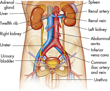


The medial aspect of each kidney is curved away from the midline; at the center of this concavity is the hilus, where the renal artery and nerves enter the kidney and where the renal vein and ureter exit the kidney. Surrounding each kidney is a tough, nearly indistensible fibrous capsule, which becomes the outer lining of the renal calyces, renal pelvis, and ureter.


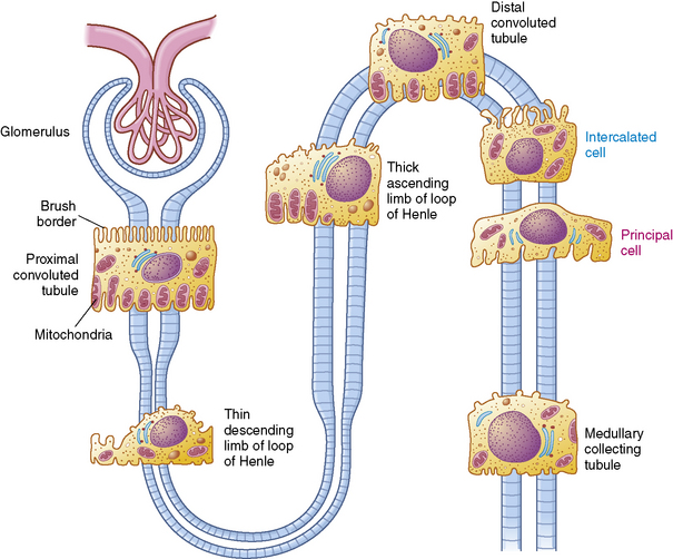
A longitudinal section of the kidney shows the three general areas of renal structure: the cortex, the medulla, and the pelvis (Fig. 13-2). The renal cortex is the outer portion of the kidney. It has a granular appearance and extends in fingerlike projections into the medullary areas. The cortex contains most of the nephrons, the smallest functioning unit of the kidney. The cortex also contains all glomeruli, the proximal and distal convoluted tubules, and the first parts of the loop of Henle and the collecting ducts.
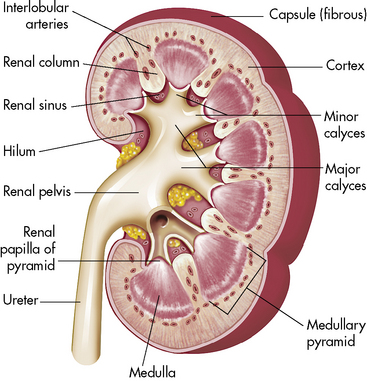


The renal medulla is composed predominately of the long loops of Henle from the juxtamedullary nephrons and the collecting ducts that grow progressively larger as they approach the renal pelvis. These structures give the medulla a striated, pyramidal appearance, with the apex of the pyramid pointing toward the renal pelvis and the base pointing toward the renal cortex.


The renal pelvis contains the outflow tract and a small amount of surrounding fat that acts as a cushion. The renal outflow tract begins with the minor calyces (Greek for cup) that receive urine from the collecting ducts. The urine flows from the minor calyces into the major calyces, into the renal pelvis, into the ureters and bladder, out of the bladder, and through the urethra to exit the body through the urethral meatus.


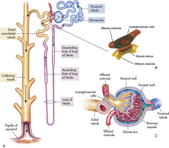
The functioning unit of the kidney is the nephron, which consists of a vascular component and a tubular component (Fig. 13-3). Each kidney contains approximately 1 million distinct nephrons. Eighty-five percent of all nephrons originate in the outermost area of the cortex. The remaining nephrons are the juxtamedullary nephrons that originate in the inner cortical area. The long loops of Henle from the juxtamedullary nephrons that extend deep into the medulla lie parallel to the medullary collecting ducts and play an important role in the concentration of urine (see Evolve Fig 13-1 in the Chapter 13 Supplement on the Evolve Website).


Renal Vasculature


In most patients, each kidney is supplied with systemic arterial blood from a single artery. The renal arteries branch from the aorta at the level of the second or third lumbar vertebrae; together they receive approximately 20% of the total cardiac output. Each artery divides into an anterior and posterior artery. These arteries continue to branch into small arterioles. Some of these arterioles will supply nutrients to the renal medulla, cortical tissue, and capsule, while other arterioles enter the glomerular capsule.


The afferent arteriole enters the glomerular capsule and divides to form the glomerulus, a tuft of capillaries that allows filtration of plasma through the capillary membranes. The glomerular capillaries do not recombine into venous channels, but instead recombine into a second arteriole called the efferent arteriole (Fig. 13-4). Because arterioles are present at either end of the glomerular capillary system, constriction or dilation of these arterioles will alter the resistance to flow through the glomerular capillaries and thus will regulate glomerular filtration.


After leaving the glomerulus, the efferent arterioles branch to form a network of capillaries that surround the convoluted tubules and the loop of Henle. These peritubular capillaries then converge into venules that will return renal venous blood to the systemic circulation via the inferior vena cava. Elements that the kidney reabsorbs from the filtrate to return to the circulation are reabsorbed into this peritubular capillary system.


Renal Tubules and Collecting Ducts


The tubular component of the nephron begins as a single layer of flat epithelial cells surrounding the glomerulus. This layer is known as Bowman’s capsule (Fig. 13-4, B). Filtered plasma from the glomerular capillaries will enter Bowman’s capsule and flow into a coiled tubule called the proximal tubule, also known as the proximal convoluted tubule (Fig. 13-5).


The structure and appearance of the proximal tubule changes as it descends toward the renal medulla. The tubular lumen narrows and the cells become flattened as the tubule makes a hairpin turn, called the loop of Henle. As the loop of Henle ascends from the medulla into the renal cortex, the tubular cells enlarge and again become cuboidal; in addition, the tubule coils, forming the distal convoluted tubule. The tubule then straightens and joins the collecting duct.


Collecting ducts are the terminus of many distal tubules; they are formed in the inner and outer renal cortex. These small collecting ducts enter the renal medulla where they form larger ducts, which in turn drain into a minor calyx in the renal pelvis. Approximately 8 to 10 minor calyces join into the major calyces, which combine to form the renal pelvis. The renal pelvis is the largest portion of the outflow tract proximal to the bladder.


The Bladder and Urethra


The urinary bladder is a hollow, muscular organ that stores urine. There are three openings in the bladder wall: the entrances of the two ureters and the exit of the urethra. These openings form the corners of a triangle, called the trigone. There is a dense area of smooth (involuntary) muscle around the neck of the bladder at the orifice of the urethra; this muscle constitutes the internal sphincter. The urethra extends from the urinary bladder to the body surface. At the point where the urethra passes through the muscles of the pelvic floor, striated (voluntary) circular muscles form an external sphincter.55


Micturition is the emptying of the stored urine from the bladder. The process normally involves both voluntary and involuntary nervous system activities in children beyond approximately 2 to 3 years of age. Once an adequate volume of urine has accumulated in the bladder, the bladder wall stretches, stimulating stretch receptors. Sensory signals are then conducted through afferent pelvic nerves to the spinal cord. Efferent nerves from the spinal cord return impulses through the parasympathetic fibers in the pelvic and hypogastric nerves to the bladder wall muscle and the neck of the bladder. Efferent nerve stimulation causes contraction of the bladder and relaxation of the internal sphincter. In addition, impulses from the central nervous system through the pudendal nerves innervate the voluntarily controlled external sphincter. If the external sphincter also relaxes, the bladder will then empty.


Appropriate contraction and voluntary intermittent emptying of the bladder require both inhibitory and facilitory impulses from the upper pons, the hypothalamus, the midbrain, and the cortex. The inhibitory centers prevent constant voiding, and the facilitory centers allow micturation to occur voluntarily (once bladder control is learned). If the inhibitory centers are injured, the patient can demonstrate an uninhibited neurogenic bladder and nearly constant urination.


Reflex bladder contraction and sphincter relaxation also require the presence of intact afferent nerves from the bladder to the second and third sacral spinal cord level and intact efferent nerves (including the hypogastric, pelvic, and pudendal nerves) from the first through the third sacral spinal level. If afferent nerves from the bladder to the spinal cord are injured or malformed, the patient can develop an atonic bladder, with loss of voluntary sphincter control. When an atonic bladder is present, the bladder fills to capacity and then overflow voiding begins.


If the spinal cord is damaged above the sacral spinal level, the patient initially loses all micturation reflexes because inhibitory and facilitory reflexes from the brain cannot be transmitted through the injured spinal cord. Later, however, simple spinal reflexes can return and the patient can void when bladder distension is sufficient. In this case, the bladder reflex will be initiated at the volume of urine that is usually present in the bladder during the patient’s convalescent period.45
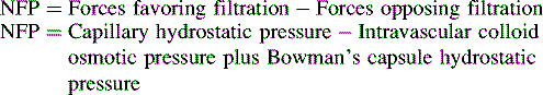


Glomerular Function


Filtration Physiology


The kidney receives its sympathetic nerve supply from the tenth through twelfth thoracic nerves and its parasympathetic nerve supply from branches of the vagus nerve. The renal blood vessels are innervated, but the renal tubules are not. Adjustment in the diameter of either or both of the afferent and efferent arterioles will affect the amount of fluid filtered in the glomerulus. As with any capillary, filtration of fluid in the glomerulus is affected by pressure gradients across the capillary bed and the intrinsic properties of the glomerular capillary membrane.


Capillary hydrostatic pressure is the pressure generated by the pumping action of the heart; it is maintained or altered by arterial resistance. Hydrostatic pressure in most capillaries is higher at the arterial end than at the venous end. This difference favors filtration of fluid out of the vascular space at the arterial end and favors reabsorption of fluid into the vascular space at the venous end. Because the glomerulus has an afferent arteriole at the proximal end and an efferent arteriole at the distal end, the glomerular capillary pressure is higher than in other capillary beds and is approximately equal to systemic arterial pressure. This high pressure favors fluid filtration out of the vascular space. The glomerular capillary pressure is altered by constriction or relaxation of the afferent or efferent arteriole.


Intravascular colloid osmotic pressure, or oncotic pressure, is the pressure opposing free water movement out of the vascular space. It is generated by dissolved proteins, ions, and other particles that are normally present in the blood. Larger particles such as proteins cannot move readily across a capillary membrane; therefore they remain in the vascular space, exerting an osmotic pressure of approximately 35   mm Hg. This oncotic pressure opposes hydrostatic filtration from the vascular space.55


The hydrostatic pressure present in Bowman’s capsule is the pressure exerted on the glomerulus by fluid in the Bowman’s capsule and collecting ducts. This pressure is normally 10 to 15   mm Hg and opposes fluid filtration from the glomerulus. Because tubular fluid is normally protein-free, the oncotic pressure in Bowman’s capsule is normally negligible. (See Evolve Fig. 13-2 in the Chapter 13 Supplement on the Evolve Website for a diagram of these pressures.)


Net filtration pressure (NFP) within the nephron is the difference between forces favoring filtration (largely resulting from capillary hydrostatic pressure) and forces opposing filtration (largely caused by intravascular colloid osmotic pressure and the hydrostatic pressure within Bowman’s capsule) as follows:


image


Any change in the hydrostatic pressure in either Bowman’s capsule or the capillaries or any changes in serum colloid osmotic pressure can change the net filtration pressure and may result in a change in the glomerular filtration rate (GFR). For example, obstruction of a ureter increases resistance to urine drainage from the renal pelvis; this increases pressure in the tubules and in Bowman’s capsule and opposes filtration. The loss of a large volume of hypotonic fluid caused by diarrhea or unreplaced insensible losses during high fever will produce dehydration and hemoconcentration; this increases the colloid osmotic pressure and opposes filtration. If the child develops severe hypotension, capillary hydrostatic pressure will fall. All these changes oppose filtration and reduce the amount of glomerular filtrate. As noted previously, capillary hydrostatic pressure is determined by cardiac output (blood flow) and resistance in the arterioles. The relationship of flow, pressure, and resistance is described by the following equation (Poiseuille’s Law):


image


image


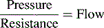
P = mean arterial pressure − venous pressure for that organ


This equation predicts that an increase in mean arterial pressure will increase blood flow if the resistance to flow remains constant. The kidney is designed to maintain glomerular hydrostatic pressure so it can create urine over a wide range of blood pressures. As a result, resistance to flow does not remain constant when the systemic arterial pressure changes. Instead, when mean arterial pressure increases, the afferent arterioles constrict; this constriction restricts renal blood flow and prevents transmission of the entire increase in arterial pressure to the glomerulus. When arterial pressure falls, sympathetic innervation to the afferent and efferent arterioles increases arterial tone and increases the resistance to flow into and out of the glomerulus; this vasoconstriction can maintain the GFR at near-normal levels despite a fall in systemic arterial pressure and renal blood flow.


The ability to respond to changes in flow into and out of the glomerulus allows the kidney to maintain solute and volume regulation at relatively constant levels, despite changes in systemic arterial blood pressure and renal blood flow; this ability is termed autoregulation. When the arterial pressure is extremely high or low, autoregulation fails and renal blood flow is proportional to arterial pressure.


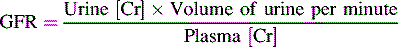
Glomerular Filtration Rate (GFR)


Renal function can be evaluated by calculating the GFR. The GFR, in turn, is roughly equivalent to the creatinine clearance; therefore it can be estimated by calculating the creatinine clearance. This estimate should not, however, be the sole means of determining renal function.56


Creatinine is a small molecule byproduct of skeletal muscle creatine metabolism. Creatinine is released at a near constant rate into the bloodstream, is filtered freely at the glomerulus, and is not broken down, reabsorbed, or synthesized by the renal tubules. Only a tiny amount of creatinine is secreted by the renal tubules. In effect, all of the creatinine that is filtered from the vascular space at the glomerulus remains in the urine and can be measured, and the creatinine clearance mirrors the GFR.62


Calculation of creatinine clearance requires collection of a urine sample for a precise period of time. A blood sample is collected during that same time period. The sampling of blood and urine enables simultaneous determination of the concentration of creatinine in the plasma and in the urine, as well as calculation of creatinine clearance.


The relationship between the plasma and urine concentrations of creatinine (Cr), the urine volume formed per unit of time, and the glomerular filtration rate is expressed as follows:


image


It is important to note that laboratory determination of serum creatinine concentration may be affected by some cephalosporin antibiotics. For this reason the blood sample for analysis of the serum creatinine level should be obtained when antibiotic drug levels are at their lowest.7


The GFR is expressed as milliliters per minute per 1.73 square meter of body surface area, which allows a comparison of renal function among children and adults. The child’s GFR is approximately 55 to 65   mL/minute per 1.73   m2 and will approach adult values (120   mL/minute per 1.73   m2) by approximately 3 years of age.


When the amount of fluid filtered by the glomerulus is expressed as a fraction (ratio) of the total renal plasma flow (600   mL/minute per 1.73   m2 in the adult), this provides an estimate of the percentage of total renal plasma flow that is filtered into Bowman’s capsule. This ratio is termed the filtration fraction and equals approximately 20% of total renal plasma flow. Plasma that is not filtered (i.e., plasma that remains in the vascular space) continues through the glomerulus into the efferent arteriole, through the peritubular capillaries, and into venules and interlobular veins.


The glomerular filtrate in Bowman’s capsule is an ultrafiltrate of blood. Its composition, like interstitial fluid from other capillaries, is usually free of proteins and cells. Animal micropuncture studies have established that all of the solutes (such as ions and amino acids) measured in the glomerular filtrate are present in virtually the same concentrations as their free, unbound concentrations in the plasma. If a substance is bound even partially to protein, that restricts its glomerular filtration because proteins normally cannot pass through the glomerular capillary membrane.


The urine that ultimately is formed by the kidneys is not merely the ultrafiltrate of plasma, because excretion of an ultrafiltrate through the urine would soon deplete the body of solutes and water. To modify the volume and content of the urine, the tubules selectively reabsorb and secrete substances.


Tubular Function


Reabsorption


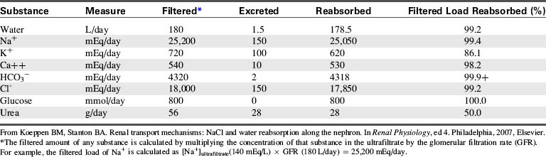
Table 13-1 summarizes the work of the renal tubular cells in the process of reabsorption. An average of 180   L of water (protein-free plasma) is filtered through the glomerulus of an adolescent per day, and yet the average urine output is 1.5   L/day. This means that 178.5   L of water is reabsorbed out of the tubular lumen back into the body’s circulation per day.


Passive and Active Reabsorption


Reabsorption of substances from the renal tubular fluid is described as passive if no energy-requiring reactions are necessary. Passive reabsorption occurs if a substance is reabsorbed as the result of an electrical or concentration gradient. An electrical gradient causes charged particles to move toward particles of opposite charge and away from particles of similar charge, or it may cause an exchange of similarly charged particles across a membrane to maintain an electrical balance.


A concentration gradient is created by the tendency of substances in solution to be distributed equally throughout that solution. Substances will tend to move across a semipermeable membrane from an area of high concentration to an area of lower concentration.


Active reabsorption or active transport of substances moves substances against a concentration or electrical gradient. Active reabsorption requires energy expenditure by the transporting cells. Both active and passive reabsorption from the renal tubules require diffusion of substances from the lumen through the tubular luminal cell membrane. Once the substances enter the cell, they traverse the cytoplasm of the tubular cell and exit through the cell membrane on the opposite side of the cell into the interstitial fluid. These substances can then pass into the adjacent peritubular capillaries for return to the systemic venous circulation. If energy is required in any of these steps, the process is considered active transport. Sodium, chloride, glucose, and bicarbonate are important substances that are reabsorbed actively, whereas water is reabsorbed passively.


Transport Maximum and Thresholds


Many of the substances that are transported actively out of the tubules can be reabsorbed only in limited quantity over time. These substances exhibit a transport maximum (Tm). This transport maximum is relatively fixed for each substance, although it can be affected by hormones or drugs. The renal threshold of a substance is the plasma and filtrate concentration at which some of the active transport tubular carriers become saturated and are unable to reabsorb all of the substance present in the filtrate. At this point, some of the substance will begin to appear in the urine because it cannot all be reabsorbed from the filtrate.


The tubular transport maximum is reached when all of the tubular carriers for that substance are saturated. Any further increase in the serum and filtered concentration of the substance beyond the transport maximum will produce a proportional increase in the urine concentration of the substance.


Glucose is a familiar substance that can be used to illustrate this concept of renal threshold and tubular transport maximum. Under normal conditions, glucose is not excreted in the urine. All the glucose filtered by the glomerulus is reabsorbed by the tubules and returned to the blood. When the serum glucose concentration exceeds approximately 180   mg/dL, some glucose tubular carriers are saturated and glucose begins to appear in the urine. The appearance of glucose in the urine indicates that the renal threshold for glucose reabsorption has been reached. If the serum glucose concentration exceeds approximately 300   mg/dL, all the tubular carriers are saturated and the transport maximum for glucose is reached. Further increase in the serum glucose concentration will produce a proportional increase in the urine glucose concentration. The difference between the renal plasma threshold and the transport maximum for glucose is caused by different transport maximums of individual nephrons and tubules.


For many substances, there is a large difference between the normal serum concentration of a substance and the renal threshold and transport maximum of that substance. This difference indicates that the kidney conserves the substance but does not regulate its serum concentrations. Once the serum concentration of the substance far exceeds the homeostatic requirements, that substance will be lost into the urine. Glucose is an example of a substance that is conserved by the kidneys, although the serum glucose concentration is not regulated by the kidneys.


If the renal threshold and transport maximum are approximately equal to the daily filtered load of a substance, then the kidneys participate in regulation of the serum concentration of the substance. In such a case, a slight increase or decrease in plasma and filtered concentration of the substance changes its rate of renal reabsorption and excretion, so the serum concentration returns to normal. The renal threshold and transport maximum for phosphate are close to the normal daily filtered load of phosphate, so the serum phosphate concentration is regulated by kidney tubular function. Phosphate transport and reabsorption also will be affected by the serum calcium concentration, parathyroid hormone (PTH), and adrenal cortical hormones.63


Reabsorption and Secretion in the Proximal Tubule


The selective reabsorption of solute begins in the proximal tubules. Approximately 67% of the filtered water, Na+, Cl, K+, and other solutes such as bicarbonate are reabsorbed in the proximal tubule. In addition, the proximal tubules normally reabsorb all filtered amino acids and glucose.63


The most important function of the proximal tubule is the reabsorption of the filtered sodium and water. The proximal tubule neither concentrates nor dilutes the urine; its primary responsibility is the reabsorption of sodium, water, and electrolytes.


Sodium


The primary mechanism for regulation of intracellular and extracellular fluid volume involves renal sodium excretion.63 Sodium is filtered freely at the glomerulus, so its concentration in the proximal glomerular filtrate is identical to its plasma concentration. Sodium is reabsorbed by an active transport mechanism; the mechanism is carrier-mediated and requires energy so the sodium can move against a gradient. Sodium is not secreted into the tubules.


Once sodium is filtered into the tubules, it moves passively through the extremely sodium-permeable brush border of the proximal tubular cell. Sodium diffuses across this cell in response to a concentration gradient to the opposite cell membrane that is impermeable to sodium. This cell membrane then actively pumps sodium out of the tubular cell into the surrounding interstitial fluid. The movement of sodium out of the tubular lumen into the interstitial fluid creates an osmotic gradient between the tubule and the interstitial fluid. Because the epithelium of the proximal tubule is highly permeable to water, water follows the movement of the sodium ion. As water moves out of the tubule, the relative concentration of the other solutes within the tubular lumen increases, establishing a concentration gradient for the solutes between the tubular lumen and the interstitial fluid. As a result, solutes such as chloride, calcium, and urea will diffuse passively out of tubules and into the tubular cells and interstitial fluid.


Diffusion and transport of the sodium ion from the tubule also creates an electrical gradient between the tubular lumen and the inside of the tubular cell; the tubular cell now contains more positively charged (sodium) ions, and the tubular lumen (which has lost positive ions), becomes more negatively charged. This electrical gradient causes passive reabsorption of negatively charged substances such as chloride.


As the ultrafiltrate reaches the end of the proximal tubule, 65% of the filtered sodium and water has been reabsorbed into the renal interstitial space, predominantly through the active transport of sodium. Because water is being reabsorbed at almost the same rate as sodium is being pumped out of the proximal tubule, the osmolality of the proximal tubular fluid will be virtually the same as the plasma osmolality (normally 275-295   mOsm/L).


Sodium and water reabsorption in the proximal tubule and in the loop of Henle varies proportionately with the glomerular filtration rate. Increases in GFR are accompanied automatically by increases in sodium and water reabsorption. This coupling between the quantity of filtrate and the amount of reabsorption is termed glomerulotubular balance. This balance means that if renal blood flow remains constant, sodium and water reabsorption will vary directly with the GFR; if the GFR increases, sodium and water reabsorption will increase. Conversely, if renal blood flow remains constant and the GFR falls, sodium and water reabsorption will decrease. This mechanism maintains sodium balance despite changes in the GFR. If there is a severe reduction in renal arterial pressure and GFR, sodium will be reabsorbed almost completely from the proximal tubule.


Bicarbonate and Hydrogen Ions


Because the kidney is responsible for bicarbonate reabsorption and is also responsible for generating new bicarbonate ions, it plays an important role in the regulation of acid-base balance. Sodium and bicarbonate ions in the glomerular filtrate enter the proximal tubule. There, as noted previously, the sodium passively diffuses by concentration gradient into the proximal tubular cell and then is actively transported out of the tubular cell. To maintain electrical balance, another positively charged ion—hydrogen—is pumped actively from the tubular cells into the tubular lumen.


Once the hydrogen ion enters the tubular lumen, it combines with the bicarbonate in the filtrate to form carbonic acid. The carbonic acid in the tubule quickly disassociates to form carbon dioxide and water. The carbon dioxide easily diffuses back through the tubular cell membrane where it recombines with water, forming carbonic acid. Subsequent disassociation of the carbonic acid within the tubular cell again forms the hydrogen ions and bicarbonate ions; and the process is repeated. The tubular cell will again actively secrete the hydrogen ions into the lumen in exchange for sodium ions, and the bicarbonate ions will then diffuse passively out of the tubule cell into the peritubular interstitial fluid in response to concentration and electrical gradients.


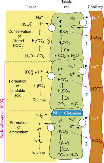
As a result of this process, for every bicarbonate ion that combines with a hydrogen ion in the lumen of the tubule, a bicarbonate ion ultimately will diffuse into the peritubular capillaries (Fig. 13-6). This secretion of hydrogen ions and reabsorption of bicarbonate ions occurs along the length of the renal tubules, but 90% of bicarbonate reabsorption occurs in the proximal tubule. (See section, Regulation of Acid-Base Balance.)


The Loop of Henle


Reabsorption of sodium and water from the proximal tubule significantly reduces the volume of the glomerular filtrate. However, because the sodium and water are reabsorbed at approximately the same rate, the osmolality of the filtrate remains unchanged as it passes through the proximal tubule; it is neither concentrated nor diluted. The function of the loop of Henle is to remove more solute and water from this filtrate.


The loop of Henle, located within the renal cortex and the medulla, provides a countercurrent mechanism for urine concentration. The descending limb of the loop of Henle does not transport sodium or chloride actively, but it is highly permeable to sodium and water. Thus, as the filtrate passes through the descending limb of the loop, it becomes progressively more concentrated. The osmolality can increase from 300 to 1200   mOsm/L between the beginning of the descending limb and the tip of the loop of Henle (Fig. 13-7).


As the filtrate begins to pass through the ascending limb of the loop of Henle, chloride is actively pumped out of the tubule, and sodium follows passively. Water, however, must remain in the tubule because the ascending limb is impermeable to water. The solute loss from the tubule produces a fall in the osmolality of the filtrate and a rise in the osmolality of the interstitial fluid surrounding the loop. Thus, the osmolality of filtrate arriving in the distal tubule is lower than that of filtrate entering the loop of Henle and lower than that of the interstitial fluid in the medulla.


The loop of Henle removes approximately 25% of filtered sodium and 15% of filtered water from the tubule, leaving approximately 10% of the filtered sodium and 20% of the filtered water to enter the distal tubule.


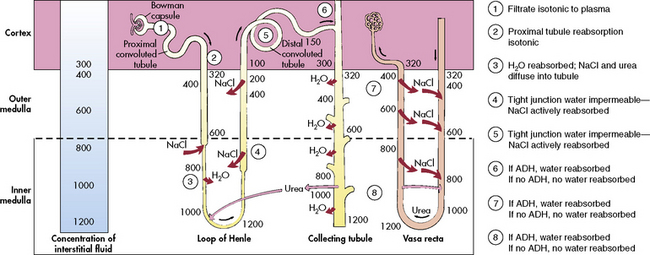
The blood vessels surrounding the loop of Henle form a hairpin loop structure, called the vasa recta. The vasa recta consists of capillaries that run parallel to the loop of Henle and the collecting ducts (for an illustration, see Evolve Fig. 13-1 in the Chapter 13 Supplement on the Evolve Website). As these capillaries follow the loop of Henle into the interstitium of the renal medulla, where osmolality is high (as the result of the tubular countercurrent mechanism), water shifts out of capillaries into the interstitial fluid, and sodium and chloride move from the interstitial fluid into the capillaries.


The vasa recta does not contribute to the creation of a concentration gradient; its content is affected by the osmotic gradients surrounding the loop of Henle. This capillary loop mechanism is termed a countercurrent exchanger; the term reflects its passive nature. By this mechanism, the solute and water in the interstitial fluid surrounding the loop of Henle and the collecting ducts are reabsorbed into the circulation while maintaining the interstitial osmolality.


The Distal Tubule and Collecting Ducts


The distal tubule arises from the ascending limb of the loop of Henle; its thick cellular structure is distinct from the thin cells of the ascending loop. Thick cuboidal cells continue up through the renal cortical area to a point where the distal tubule is in direct contact with the afferent arteriole of its glomerulus. At this junction, the distal tubule cells become more densely packed and more columnar, and the muscle cells of the arteriole enlarge and take on a granular appearance. This point of contact between the distal tubule and the glomerular afferent arteriole is called the juxtaglomerular apparatus (see Fig. 13-3).


The juxtaglomerular apparatus consists of the columnar cells of the distal tubule (called the macula densa because of their prominent nuclei) and large cells of the afferent arteriole (called polkissen or polar cushion). The term juxtaglomerular cells most commonly refers to the cells of the afferent arteriole; these cells are able to sense pressure and secrete the hormone renin.


Beyond the juxtaglomerular apparatus, the distal tubule joins the collecting duct. The collecting duct will in turn descend from the renal cortex through the medulla and into the renal calyces. The filtrate present in the early distal tubule has a lower osmolality and lower sodium concentration than the plasma and the surrounding interstitial fluid. As the urine filtrate passes through the distal tubule and the collecting ducts, more water will be removed to further concentrate the urine. The final concentration of urine in the distal tubule and the collecting ducts is adjusted by the active transport of sodium out of the distal tubule and changes in the relative permeability of the collecting ducts to water (e.g., under the influence of antidiuretic hormone).


The distal tubule is the site of final adjustments in the urine sodium and potassium content. The distal tubule actively reabsorbs approximately 10% of the filtered sodium. This active transport process occurs against a high electrical and concentration gradient and is influenced by the volume and character of the fluid arriving from the loop of Henle, as well as by hormones, especially aldosterone.


Renin, Aldosterone, and Antidiuretic Hormone


Renin is secreted from the polkissen cells of the afferent arteriole in the juxtaglomerular apparatus. In turn, renin forms angiotensin I from renin substrate (a circulating peptide from the liver). The amounts of renin released and angiotensin formed are determined by the renal perfusion pressure, sympathetic nervous system stimulation, circulating vasoactive substances, and changes in electrolyte concentration.55


Angiotensin I circulates to the lung and is converted enzymatically to angiotensin II. Angiotensin II produces peripheral vasoconstriction and an increase in aldosterone secretion, which increases renal sodium and water reabsorption. These effects should increase intravascular volume (Fig. 13-8). Angiotensin I and II are destroyed by angiotensinase, an enzyme that is present in plasma and secreted by a variety of organs, such as the kidney, intestine, and liver.
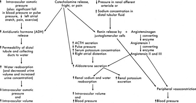


The quantity of sodium that is excreted in the urine when aldosterone is absent totals approximately 2% of the total filtered sodium. If aldosterone is absent (e.g., in patients with untreated adrenal insufficiency), excretion of that sodium will be associated with excretion of a large volume of water that can produce hypovolemic shock. Thus, aldosterone is responsible for the reabsorption of a very small but significant portion of the filtered sodium.


Aldosterone is secreted by the adrenal cortex in response to pituitary adrenal corticotropic hormone (ACTH) secretion and a variety of other stimuli. A fall in the pulse pressure, decreased stretch of the right atrium, and an increased serum potassium concentration all stimulate aldosterone secretion.63 An important stimulus for aldosterone is formation of angiotensin from renin released by the juxtaglomerular apparatus. Aldosterone stimulates epithelial cell transport of sodium in the renal tubular epithelium, along the intestinal lumen, and in sweat and saliva. Increased aldosterone levels increase the active reabsorption of sodium and decrease potassium reabsorption. The increased sodium reabsorption produces water reabsorption; this increases intravascular volume and reduces the juxtamedullary secretion of renin. The reduction in potassium tubular reabsorption increases potassium excretion in the urine and should result in a fall in the serum potassium concentration. These responses to aldosterone should in turn reduce the stimulus for aldosterone secretion (see Fig. 13-8).


Antidiuretic hormone (ADH), or arginine vasopressin (AVP), secretion also affects the final concentration of urine. ADH is produced by the supraoptic and paraventricular nuclei in the hypothalamus and is transported to the posterior lobe of the pituitary, where it is released in response to an increase in serum osmolality. ADH secretion is stimulated by serum osmolality greater than 280 to 285   mOsm/L (or a rise in serum osmolality of 2% or more). It also is secreted in response to significant (10%-15%) volume depletion, a fall in blood pressure, painful stimuli, fear, and exercise. Hemoconcentration, diabetic ketoacidosis,90 and mannitol administration increase ADH secretion, and administration of hypertonic glucose often inhibits ADH secretion.34,54 The predominant stimulus for ADH secretion is a rise in serum osmolality sensed by osmoreceptors in and around the supraoptic nucleus of the hypothalamus.


If ADH is present, the renal distal tubule and collecting ducts become highly permeable to water. As the collecting ducts descend through the hypertonic interstitium in the renal medulla, water will move from the collecting ducts into the medullary interstitium to be reabsorbed into the circulation. Thus, ADH secretion reduces urine volume and increases urine concentration.


If ADH levels are low, ADH secretion is absent (i.e., neurogenic diabetes inspidus [DI]), or the kidney is unresponsive to ADH (i.e., nephrogenic DI), the distal tubule and collecting ducts remain relatively impermeable to water, so water will remain in the filtrate that flows into the renal calyces. Large quantities of dilute urine will then be excreted.


Regulation of Acid-Base Balance


The kidney plays a critical role in balancing serum acids and bases. A substance is labeled as an acid or a base according to its ability to lose or gain a hydrogen ion (a proton). Strong acids dissociate freely in solution, readily yielding a hydrogen ion; therefore they will contribute to the development or progression of acidosis. Weak acids only partially dissociate into a solution that will then contain both acid and base; thus, they do not contribute to changes in acidity. Bases are substances that will accept a free hydrogen ion; they reduce the hydrogen ion concentration, increasing the pH.


The pH is the inverse of the logarithm (log) of the hydrogen ion concentration; as the hydrogen ion concentration rises, the pH falls (the serum becomes more acid). The normal range of pH is 7.35 to 7.45. If the pH is less than 7.35, acidosis is present; if the pH exceeds 7.45, alkalosis is present. Even slight changes in hydrogen ion concentration or serum pH can alter metabolic and cell functions.


Buffering Systems


All body fluids contain buffers. These buffers are compounds that combine with any acid or base so the acid or base does not significantly alter the serum or tissue pH. Effective buffering requires interaction of serum and cell buffers. When the hydrogen ion concentration changes significantly, plasma, respiratory, and renal buffering systems are activated.


The Bicarbonate-Carbonic Acid Buffering System

The bicarbonate-carbonic acid buffering system operates in both the lung and the kidney and is the most important plasma buffering system. It consists of the buffer pair of carbonic acid (H2CO3—a weak acid) with sodium, potassium, or magnesium bicarbonate. Because two end products of the system (carbon dioxide and bicarbonate) are closely regulated, this buffering system maintains the serum pH within a narrow range.


Carbon dioxide (CO2) is produced by tissue metabolism and is dissolved in plasma. The plasma concentration of CO2 is proportional to the partial pressure of carbon dioxide in the gas phase with which the solution is equilibrated (dissolved CO2 = 0.003 × PaCO2). Under normal conditions, CO2 is eliminated readily through the lungs, and dissolved CO2 does not contribute to hydrogen ion accumulation.


If CO2 accumulates, it combines with water to form carbonic acid; this reaction is catalyzed by carbonic anhydrase. Carbonic acid then dissociates into equal amounts of bicarbonate and hydrogen ion as follows:


image


The increase in hydrogen ion concentration will result in a fall in serum pH unless or until CO2 elimination by the lungs is enhanced and/or hydrogen ion excretion and bicarbonate ion reabsorption by the kidneys is increased (see section, Interpretation of Blood Gas Values).


When hydrogen ions accumulate, they combine with and are buffered by hemoglobin that has released its oxygen. Hydrogen ions readily combine with and are buffered by bicarbonate, resulting in the formation of carbonic acid; carbonic acid ultimately dissociates into CO2 and water, and the CO2 is normally eliminated through the lungs.


Renal Hydrogen Ion Excretion and Bicarbonate Reabsorption

The kidneys regulate serum pH and image concentration through hydrogen ion secretion and bicarbonate reabsorption and reclamation. Renal compensation for respiratory acidosis requires several hours to begin and will not be fully effective for several days; it requires reabsorption of all filtered bicarbonate and generation of new bicarbonate through the formation of titratable acids.


The major stimulus for increased bicarbonate reabsorption or reclamation in the proximal tubule is the presence of increased hydrogen ion concentration in the cells of the proximal tubule, as occurs with the development of metabolic acidosis. It is important to note, however, that bicarbonate reabsorption is also affected by changes in serum potassium and chloride concentrations. Both hypokalemia and hypochloremia increase hydrogen ion concentration in the renal tubular cells, so that hydrogen ion secretion into the proximal tubule and bicarbonate reabsorption are enhanced. This process is the mechanism for development of alkalosis with hypokalemia or hypochloremia (i.e., hypokalemic or hypochloremic metabolic alkalosis).


A hydrogen ion is secreted into the proximal renal tubule in exchange for a sodium ion. Once in the tubule, the hydrogen ion combines with filtered bicarbonate to form carbonic acid and then quickly dissociates into CO2 and water. The CO2 diffuses back into the renal tubular cell, where it recombines with water to form carbonic acid, and then quickly dissociates into hydrogen ion and bicarbonate. The bicarbonate diffuses out of the tubular cell into the interstitial fluid and ultimately into the plasma while the hydrogen ion is again secreted into the renal tubule. This method of reclaiming bicarbonate ions results in a net reabsorption of filtered bicarbonate ions from the renal tubule, without any net reabsorption of hydrogen ions.


New bicarbonate can be formed when CO2 combines with water, yielding carbonic acid. The carbonic acid then dissociates into hydrogen ions and bicarbonate; the hydrogen ion is bound to phosphate buffers or ammonia to form hydrogen phosphate or ammonium (image). Hydrogen phosphate and ammonium are nonreabsorbable, and they are excreted unchanged in the urine. When hydrogen ions are excreted in this way, a quantity of acid can be measured in the urine; this buffering mechanism results in the formation of titratable acids (see Fig. 13-6). The amount of hydrogen ion excreted in the urine is limited, because the kidney cannot secrete urine with a pH lower than approximately 4.4. In addition, the formation of titratable acid will be limited by the amount of ammonia, phosphate, and other inorganic buffers available.


To determine the quantity of hydrogen ions present in the urine in combination with buffers, sodium hydroxide (NaOH) is titrated into the urine sample. The number of milliequivalents of NaOH needed to restore the pH to 7.4 will equal the number of milliequivalents of hydrogen ions present in the urine in combination with buffers. This quantity of hydrogen ion is referred to as the titratable acid in the urine.


Interpretation of Blood Gas Values


When evaluating acid-base disturbances, it is important to identify the effects of the primary disorder and the results of respiratory or renal compensation. If an acute problem is present, treatment must focus on the underlying disorder, while supporting whatever compensation is occurring. By definition, compensatory mechanisms will strive to restore the pH to near-normal levels; therefore compensation will never result in overcorrection or a change in the pH in a direction opposite the initial stimulus. For example, renal compensation for chronic respiratory acidosis can restore the pH to near the 7.35 to 7.45 range, but will not create an alkalotic condition (pH above 7.45). If a patient with chronic respiratory acidosis has an alkalotic pH, that patient has an additional condition causing the metabolic alkalosis.


Treatment of acid-base disorders often complicates the interpretation of acid-base imbalance. For example, if the patient with metabolic acidosis arrives in the pediatric critical care unit breathing spontaneously, with appropriate respiratory compensation, the patient’s pH may be near normal (e.g., 7.31). If aggressive treatment of the metabolic acidosis is provided, spontaneous hyperventilation can continue for several hours after effective treatment of the acidosis, because it takes several hours for ventilatory response to pH changes to be maximal. Continued hyperventilation can produce a transient alkalosis that results not from respiratory overcorrection of the acidosis, but from combined intrinsic respiratory compensation coupled with extrinsic buffering of the patient’s pH (therapy).


Evaluation of the pH and PaCO2

Blood gas analysis requires evaluation of the pH, the PaCO2, the calculated base deficit or excess, and the serum bicarbonate. The first step is evaluation of the pH. If the pH is less than 7.35, acidosis is present; if the pH is greater than 7.45, alkalosis is present. The second step is evaluation of the PaCO2 in light of the pH to determine whether any existing change in pH can be explained by the alteration in PaCO2. For every uncompensated torr unit rise in PaCO2 above 45, the pH should fall 0.008 units below 7.35, and for every uncompensated torr unit fall in PaCO2 below 35, the pH should rise 0.008 units above 7.45. Acidosis or alkalosis in excess of that predicted from the PaCO2 must be metabolic in origin (Box 13-1).


Dec 3, 2016 | Posted by in NURSING | Comments Off on Renal Disorders

Full access? Get Clinical Tree

Get Clinical Tree app for offline access