11 Pneumothorax/hemothorax
PneumothoraxOverview/pathophysiology
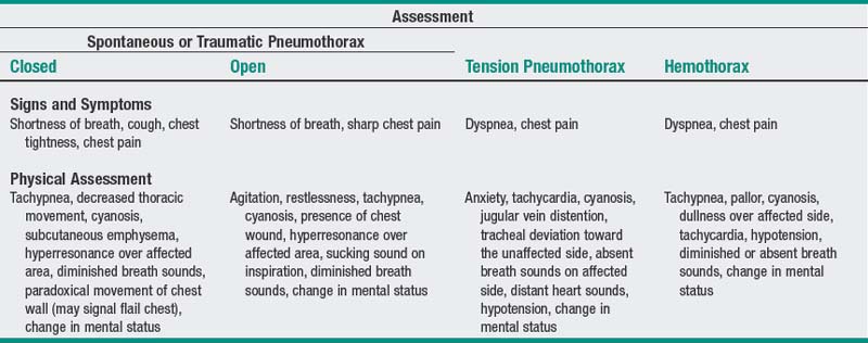
Pneumothorax is an accumulation of air in the pleural space that leads to increased intrapleural pressure. Risk factors include blunt or penetrating chest injury, chronic obstructive pulmonary disease (COPD), previous pneumothorax, and positive pressure ventilation. The three types of pneumothorax are as follows.
Spontaneous:
Also referred to as closed pneumothorax because the chest wall remains intact with no leak to the atmosphere. It results from rupture of a bleb or bulla on the visceral pleural surface, usually near the apex. Generally, the cause of the rupture is unknown, although it may result from a weakness related to a respiratory infection or from an underlying pulmonary disease (e.g., COPD, tuberculosis, malignant neoplasm). The affected individual is usually young (20-40 yr), previously healthy, and male. Generally, onset of symptoms occurs at rest rather than with vigorous exercise or coughing. Potential for recurrence is great, with the second pneumothorax occurring an average of 2-3 yr after the first.
Traumatic:
Can be open or closed. An open pneumothorax occurs when air enters the pleural space from the atmosphere through an opening in the chest wall, such as with a gunshot wound, stab wound, or invasive medical procedure (e.g., lung biopsy, thoracentesis, or placement of a central line into a subclavian vein). A sucking sound may be heard over the area of penetration during inspiration, accounting for the classic wound description as a “sucking chest wound.” A closed pneumothorax occurs when the visceral pleura is penetrated but the chest wall remains intact with no atmospheric leak. This usually occurs following blunt trauma that results in rib fracture and dislocation. It also may occur from use of positive end-expiratory pressure (PEEP) or after cardiopulmonary resuscitation.
Tension:
Generally occurs with closed pneumothorax; also can occur with open pneumothorax when a flap of tissue acts as a one-way valve. Air enters the pleural space through the pleural tear when the individual inhales, and it continues to accumulate but cannot escape during expiration because the tissue flap closes. With tension pneumothorax, as pressure in the thorax and mediastinum increases, it produces a shift in the affected lung and mediastinum toward the unaffected side, which further impairs ventilatory efforts. The increase in pressure also compresses the vena cava, which impedes venous return, leading to a decrease in cardiac output and, ultimately, to circulatory collapse if the condition is not diagnosed and treated quickly. Tension pneumothorax is a life-threatening medical emergency.
HemothoraxOverview/pathophysiology
Hemothorax is an accumulation of blood in the pleural space. Hemothorax generally results from blunt trauma to the chest wall, but it can also occur following thoracic surgery, after penetrating gunshot or stab wounds, as a result of anticoagulant therapy, after insertion of a central venous catheter, or following various thoracoabdominal organ biopsies. Mediastinal shift, ventilatory compromise, and lung collapse can occur, depending on the amount of blood accumulated.
Chest x-ray examination:
Will reveal presence of air or blood in the pleural space on the affected side, pneumothorax/hemothorax size, and any shift in the mediastinum.
Arterial blood gas (ABG) values:
Hypoxemia (Pao2 less than 80 mm Hg) may be accompanied by hypercarbia (Paco2 greater than 45 mm Hg) with resultant respiratory acidosis (pH less than 7.35). Arterial oxygen saturation may be decreased initially but usually returns to normal within 24 hr.
Complete blood count (CBC):
May reveal decreased hemoglobin proportionate to amount of blood lost in a hemothorax.
Nursing diagnosis:
Ineffective breathing pattern
related to decreased lung expansion occurring with pneumothorax/hemothorax, pain, or malfunction of chest drainage system
Desired Outcome: Following intervention, patient becomes eupneic; lung expansion is noted on chest x-ray.