Inspection
During the inspection component of the examination, the child should be sitting, if possible. Keep in mind that a younger child may be more comfortable sitting on the parent’s lap. Inspection of the chest should occur with little or no clothing depending on the age and gender of the child. A gown should be available to school-age and adolescent children. Additionally, the space should be private, lighting should be of good quality, and the environmental temperature should be comfortable.
The shape and symmetry of the chest should be noted anteriorly, laterally, and posteriorly. The anteroposterior (AP) diameter of the chest should be approximately half the transverse diameter (Seidel et al., 2003). A barrel chest is characterized by horizontal ribs, a somewhat kyphotic spine, and a prominent sternal angle. Pectus carinatum (pigeon chest) is depicted by a prominent protrusion of the sternum. Pectus excavatum (funnel chest) is illustrated by an indentation of the sternum (Lippincott, 2007; Seidel et al., 2003).
It is helpful to record a respiratory rate during the inspection portion of the examination. For infants and toddlers, it is useful to measure the respiratory rate before beginning the hands-on portion of the examination because the child is more likely to be fussing or crying during that portion of the examination. For school-age and adolescent children, it is useful to measure the respiratory rate when they do not know you are counting because the child may have a self-conscious response to your measurement; this response may alter the results (Seidel et al., 2003). The respiratory rate should be counted for a full minute. Normal respiratory rates based on age are as follows:
| Age | Rate (breaths/min) |
| Infants | 30–40 |
| Toddlers | 24–26 |
| Preschoolers | 24 |
| School-age children | 20 |
| Adolescents | 20 (Custer & Rau, 2009) |
Next, inspect respiratory effort, respiratory pattern, and chest expansion. Breathing should be relaxed and regular, and chest expansion should be symmetrical. The inspiratory-to-expiratory ratio (I : E ratio) should be 1:2. A list of respiratory patterns along with implications of the patterns are presented in Table 2.1 (Hilman, 1993; Lippincott, 2007; Seidel et al., 2003).
Table 2.1 Breathing patterns.
| Pattern | Description | Potential implications |
| Tachypnea | Abnormally high respiratory rate | Respiratory disease with decreased compliance, fever, anxiety, metabolic acidosis |
| Bradypnea | Abnormally low respiratory rate | Depression of central nervous system, metabolic alkalosis |
| Hyperpnea | Unusually deep respirations | Anxiety, exercise, fever, metabolic acidosis |
| Hypopnea | Shallow breathing | Central nervous system depression, metabolic alkalosis, pain |
| Apnea | Cessation of respirations for more than 15 seconds or for less if accompanied by bradycardia or cyanosis | |
| Periodic breathing | At least three respiratory pauses of 3- to 10-second duration with less than 20 seconds of respiration between pauses | Common in preterm infants and sometimes seen in normal full-term infants under 3 months of age |
| Paradoxical breathing | Seesaw breathing/thoracoabdominal asynchrony | Can be common in newborns (especially preterm infants) due to the compliant nature of their chest wall; it can also be due to respiratory distress |
| Kussmaul’s breathing | Deep, slow, regular respirations with a prolonged expiratory phase | Ketoacidosis |
| Cheyne–Stokes breathing | Cycles of increasing and decreasing depths of tidal volumes separated by periods of apnea | Increased intracranial pressure, congestive heart failure |
| Biot’s breathing | Cycles of irregular respiration at variable tidal volumes associated with periods of apnea of varying lengths | Severe brain damage |
The chest should be inspected for the presence of retractions. The nurse should remember that newborns and infants have a chest wall that is more compliant than older children and adults. Therefore, some mild retractions may be present at baseline at this age. The location of retractions, if present, should be noted and documented. Retractions can be subcostal, substernal, intercostal, suprasternal, or supraclavicular; the higher up on the chest the retraction, the more severe the respiratory distress and the degree of airway obstruction.
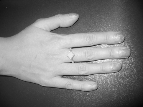
Inspection of the respiratory system includes more than an evaluation of the chest. The child’s head should be assessed for signs of respiratory distress, such as head bobbing, nasal flaring, an anxious facial expression, or pursed lip breathing (O’Hanlon-Nichols, 1998). Grunting or mouth breathing should be noted. Additionally, skin, nail bed, and lip color should be assessed for cyanosis and pallor. Extremities should be assessed for digital clubbing. Clubbing is an abnormal enlargement and an increase in the angle of the finger (see Figure 2.2) and toe bases. To check for clubbing, ask the child to place both thumbs together (nail beds facing each other). Normally, the nurse will see a diamond shape space between the fingers when the thumbs are in this position. Clubbing eliminates this space because the angle of the nail base is greater than 180° (Cox, 2001). Clubbing may be due to chronic hypoxia. However, it can be hereditary, so the nurse should always evaluate the parents’ fingers if he/she suspects clubbing.
Figure 2.2 Clubbing in a 20-year-old cystic fibrosis patient.

Stay updated, free articles. Join our Telegram channel

Full access? Get Clinical Tree


