CHAPTER 37 Orthopedic Trauma
I. GENERAL STRATEGY
The aim in caring for the patient with an orthopedic emergency is to restore and preserve function.
A. Assessment
1. Primary and secondary assessment/resuscitation (see Chapters 1 and 31)
Table 37-1 GRADING OF PULSE QUALITY
| Grade | Description |
|---|---|
| 0 | No pulse |
| 1 | Weak and easily obliterated with pressure |
| 2 | Difficult to palpate but easy to feel once located |
| 3 | Easily palpated and considered normal |
| 4 | Strong and bounding |
Box 37-4 MUSCLE STRENGTH
Table 37-2 DEEP TENDON REFLEXES*
| Grading of Reflexes | |
|---|---|
| Grade | Description |
| 0 | No reflex |
| +1 | Less than normal |
| +2 | Average |
| +3 | Stronger than average |
| +4 | Intense (clonus) |
* Assess major reflexes: biceps, brachioradialis, triceps, patellar knee jerk, Achilles tendon.
Table 37-3 LABORATORY STUDIES TO AID DIAGNOSIS OF ORTHOPEDIC CONDITIONS
| Test | Examples |
|---|---|
| Alkaline phosphatase level | Increased with healing fractures, metabolic bone disease, osteoporosis, and metastatic tumors of bone |
| Calcium level | |
| Creatine kinase level | Increased in dehydration, hyperthyroidism, renal failure, and rhabdomyolysis |
| Phosphorus level | Increased in bone metastases and hypoparathyroidism |
| Alanine aminotransferase (ALT), aspartate aminotransferase (AST) | Increased in myositis |
| Uric acid level | Increased in gout, multiple myeloma, and acute tissue destruction as a result of starvation or excessive exercise |
| C-reactive protein | Increased in acute inflammatory changes and rheumatoid arthritis |
| Antinuclear antibodies | Positive in rheumatoid arthritis, systemic lupus erythematosus, and polymyositis |
| Serum rheumatoid factor | Positive in rheumatoid arthritis and some chronic inflammatory diseases |
C. Planning and Implementation/Interventions
F. Age-Related Considerations

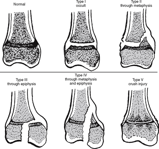
FIGURE 37-1 Salter-Harris classification of pediatric fractures.
(From Simon, R. R., & Koenigsknecht, S. J. [2001]. Emergency orthopedics (4th ed.). New York: McGraw-Hill.)
II. SPECIFIC SOFT TISSUE INJURIES
A. Contusions/Hematomas
2. Analysis: differential nursing diagnoses/collaborative problems
3. Planning and implementation/interventions
4. Evaluation and ongoing monitoring (see Appendix B)
Box 37-5 SKIN DISCOLORATION REFLECTING AGE OF CONTUSION
24 to 48 hours after injury: Area tender and swollen; ecchymosis may not appear; reddish blue or purple color may take up to several days to appear, depending on location of injury, distance of injured blood vessels from skin surface, and amount of bleeding
5 to 7 days: Color begins changing on periphery, proceeding toward center; takes on greenish tint
10 to 14 days or longer: Brown
B. Strains and Sprains
Injuries to the structures around a joint are usually caused by excessive stretch or sudden force. This results in pulling on the structures, which causes tears in muscle and/or tendon. A sprain is the stretching, separation, or tear of a supporting ligament, and a strain is the separation or tear of a musculotendinous unit from a bone. Injury may result in pain, inability to weight bear fully, and swelling of the affected area. Sprains and strains are rare in small children, whose epiphyseal plates are still open and more vulnerable to forces. Athletes and obese patients resuming physical fitness are at risk for these types of injuries. Both strains and sprains are classified based on the amount of damage (Box 37-6).
Box 37-6 DEGREE OF SPRAINS/STRAINS
First degree: Minor tear in the fibers; minimal swelling, minor discomfort, absent or minor ecchymosis
Second degree: Partial tear; joint intact; more severe swelling, visible ecchymosis
Third degree: Complete disruption of ligament; joint may be open; minimal to severe swelling; resultant separation of muscle from muscle, muscle from tendon, or tendon from bone


