Chapter 18 Ophthalmology
Basic concepts
1 Describe the course of visual information arriving from the left and right visual fields
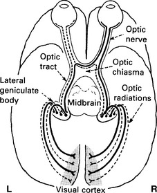
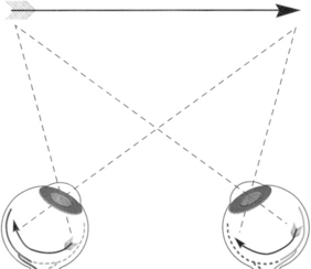
Light from the left visual field encounters the right half of each retina, temporally in the right eye and nasally in the left. Fibers from the temporal retina of the right eye travel in the right optic nerve and pass on the outside of the optic chiasm without any crossing. Fibers from the nasal retina of the left eye travel in the left optic nerve to the optic chiasm, where they cross to the other side and join the temporal fibers from the right eye to form the right optic tract. The right optic tract, which compromises the nasal fibers from the left eye (left visual field) and temporal fibers from the right eye (left visual field), synapses primarily in the right lateral geniculate nucleus (LGN) and in the Edinger-Westphal nucleus for pupillary reactions. Projections from the right LGN will then divide so that the left upper visual field information will travel through the temporal lobe and left lower visual field information will travel through the parietal lobe. These optic projections synapse in the right visual cortex within the occipital lobe. The visual information coming from the right visual field follows the same concepts as described for the left visual field but encounters the left half of each retina. The input from the two eyes is combined at the chiasm and travels to the left side of the brain (Figs. 18-1 and 18-2).
2 What visual field defect results from midline sectioning of the optic chiasm? Explain
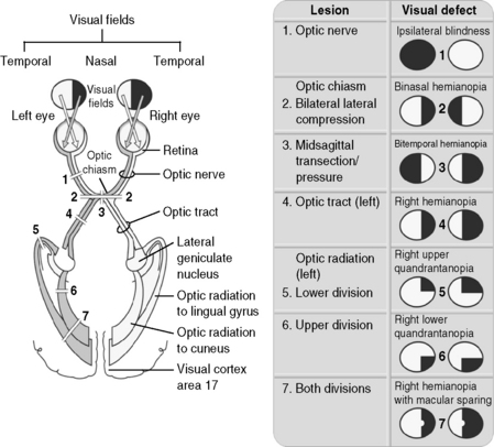
Bitemporal hemianopia is a loss of the temporal (lateral) fields of vision in both eyes, resulting in “tunnel vision.” Fibers from the nasal retina, which receive visual input from the contralateral temporal visual field, cross over in the optic chiasm and would be severed by a midline section of this structure or by a pituitary tumor. Figure 18-3 shows the visual field deficits resulting from lesions at various points in optic pathways.

Figure 18-3 The visual pathways, showing the consequences of lesions at various points.
(From Brown TA: Rapid Review Physiology. Philadelphia, Mosby, 2007.)
3 What visual field deficit will occur with sectioning of the left optic tract and why?
Right homonymous hemianopia results from the loss of the right field of vision in both eyes. The left optic tract receives input from the right nasal retina and left temporal retina, both of which receive information from the right visual space (see Figs. 18-1 and 18-2).
5 A physician shines a light into a patient’s right eye and notes bilateral constriction of both pupils (normal response). Describe the pupillary light reflex
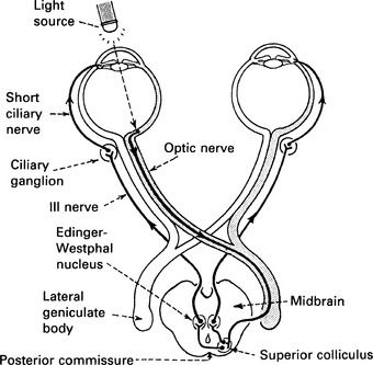
When light is shone into the right eye, the photosensitive retinal ganglion cells of the right eye convey this information to the right optic nerve, which connects to the right pretectal nucleus of the upper midbrain. Axons then connect from the pretectal nucleus to the neurons in the right Edinger-Westphal nucleus, whose axons run along both right and left oculomotor nerves. The oculomotor nerves synapse on the ciliary ganglion neurons of each respective eye, which innervate the constrictor muscles of the irises. This stimulates bilateral pupillary constriction (Fig. 18-4).


