Exposed vital organs (treatment may proceed after the organ has been covered by vicryl absorbable mesh).
Inadequately debrided wounds; granulation tissue that will not form over necrotic tissue.
Untreated osteomyelitis or sepsis within the vicinity of the wound.
Presence of untreated coagulopathy.
Necrotic tissue with eschar.
Malignancy in the wound (negative pressure therapy may lead to cellular proliferation).
Allergy to any component required for the procedure.
A6550 | Wound care set, for negative pressure wound therapy electrical pump, includes all supplies and accessories | ||
E2402 | Negative pressure wound therapy electrical pump, stationary or portable | ||
K0743 | Suction pump, home model, portable, for use on wounds | ||
K0744 | Absorptive wound dressing for use with suction pump, home model, portable pad size 16 square inches or less | K0745 | Absorptive wound dressing for use with suction pump, home model, portable pad size more than 16 square inches but less than or equal to 48 square inches |
K0746 | Absorptive wound dressing for use with suction pump, home model, portable, pad size greater than 48 square inches | ||
Other related codes to consider: | |||
A7000 | Canister, disposable, used with suction pump, each | ||
 |
Contraindicated where there is evidence of exposed arteries or veins in wound, fistula—unexplored or nonenteric, untreated osteomyelitis, malignancy, or necrotic tissue with eschar.
Contraindicated for application directly to exposed blood vessels, organs, or nerves. Do not apply the Bio-dome wound dressing directly to exposed bowel surfaces.
Contraindicated for any suction application that requires more than 10 LPM of free airflow.
Warning indicated for patients at risk for bleeding or who are on an anticoagulant; care must be taken when applying suction in the vicinity of weakened
blood vessels or organs (e.g., sutured blood vessels, infected blood vessels, or blood vessels that have been exposed to radiation); exposed blood vessels, organs, tendons, ligaments and nerves should be covered with multiple layers of fine mesh non-adherent dressings.
Note: Negative pressure should be applied for at least 22 of 24 hours per day. If suction will not be maintained for a period greater than 2 hours, the Wound Cover and Bio-dome Wound Dressing should be removed and a traditional dressing applied in its place. The hours meter on the unit only counts time when therapy, at the applied pressure setting, actually occurs using the Compliant Hours Monitoring Technology. A visual indicator will also show the status of the unit when switched on: Green (normal operation), Blue (possible occlusion), Red (High Leak) and Yellow (Low Leak—however therapeutic pressure is being delivered)
Carefully inspect the wound, and treat per the order of the patient’s physician and according to the institution’s protocol and standards of practice for wound care. This should include proper hand washing and gloving practices. An appropriate skin preparation should be used to preserve the wound margins and prevent epithelial stripping. When bridging two wounds, be sure to protect the undamaged skin under the bridge with a cover dressing.
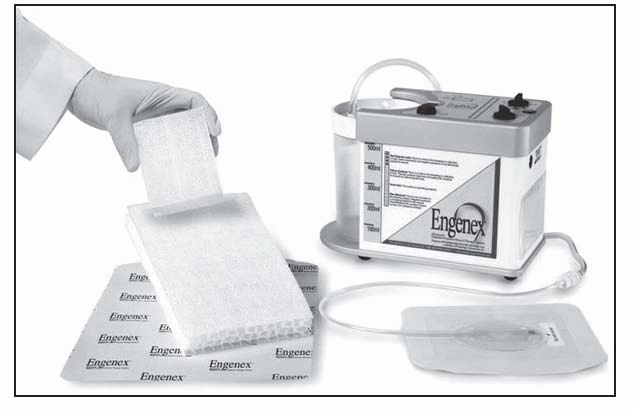
Select the appropriate Bio-dome dressing, and cut the wound contact dressing just smaller than the size of the wound. The Bio-dome Surface must face the wound (“dimples down”). Carefully place the dressing in the wound. Do not force the wound dressing into cavity wounds. Always count and record the number and types of dressings used.
When using multiple dressings, including the tunnel dressings and bridging, the neighboring dressings need to be in communication to allow optimal fluid movement from the wound. This communication is achieved by ensuring that there is sufficient overlap (approximatelyttis recommended) of dressings situated directly next to each other (consider extra overlap if patient movement/range of motion may cause dressings to lose communication).
Prior to application of the cover, ensure that the skin around the wound is clean and dry. Use of a skin preparation layer may protect periwound skin and promote and prolong cover adhesion. Following the instructions for use, position the dressing over the wound and carefully apply, taking care to minimize folds and wrinkles. Use only the amount of film necessary to secure the dressing in place and maintain an optimal environment for NPWT.
Cut a hole approximately 3 cm in size in the film cover at the location of the wound where the Tube Attachment Device (TAD) will be secured. It is permissible to cut into the wound packing when creating the hole.
Remove the backing from the TAD. Place the screened portion of the TAD over the 3-cm hole that was cut in the cover. Make sure that the TAD is adequately secured to cover. Do not cover the vent on the TAD.
Connect the TAD to the suction tubing of the Engenex canister. Connect the Engenex Canister to the Engenex Therapy Unit.
Set the vacuum level per physician’s orders, typically 75 mm Hg. Turn the pump to ON for continuous operation or INTER for intermitting suction per physician’s orders.
The canister should be checked periodically and should be replaced whenever full or nearly full. Replace the canister after 5 days of use even if not full.
The Bio-dome Wound Dressing, Wound Cover, and TAD should be replaced every 48 hours. For infected wounds, dressings should be replaced every 12 to 24 hours.
Turn Off the Engenex Therapy Unit and disconnect the Engenex Canister from the TAD.
Carefully remove the wound cover and all dressing materials from the wound.
Cleanse the wound and perform any necessary procedures as directed by the physician. Inspect the wound for the presence of infection, osteomyelitis or other potentially abnormal conditions, and report to the physician.
Dress the wound with fresh, sterile dressing materials as previously described, and set the Engenex Therapy Unit at the prescribed settings. If evidence suggests that there is an infection in the wound, pay strict adherence to physician orders and hospital policy. Discontinue use if appropriate.
If the wound dressing is sticking to the wound or the patient is experiencing serious pain upon removal, try moistening the wound bed with saline to reduce adhesion or use saline-moistened, sterile cotton-tipped applicators to remove dressings that are sticking to the wound bed. Contact the physician to consider changing the orders to include more frequent dressing changes or titrating down the level of suction.
Dispose of used dressing materials and canisters as medical waste.
Dressing Sets | |
Small: | 1 dressing (10 × 8 × 3 cm), 1 suction tubing with SpeedConnect, |
1 polyurethane drape; A6550; CPT 97605/97606 | |
Medium: | 1 dressing (20 × 12.5 × 3 cm), 1 suction tubing with SpeedConnect, |
2 polyurethane drapes; A6550; CPT 97605/97606 | |
Large: | 1 dressing (25 _ 15 _ 3 cm), 1 suction tubing with SpeedConnect, |
2 polyurethane drapes; A6550; CPT 97605/97606 | |
Extra Large: | 1 dressing (58.5 _ 33 _ 3 cm), 1 suction tubing with SpeedConnect, |
5 polyurethane drapes; A6550; CPT 97605/97606 | |
ITI Black Foam Dressing: A hydrophobic, open-cell reticulated polyurethane foam evenly distributes negative pressure across the wound base and removes exudate and fluids.
ITI Suction Tubing with SpeedConnect: An 8-foot (2.4 m) tube securely delivers negative pressure to the wound.
ITI Polyurethane Drape: A clear, semiocclusive polyurethane film with adhesive covers the foam-filled wound.
Do not place black foam on intact skin or wound edges.
Do not cut black foam while holding it directly over the wound.
Do not apply directly over bone, tendon, or ligaments.
Do not use in wound tunnels.
Carefully remove any previously applied dressings.
Carefully inspect wound—visually and manually—to ensure complete foam removal. Consult prior documentation of number of pieces inserted.
Thoroughly cleanse wound and all dead space. Flush a generous amount of irrigation solution across the surface and any dead space.
Apply wound contact layer or ITI White Foam if indicated.
Apply ITI Black Foam (designed to minimize fraying, can be cut, thin, and layered as needed). Loosely fill all dead space, but do not pack or tightly fill; extend black foam slightly higher than skin level; when filling undermined spaces, always leave a significant portion of the foam visible in the wound base so it is found during dressing removal.
Document the number of pieces of foam used in the wound.
Cover with ITI Polyurethane Drape.
Apply ITI Suction Tubing with SpeedConnect to wound and attach to SVED Canister. (If prescribed, also apply ITI Irrigation Tubing with SpeedConnect.)
Select SVED device therapy settings and begin therapy.
Change dressings every 48 to 72 hours (more frequently for infected wounds) and replace SVED Canister at least twice a week.
Gently stretch and lift the ITI Polyurethane Drape while using a gloved index finger to hold down intact skin.
Remove all foam. To ensure all components have been removed from the wound, after removing the dressing: Carefully observe the visible wound base; gently sweep undermined or tunneled areas with a gloved finger if possible to manually check for a clean wound base; consult prior documentation and then count pieces of foam to be certain all previously inserted pieces have been removed.
ITI Bridge Foam Dressing should never touch intact skin; always drape the skin before placing a foam bridge over it
Bridge foam should never directly contact the wound base; dressing must directly contact the foam covering/filling the wound
Do not increase negative pressure in an effort to move more fluid across the bridge; increased pressure does not fi× chronic blockage (consider, if medically advised, setting a lower pressure, setting an intermittent pressure, or adding irrigation)
Place the ITI polyurethane drape over periwound area.
Measure length between two wounds or two wound areas.
Cut, peel, and adhere ITI Bridge Foam Dressing to drape.
Drape over and seal.
Gently stretch and lift the ITI Polyurethane Drape while using a gloved index finger to hold down intact skin.
Remove the ITI Bridge Foam Dressing and all other foam. Ensure all components have been removed from the wound.
Unstable thoracic wounds or conditions where the fluid temperature could cause an adverse reaction
Not for use with enzymatic debridement ointments, solutions containing hydrogen peroxide or alcohol, or canisters with a capacity over 500 mL
In addition to applying the ITI Suction Tubing with SpeedConnect, cut an additional 1.5 cm hole in the ITI polyurethane drape to accommodate irrigation therapy.
Remove the paper backing from the ITI Irrigation Tubing with SpeedConnect and press the flange firmly onto the drape—centered over the hole.
Connect the distal end’s non-Luer lock to the irrigation bag.
On the SVED, select irrigation mode (continuous or intermittent) and rate (typically 25 to 30 mL/hour).
Before beginning NPWT, open clamps for suction tubing and irrigation tubing.
When changing the SVED Canister, put a tubing cap on the ITI Irrigation Tubing with SpeedConnect to prevent leaking.
Close tubing clamps.
Turn off SVED pump.
Remove flange from the ITI polyurethane drape.
Remove the distal end’s non-Luer lock to the irrigation bag.
Polyurethane drape: | 33 × 25.5 cm; A6550; CPT 97605/97606 |
SensiSkin drape: | 33 × 25.5 cm; A6550; CPT 97605/97606 |
For sensitive skin, consider ITI SensiSkin Drape for Sensitive Skin; other interventions for sensitive skin include skin barrier wipe, antifungal powder, ostomy powder, medical adhesive, steroid spray, therapy holiday
After applying foam to wound, cut a 1.5 cm hole in the drape
Cover all foam plus approximately 2” of surrounding skin.
Remove the paper backing from the ITI Suction Tubing with SpeedConnect and press firmly onto the drape—centered over the hole. If irrigation prescribed, use same procedure with ITI Irrigation Tubing with SpeedConnect.
The drape can be applied as a single piece or in strips. Strips can be overlapped. Use as needed to patch air leaks. It is highly recommended to frame SpeedConnects with the ITI Framing Drape.
Gently stretch and lift drape while using a gloved index finger to hold down intact skin.
Remove all foam. To ensure all components have been removed from the wound, after removing the dressing: Carefully observe the visible wound base; gently sweep undermined or tunneled areas with a gloved finger if possible to manually check for a clean wound base; consult prior documentation and then count pieces of foam to be certain all previously inserted pieces have been removed.
If the drape does not release easily, or if the skin is very fragile, try one of the following: Apply a warm wet cloth to the skin as the drape is gently lifted; use a medical adhesive remover, then wash and rinse the skin thoroughly to remove any residue that might prevent the new drape from adhering; if the skin is extremely fragile, carefully cut away the drape only over the open wound to enable the dressing change, and layer a new drape over the old drape—by the next dressing change, the old drape should more readily release from the skin; consider premedication, use of a nonadherent prior to foam placement, or irrigation with a topical anesthetic agent such as 1% Lidocaine prior to dressing removal.
Do not use any other manufacturer’s power supply, which could result in serious electrical damage.
Plug the ITI Power Supply into a suitable 120 VAC, 60 Hz outlet.
Insert the other end of the ITI Power Supply into the AC adaptor connector on the side of the SVED.
Unplug the ITI Power Supply from the side of the SVED and then from the wall outlet.
Stay updated, free articles. Join our Telegram channel

Full access? Get Clinical Tree


