Medical Emergencies

CHAPTER 20 Medical Emergencies


I. GENERAL STRATEGY


A. Assessment


1. Primary and secondary assessment/resuscitation (see Chapter 1)


2. Focused survey


3. Diagnostic procedures


II. SPECIFIC MEDICAL EMERGENCIES


A. Reye’s Syndrome


Reye’s syndrome is an acute noninflammatory encephalopathy characterized by the triad of hepatic, metabolic, and neurologic dysfunction. Although the origin of Reye’s syndrome is unknown, the condition typically occurs in children following a viral illness such as influenza B, gastroenteritis, or varicella and is associated with the administration of aspirin during the illness. Parainfluenza, adenovirus, coxsackieviruses A and B, echovirus, Epstein-Barr virus (EBV), rubella, measles, cytomegalovirus, herpes simplex virus, parainfluenza viruses, and poliomyelitis viruses are less commonly involved than the other pathogens listed but can be the cause of Reye’s syndrome. The peak ages of developing Reye’s syndrome are 6 and 11 years. Six-year-old children are more likely to develop Reye’s syndrome following a varicella illness, whereas 11-year-old patients tend to develop the syndrome following influenza B illnesses. The greatest incidence occurs in late winter and early spring; more cases are reported in white patients living in rural or suburban areas.


The primary defect is mitochondrial injury. Glycogen stores are depleted as a result of starvation from prolonged vomiting and dysfunction of gluconeogenesis enzyme pathways. An alternative energy source is not available because fatty acid breakdown does not occur normally. Consequently, acids build up because the body cannot eliminate waste products effectively. The liver enzyme system that converts ammonia to urea is damaged, so ammonia also accumulates. Additionally, the liver and renal tubules become edematous and infiltrated with fat droplets. Fatty infiltrates develop in the heart, with petechiae throughout the epicardium. Hyperammonemia, hypoglycemia, and acidosis contribute to neurologic dysfunction, cerebral edema, and coma.


The manifestations of Reye’s syndrome are not unique to this syndrome. There is no test specific for Reye’s syndrome; therefore, the diagnosis must be one of exclusion. Signs and symptoms of Reye’s syndrome correlate with illness stages and include protracted vomiting with or without clinically significant dehydration, encephalopathy in afebrile patients with minimal or absent jaundice, and hepatomegaly in 50% of patients (Table 20-1). Some authorities postulate that antiemetics mask early symptoms, whereas others propose that antiemetics may further predispose the individual to the disease. Although it is rare, Reye’s syndrome needs to be considered in any child presenting with altered mental status and vomiting. An appropriate history should be obtained for all children presenting with symptoms similar to those of Reye’s syndrome to determine whether an inborn error of metabolism (IEM) should be considered. Diarrhea and hyperventilation may be the first signs in children younger than 2 years of age.


Table 20-1 STAGES OF REYE’S SYNDROME


Stage Signs and Symptoms
Stage 0 Awake and alert
Stage I
Stage II Delirium, combativeness, hyperreflexia, dilated pupils with sluggish response, tachycardia, appropriate response to noxious stimuli, hepatic dysfunction, EEG abnormalities
Stage III Hyperventilation, obtunded, coma, decorticate posturing, normal pupillary and oculovestibular responses, continued hepatic changes, EEG changes
Stage IV Deeper coma, decerebrate rigidity, fixed and dilated pupils, loss of oculocephalic reflex, abnormal oculovestibular reflex, minimal liver dysfunction, abnormal EEG findings
Stage V Seizure, flaccidity, areflexia, dilated, nonreactive pupils, apnea, isoelectric EEG findings
Stage VI Patients who cannot be classified because they have been treated with medication that alters level of consciousness

EEG, Electroencephalogram.


1. Assessment


2) Diagnostic procedures


2. Analysis: differential nursing diagnoses/collaborative problems


3. Planning and implementation/interventions


4. Evaluation and ongoing monitoring (see Appendix B)


B. Gout/Pseudogout


Gout and pseudogout are the two most common crystal-induced arthropathies. They are debilitating illnesses in which pain and joint inflammation are caused by the formation of crystals within the joint space. Gout is inflammation caused by monosodium urate monohydrate (MSU) crystals and results from the overproduction or reduced secretion of uric acid. Thiazide diuretics and foods that are rich in purines increase the frequency of attacks. The first bout of gouty arthritis usually occurs at the first metatarsophalangeal joint. Pseudogout is inflammation caused by calcium pyrophosphate (CPP) crystals and is sometimes referred to as calcium pyrophosphate disease (CPPD). Many cases of pseudogout are idiopathic and are clinically indistinguishable from gout. Gout and pseudogout can be caused by lead poisoning, hemoproliferative disorders, and renal disease. Pseudogout has been associated with aging, trauma, and many different metabolic abnormalities, the most common of which are hyperparathyroidism and hemochromatosis. Gout has been associated with a large number of different autoimmune and metabolic disorders. Specific therapies and prophylactic measures have been developed to address the underlying problems. Treatment of the acute phase of pseudogout is identical to that of gout. Unlike gout, however, no specific therapeutic regimen exists to treat the underlying cause of pseudogout, and no known prophylactic therapy exists.


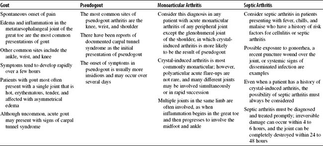
Three stages of gout occur: asymptomatic hyperuricemia, acute gout, and tophaceous gout. Nephrolithiasis may occur at any time. For gout, the male-to-female ratio is 9:1. Extra-articular deposits of MSU, known as tophi, may be seen along the Achilles tendon or on the ear helix, olecranon bursa, or prepatellar bursa. Acute monoarticular arthritis, septic arthritis, gout, and pseudogout can manifest in very similar ways (Table 20-2).


Stay updated, free articles. Join our Telegram channel

Nov 8, 2016 | Posted by in NURSING | Comments Off on Medical Emergencies

Full access? Get Clinical Tree

Get Clinical Tree app for offline access