Management of Patients Undergoing Neurosurgical Procedures


Management of Patients Undergoing Neurosurgical Procedures


Lori K. Madden

Peter K. Tham

Kiarash Shahlaie


THE PREOPERATIVE PHASE

For the patient undergoing a neurosurgical procedure, the circumstances of admission influence preoperative preparation. Unplanned emergency hospital admissions are often related to trauma or a sudden, life-threatening event such as an aneurysmal rupture. Immediate surgery may be a necessary life-saving measure. The urgency of the surgery compresses all preoperative preparations. With elective or delayed surgery, there is time for preoperative teaching. For the patient who is conscious and oriented, the prospect of a neurosurgical procedure is often associated with overwhelming anxiety and fear. Paramount is the fear of loss of life, cognitive and physical abilities, self-control, personality, and independence. In addition, concerns about permanent disability and chronic illness create concerns about their effects on relationships with family and friends and about burden on loved ones. In contrast, patients with an altered level of consciousness or impaired cognitive function may be unable to grasp the seriousness of the situation and appear apathetic. The crisis and the related high-stakes uncertainty are experienced by the patient’s family and friends. The impact on each family member and friend will vary depending on the pre-event relationship with the patient. A family member or significant other may become the surrogate decision maker for the unconscious or impaired patients.


Informed Consent

When obtaining informed consent, the physician discusses the purpose of surgery, alternative treatments, potential risks, and expected outcomes. Discussing these points honestly and answering all questions can reduce the possibility of misunderstanding and litigation. Although obtaining consent is required to meet legal and ethical obligations, the process also should be embedded in the development of a positive professional relationship between providers and patients. In practice, patients frequently do not perceive the process this way, seeing it as an overwhelming bureaucratic hurdle that leaves them feeling disempowered. Because altered consciousness, impaired cognition, or both can significantly influence comprehension, a responsible family member should be present during the discussion. In some instances, it will be necessary for the next of kin or guardian to consent for surgery as the patient’s surrogate. This is an awesome responsibility for which most people are not prepared. The nurse has an important role as a patient advocate and coach for the decision maker.

Where does the nurse begin in supporting the surrogate decision maker? First, frame the responsibility being assumed. The surrogate decision maker is asked to make decisions that he/she believes the patient would make if he/she were able to speak for him or her. The nurse can help to ask probing questions such as, “Has your loved one ever made statements about what he/she would want done if he/she was critically ill?” “What was the quality of life that your loved one expected?” and “What quality of life would not be acceptable?” Probe questions help the surrogate decision maker to recall conversations that provide insights about the patient’s wishes.

Second, help the surrogate decision maker gather necessary information to make an informed decision. Helping the person ask questions of the physician is critical. Such questions include, “What is the best- and worse-case scenario?” “What can be expected if the surgery is performed?” “What is the best- and worst-case scenario if the surgery is not performed?” “How will the patient be on a day-to-day basis in terms of independence in activities of daily living and cognitive functions?” and “Are there other options to surgery and, if so, what are the advantages and disadvantages?” With sufficient information, the surrogate decision maker can better weigh what he/she believes would be the patient’s wishes against each option.

Third, the nurse supports the decision maker in whatever choices are made. He/she also supports the decision maker in sharing that decision with care providers, family, and friends. The support continues through the postdecision time period when the decision maker may have second thoughts about the soundness of the decision made. The advocate, coach, and supportive role of the nurse provide the professional care and healing that has ramifications not only for the present but also for how the decision maker feels about the experience in the future.


Preoperative Teaching and Support

The nurse is tasked with the major responsibility for patient and family preparation and for provision of emotional support throughout the experience. The teaching plan is individualized and directed toward providing information and anticipatory guidance about activities associated with the neurosurgical procedure (Table 14-1). In addition, printed material is helpful and is a tangible resource that can be reviewed. If slight to moderate cognitive deficits are present, provide a more simplified explanation. With severe deficits, the
family assumes a greater decision-making role and often becomes the major focus of teaching. The expected outcomes of the teaching plan include control of fear and anxiety and maximization of patient and family coping strategies. Specific attention review of the precautions that will be taken by the surgical team to minimize the risks of surgery (prophylactic antibiotics prior to incision, neuronavigation and neuromonitoring to reduce the risk of neurological injury, intraoperative imaging to confirm surgical site, etc.) may help to reduce anxiety and strengthen the relationship between the patient/family/friends and the nurse/physician team. Common related patient problems include knowledge deficit, anxiety, fear, and family dysfunction.


TABLE 14-1 GENERAL PREOPERATIVE TEACHING PLAN FOR NEUROSURGICAL PROCEDURES


Patient and Familya


  • Clarify and reinforce information provided by the physician.


  • Provide printed material about the specific type of surgery and review it with the patient/family.


  • Describe preparatory events before surgery (e.g., blood work, electrocardiogram, chest radiograph, application of SCDs, visit from anesthesiologist, NPO before surgery).


  • Discuss the need to clip some hair from the scalp for the procedure.


Patient


  • Teach any special activities, such as leg exercises or deep breathing exercises.


  • Review what to expect throughout hospitalization and after.


Family


Discuss:


  • Location of the waiting area and amenities (e.g., telephones, restrooms, food)


  • Location of where the neurosurgeon will talk to the family after the surgery


  • If not physically present, how the family can be reached by telephone


  • Provisions for periodic updates if the surgery time is extended


  • Unit to which the patient will go after surgery (e.g., recovery room; intensive care unit [ICU])


  • Visiting hours before surgery and in the ICU


  • Estimated length of intracranial surgery (may take several hours and this is not unusual)


  • What to expect in the ICU (e.g., tubes, monitors, IV lines, change in appearance for dressings, or ecchymosis)


a Be prepared to repeat and reinforce information (people under stress often have difficulty retaining information).


Preoperative Preparations

For planned admissions, routine tests for any surgical patient are often completed on an outpatient basis (including laboratory tests which require review and possibly treatment prior to surgery, such as a urinalysis indicating urinary tract infection). Specific diagnostics that lead to the decision for neurosurgical intervention are typically performed prior to admission. Additional tests may be required upon admission. Because many patients are admitted on the day of surgery, any preoperative home preparation must be discussed ahead of time and expectations clearly specified (i.e., nothing by mouth after a specified hour, arrival time at the admissions or registration desk prior to procedure, which home medications to take on the morning of surgery and which should be withheld). Other preoperative activities are completed on admission such as verifying nothing by mouth (NPO) status, review of procedure and permit completion, obtaining intravenous (IV) access, and preoperative antibiotic administration.


Additional preoperative preparations are related to the specific surgical procedure and operative site. For intracranial surgeries, hair should be clean and long hair neatly secured in a manner that will not cause pressure or tension, such as loosely braided. Skin preparation, including hair clipping, is completed in the OR. Because neurosurgical procedures are often long, the risk of deep vein thrombosis (DVT) and pulmonary embolus (PE) is high.1 Sequential compression devices (SCDs) should be applied preoperatively and used intraoperatively with most neurosurgical patients. The final preoperative neurological assessment and vital signs are documented in EMR.

Depending on the circumstances, the family may visit briefly before surgery (Table 14-1 for family support and needs). Any other special preoperative orders are completed, and the patient is transported to the OR.


THE PERIOPERATIVE PHASE


Arrival in the Preoperative Area

Perioperative neuroscience nursing includes preoperative review of issues directly related to the planned surgical procedure (Table 14-2 for a perioperative checklist). Although the perioperative nurse works collaboratively with other professionals to meet the needs of the patient during the procedure, he/she is accountable for patient outcomes resulting from best nursing practice provided during the invasive surgical procedure. The perioperative nurse plans, coordinates, delivers, and evaluates nursing care to the patient whose reflexes and self-care abilities are temporarily compromised due to invasive procedure and anesthesia/sedation. The nursing plan addresses the physical, psychological, and spiritual needs for the patient perioperatively. The main goals are to assist the patient to achieve a level of wellness equal to or greater than that which they had before the surgery, and to prevent any complication such as pressure sore, and infection.

When the patient and his/her family and/or friends arrive the morning of surgery, it is important that the entire team provide sufficient information in a direct, confident, and compassionate way. The patient’s family should be advised of the expected length of surgery, and they should be told that surgical times are only estimates and that shorter or longer durations are normal and expected. It is critical for the perioperative nurse to obtain telephone contact information form at least one family member that will be available throughout the surgery; the operative team may need to communicate with the family should there be a reason during the surgery to give them an update on the duration of surgery or any possible changes in the operative plan.


TABLE 14-2 PERIOPERATIVE CHECKLIST


Identification


Name, age, handedness, gender, next of kin/family/other contact person


H and P Summary


Presentation, symptoms, signs, impairments, treatment, medical and surgical history


Pre-op test results


Imaging, physiologic, hematologic, coagulation, chemistries, cultures, intoxication screening


Diagnosis and allergies


Pre-op diagnosis, allergies


Pre-op medications


Antibiotics, steroid, anticonvulsant, diuretic, concurrent diagnosis and treatment


Consent


Informed consent, phone consent if patient is incompetent, two-physician consent in case of emergency


Surgical goals


Relieve Intracranial pressure, stop bleeding, monitoring, removal of space occupying lesions, insertion of electrodes or implanting chemotherapy, treating epilepsy.


Approach and positioning


Surgical site and side (right or left, midline), landmarks, maneuvers, intentions, strategies, risks


Specific risks


Focal, remote, neurological, vascular, cardiac, respiratory, infections, skin, urinary tract, cosmetic


Resources needed


OR room equipment and instrument checklist


  • OR bed, patient transferring roller, safety belt, arm boards


  • Surgical supplies: instruments, implants, sutures sponges


  • Positioning devices and supplies: doughnut, horseshoe, Mayfield skull clamp, skull pins, pillows, gel bolsters


  • Bipolar and irrigator, Bovie, suction ×2, suction regulator


  • Operative site preparation: shaving supplies (hair clipper, shave kit, razors, skin marker, zip closure bag, Mayfield skull clamp with pins, Bacitracin ointment); skin prep table (skin prep kit, local anesthetic


  • Back table


  • Medications (local anesthetic, antibiotic irrigation, Surgicel, Surgifoam, Thrombin, Gel foam)


  • Bair hugger, room temperature


  • Other equipment (microscope, sitting chair, headlight)


Operative team


Surgeon, assistants, observer, nursing, anesthesia, hospital assistants


Specimen management


Permanent specimen (fresh or fixed), microbiology, cranial flap, evidence (e.g., bullet), frozen section


Blood product


Packed red blood cells, fresh frozen plasma, cryoprecipitate, platelets


Perioperative and postoperative monitoring


ICP, vital signs, neurological signs, spinal signs, ECG, arterial pressure, CVP, blood gases, fluid and electrolyte balance, urinary output, pain and comfort, safety, progress/regression, hematology and chemistry, coagulation (DIC)


Communication plan


Update the family with the process, with other professionals (Recovery Room, ICU, respiratory therapist, CT scan, etc.)


Documentation


Operative record, bone flap documentation, pathology form, evidence form


Transport of the patient from the preoperative area to the OR can be a stressful event for the patient and his/her family and/or friends accompanying on the day of surgery. The perioperative nursing staff plays an important role in facilitating this important event on the day of surgery. Once the patient arrives in the OR, his/her care is directed by the anesthesia team. The patient is moved to the OR table, and positioned in a manner that is comfortable and relaxing for the patient. In the pre-op holding room, the perioperative nurse meets with the patient and family. His/her main tasks are to


  • validate the patient’s identification with patient, patient’s significant other(s), identification bracelet, and medical records. Once the patient receives anesthesia, the surgical team depends on the identification bracelet for verifying blood, medication, specimen, etc.


  • review the medical records to verify operative procedure, informed consent notes, medical history, and physical examination.


  • verify the presence of consent form with surgeon’s and patient’s signature. If the patient’s cognitive level is impaired or the patient is a minor, then the next of kin, conservator, or guardian signs the consent.


  • validate the operative site and ensures site marking, if appropriate.


  • assess NPO status and appropriate studies, reporting any deviations.


  • verify allergies.


  • determine the availability of blood and blood products. In some cases, packed red blood cells (RBCs) or other blood products may be sent to the OR suite “standing by” for any emergency needs.


  • assess the physical status including vital signs, respiratory status, cardiovascular, renal, skin, sensory and motor, and nutritional status.


  • assess presence of communicable disease.


  • note any physical abnormalities, injuries and previous surgery. Identify prosthesis, implants, and external fixator.


  • screen for substance abuse.


  • identify any personal belongings of the patient, including dentures, jewelry, clothing, glasses, hearing aids, etc. If possible, ask the patient’s family to keep them.


  • assess the psychosocial status of patient/significant others, including perception of surgery, expectation of surgical care and postoperative care, cultural and religious practices, and cognitive level.


  • check preoperative orders and administer premedication per prescriptions.


Arrival in the Operating Room Suite

Before the patient enters the OR suite, the perioperative nurse and the scrub nurse/technician prepare the OR suite, equipment, supplies, and instruments according to the surgeon’s preference. They continue to provide high quality nursing care to the patient and support for the surgical team and anesthesiologist when the patient is undergoing surgery. Duties of a perioperative nurse before the patient enter the OR suite include the following:


  • Check environment status: room temperature, humidity, and electrical.


  • Ensure cleanliness and safety of general environment.


  • Check proper functioning of basic equipment: electrocautery (Bovie), suction, OR lights, computer, appropriate OR bed, and other basic furniture.


  • Ensure the readiness of specific equipment requested by the surgeon: microscope, laser, ultrasonic aspirator, navigation system, intraoperative CT scan and ultra-sonogram, C-arm or O-arm, neuromonitoring, EEG monitoring, and deep brain recorder.


  • Gather all positioning devices: Mayfield skull clamp and pins or horseshoe, head holder for portable CT or MRI, bolsters (if prone), bean bag with extra pillows and multi-task armboard (if lateral).


  • Ensure the availability of implants and specific instrumentation.


  • Obtain and dispense medications/solutions for the sterile back table.


  • Do the initial count (all sponges, Raney clips, needles, cottonoids, needles and all small items) with the scrub nurse/technician prior to patient entering the OR suite.

Intraoperatively, the perioperative nurse continues to observe and monitor the sterile field. Specific procedures should be done according to institutional policy. Generally, the perioperative nurse reports and corrects break in sterile technique, and provides support to the surgical team and patient care to the anesthetized patient including the following responsibilities.


  • Clearly post case information (patient’s names and IDs, procedure, counts, medications, allergies, availability of blood products, and all personnel) on a white board that is visible to all members in the room.


  • Ensure safe transfer of patient to OR bed from either a Gurney or regular patient’s bed. Ensure adequate help, if patient is overweight or difficult positioning is performed.


  • Assist and support anesthesiologist during induction, intubations, and other pre- and postinduction procedures (such as peripheral lines, central line, and arterial line).


  • Protect the eyes from corneal abrasions; this is often accomplished with application of a bland eye ointment and taping the eyelids closed; sterile eye pads may be applied.


  • If not already present, insert an indwelling urinary catheter as indicated.


  • Apply lower extremity SCDs to prevent pooling of blood in the lower extremities (can lead to DVT).


  • Cover the patient with blanket of an air pump warmer. If hypothermic therapy is required, the temperature of the air pump can be adjusted to room temperature or cooler.


  • Initiate the surgical time out accordingly to institutional policy. Each member (perioperative nurse, scrub nurse/technician, surgeon, anesthesiologist) of the team takes turn, and verbally announces and confirms the patient’s identification, procedure, and the surgical site. The nurse announces the time out is done when it is completed.


  • Perform skin preparation with prep solution per surgeon preference. Prevent prep solution, especially alcoholic solution, from entering eyes and ears, which causes irritation.


  • Preferably, keep hair in place. However, for craniotomy, if hair is removed, it should be removed by clipping only. Shaving is considered the least acceptable way of hair removal because studies show that it increases the infection rate.2, 3


  • Monitor the patient’s general condition including vital signs, blood loss, and irrigation used.


  • Deliver specimens/cultures to the appropriate laboratory with patient identifier and name of the specimen. Send frozen section/cultures immediately.


  • Communicate with patient’s family regularly and provide appropriate updates.


  • Perform two final counts of all countable items with the scrub nurse/technician. Inform the surgeon if there is anything missing, and start searching for the missing item immediately.


  • Keep an accurate OR record, including time, personnel, procedure, used supplies and instrument, implants and explants, medications, patient care plan, specimens collected, counts, timeout procedure, etc.


  • Give report to Postanesthesia Care Unit (PACU) nurse 30 minutes before the incision is closed. Assist the surgeon to apply dressing.


  • Assist and support anesthesiologist to wake the patient up from anesthesia and extubate if indicated.


  • Transfer the patient to the PACU safely on a gurney/ICU bed.


Positioning During Surgery

Once the anesthesiology team has placed all vascular access lines and stabilized the patient for surgery under anesthesia, patient positioning is complete. To obtain optimum exposure and facilitate surgery, the surgeon places the patient in an optimal position. For cranial neurosurgery, the head typically rests on a cushion or is secured to the OR table using a clamp device. Cranial procedures are typically performed in the supine, lateral, prone, or sitting positions. Spine operations also require careful positioning by the surgeon, and are typically performed in supine or prone positions. The anesthetized patient may be susceptible to pressure ulcers if prolonged surgery or improper padding or positioning occurs. In addition, nerve injury may be resulted if the positioning of extremities is improper. In order to prevent or minimize these complications, the perioperative nurse is tasked to


  • observe pressure points before or after the surgery, and protect those areas with extra padding, such as foam or gel pads, intraoperatively.


  • place and support all extremities in natural position, if possible, to avoid peripheral nerve injury.


  • check the proper function of the Mayfield clamp before applying on the patient. Improper application of the skull clamp and pressure of the pins may cause slipping which results in laceration and bleeding at the pin sites.


  • Prone position may increase bleeding in spine procedure due to high abdominal venous pressure. Check PEEP pressure because prone can restrict respiratory function, especially overweight patient. If Mayfield skull clamp is used, make sure nose and chin are not pressed on the head holder and mattress. If prone pillow is used, make sure both eyes are free from pressure. It has been frequently reported that blindness can be the result of prolonged high intraocular pressure during surgery.4, 5 To protect the brachial plexus, soft padding should be placed under axilla and both upper extremities should be flexed with an angle less than 90 degrees, if they are placed on armboard.


  • Supine position has less complication. Observe all pressure points and protect them with foam or gel pads.


  • Align the head and neck with the axis of the body when patient is in lateral position. Arms are supported with armboard/pillows and adequate padding. Place an axillary roll, made of foam pads and Webril, under the axilla for protection of the axillary nerve. Lower extremities should be positioned with the upper leg extended and the lower leg flexed. Adhesive tape may be used to secure the patient.


  • Monitor for venous air embolism (VAE) when the patient is positioned with the head above 20 degrees. VAE, a potentially life-threatening problem associated with the sitting operative position, is uncommon when the head is raised 20 degrees or less. The head is higher than the heart in the sitting position, and negative pressure is created in the dural venous sinuses and veins draining the brain and head. If air is introduced into the venous system, through the venous sinuses and/or cerebral veins, it is quickly carried to the right side of the heart, resulting in transient cardiovascular deficits or insufficiencies. Early signs of VAE are precordial Doppler sounds, increased end-tidal nitrogen (ETN2), and increased end-tidal carbon dioxide (ETCO2). Late signs include a rise in CVP, pulmonary artery pressure, and pulse oximetry desaturation. Final signs are hypotension, tachycardia, cyanosis, and a mill-wheel murmur.6 When an air embolus is suspected, the surgeon is notified so that an
    attempt can be made to identify and occlude the possible site of air entry. When the problem site has been occluded, the anesthesiologist can aspirate air through the central venous catheter using a 20-mL syringe and an airtight stopcock. If the entry site cannot be located, the patient is placed in the supine position, and the surgery is terminated.


Neuroanesthesia

Preoperative assessment is important to determine the general physical and neurological status of the patient. The American Society of Anesthesiologists has developed a five-point grading scale to estimate anesthetic risk for any type of surgery based on physical status.8 Class 1 includes healthy people without any organic disturbances whereas Class 5 includes moribund patients for whom surgery is a last effort for survival. Physical status and acuity are factors considered in providing anesthesia.

The anesthesia team intubates the patient and connects respiratory support equipment. Various devices for ongoing intraoperative monitoring are connected. Such equipment may include continuous electrocardiogram, esophageal or tympanic temperature probe to gauge targeted temperature management, arterial line for continual monitoring of arterial blood pressure, central venous catheter, precordial Doppler placed over the right atrium (to detect venous air emboli, especially if the sitting position is used), pulse oximeter, and other devices as necessary. In addition, ETCO2 and other respiratory parameters may be monitored.

Anesthetic management of neurosurgical patients is based on how the selected agents affect CNS. Neuroanesthesia combines agents that favorably affect cerebral hemodynamics, cerebral metabolism, and ICP with goals of providing optimal operating conditions (i.e., hemostasis and adequate brain relaxation) and promoting the best possible outcome. Of particular interest when choosing an anesthetic agent is its effect on CBF, cerebrospinal fluid (CSF) volume, and cerebral metabolic oxygen rate of consumption (CMRO2) in addition to the ICP (Table 14-3). These criteria are used for evaluating the usefulness and safety of new drugs.9


TABLE 14-3 EFFECTS OF ANESTHETIC AGENTS ON CEREBRAL BLOOD FLOW, CEREBRAL METABOLIC OXYGEN CONSUMPTION, AND INTRACRANIAL PRESSURE13


ANESTHETIC


CBF


CMRO2


ICP


Thiopental


Decrease


Decrease


Decrease


Etomidate


Decrease


Decrease


Decrease


Propofol


Decrease


Decrease


Decrease


Fentanyl


0/Decrease


0/Decrease


0/Decrease


Alfentanil


0/Decrease/increase


0/Decrease


0/Decrease/increase


Sufentanil


0/Decrease/increase


0/Decrease


0/Decrease/increase


Ketamine


Increase


0/Increase


Increase


Midazolam


Decrease


Decrease


0/Decrease


Nitrous oxide


Increase


0/Increase


Increase


Isoflurane


Increase


Decrease


Increase


Desflurane


Increase


Decrease


Increase


Sevoflurane


Increase


Decrease


Increase


CBF, cerebral blood flow; CMRO2, cerebral metabolic oxygen consumption; ICP, intracranial pressure.


For neurosurgical procedures, a combination of inhalants and IV drugs are used. Common combinations of drugs include the following.9, 10


  • IV agents


    • Barbiturates (thiopental)


    • Benzodiazepines (midazolam, lorazepam)


    • Sedatives (etomidate primarily for induction; dexmedetomidine)


    • Sedative hypnotic (propofol)


    • Narcotics (fentanyl, sufentanil)


    • Paralytics (succinylcholine primarily for induction; vecuronium, pancuronium)


    • Other (e.g., lidocaine suppresses laryngeal reflexes during intubation to blunt increases of ICP)


  • Inhalation agents that may impart some degree of cerebral protection


    • Isoflurane


    • Desflurane


    • Sevoflurane


    • Oxygen

IV mannitol is sometimes given during surgery to reduce brain volume. CSF may be removed to facilitate an optimal operative site using a ventricular or lumbar drain.7 Other drugs administered during the procedure may include dexamethasone to control cerebral edema, antiepileptic drugs like phenytoin or levetiracetam to control seizure activity, and antibiotics as prophylaxis against infection. Vasoactive medications are often required to maintain strict blood pressure parameters.


Cerebral Protection

During surgery, goals include preservation of CBF, as well as avoiding hypoxemia (insufficient oxygenation of arterial blood) and hypoxia (inadequate oxygen supply to the tissues). Interventions are directed at maximizing oxygenation by increasing oxygen supply and decreasing oxygen demand.11 Controlled moderate hypothermia, induced hypotension, and mild hyperventilation are useful.


Hypothermia. Many neurosurgical patients are candidates for hypothermia therapy. However, routine use of intraoperative hypothermia is controversial and not recommended.12 Candidates for hypothermia therapy may include patients with11


  • space-occupying lesions with or without increased ICP.


  • cerebral aneurysm, arteriovenous malformation, or cavernous angioma requiring clipping or excision.


  • carotid occlusion requiring extracranial vascular procedures, such as carotid endarterectomy or superficial temporal artery to middle cerebral artery bypass.


  • traumatic brain injury (TBI).


Hypothermia is useful to decrease metabolic and functional activities of the brain by reducing the cerebral metabolic rate for oxygen consumption (CMRO2) by 6% to 7% for each 1°C decline. Small decreases in temperature result in substantial reductions in the tissue effects of cerebral ischemia. Thus far, no drugs are available that mimic hypothermic ability to reduce neuronal maintenance energy. Current therapeutic recommendations for hypothermic protection are mild to moderate hypothermia with a brain temperature of 32 to 35°C, or the more commonly measured core temperature of as low as 32°C. If the temperature drops below 28°C (82.4°F), cardiac irritability increases and extracorporeal support of the systemic circulation becomes necessary. Equally important, hyperthermia/fever must be avoided to avert increases in cerebral oxygen consumption and exacerbation of ischemic injury.10, 11

Hypotension. Induced hypotension is the intentional reduction of mean arterial blood pressure (MAP) to a level of 50 to 65 mm Hg in baseline normotensive patients. Lowering the MAP during neurosurgery decreases blood loss, optimizing visualization of the surgical field, and decreases the need for blood transfusions. In cerebral aneurysm clipping procedures, induced hypotension is employed after dissection and identification of the aneurysm, just prior to clip application. By decreasing systemic pressure, hypotension within the aneurysm itself is induced, thereby decreasing the risk of rupture during manipulation as well as facilitating clip application. Significant risks associated with induced hypotension include myocardial and cerebral ischemia, which are exacerbated if underlying anemia or hypovolemia are not corrected prior to MAP manipulation. A uniform safe level of hypotension has not been established and is case specific. Close monitoring guides individual response and optimal parameters.10, 13

Hyperventilation. Controlled hyperventilation is often maintained during neurosurgical procedures. Mildly reduced PaCO2 blood levels (30 to 35 mm Hg) result in vasoconstriction and a reduction in cerebral blood volume, causing a reduction in brain bulk and ICP. These effects are self-limited until CSF pH metabolically normalizes. Hyperventilation for prolonged periods or excessive PaCO2 reduction results in diffuse cerebral oligemia and hypoxia as well as lactic acid buildup, which are counterproductive. Reductions in coronary artery perfusion, venous blood return, and hypokalemia also occur with hyperventilation.11, 14


NEUROSURGICAL PROCEDURES AND OTHER RELATED THERAPEUTIC TECHNIQUES

Refinement of instrumentation, lasers, and radiation therapy has increased the options offered to neurosurgical patients. A craniotomy may be combined with laser treatment for some conditions previously managed by craniotomy alone. Minimally invasive procedures such as smaller, keyhole craniotomies or endoscopic surgeries (1) directly into the brain (intraventricular neuroendoscopy) or (2) through the sinuses (endonasal) and (3) skull base (endoscopic) approaches, along with other treatment modalities have been introduced and have decreased the necessity for conventional surgery. Below are the explanations of several common terms and neurosurgical procedures.


  • Surgery can be classified by anatomic location. Two terms differentiate the areas of the brain on which surgery is performed (Fig. 14-1):


    • The supratentorial area is above the tentorium and includes the cerebral hemispheres. The tentorium cerebelli is a double fold of dura mater that forms a partition between the cerebral hemispheres and the brainstem and cerebellum. The supratentorial approach is used to gain access to lesions of the frontal, parietal, temporal, and occipital lobes.


    • The infratentorial area is below the tentorium in the posterior fossa and includes the brainstem (midbrain, pons, medulla) and cerebellum. The infratentorial approach is used to gain access to lesions of the brainstem or cerebellum. Occasionally, temporal or occipital lobe lesions located close to the tentorial margin may be excised through the infratentorial approach.


  • A burr hole is created in the skull with a special drill that permanently removes a small circular area of bone. The burr hole may be
    used to evacuate an extracerebral clot, to introduce a biopsy probe for isolated tissue sampling, or in preparation for craniotomy. In the case of a craniotomy, a series of burr holes are made for introduction of a special saw that cuts between the holes, allowing removal of the piece of bone or creation of a flap.


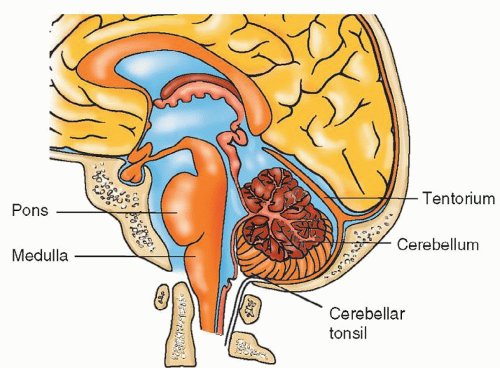
    Figure 14-1 ▪ Surgery on the area of the brain above the tentorium is called supratentorial; surgery below the tentorium is infratentorial.


  • A craniotomy is a surgical opening of the skull to provide access to the intracranial contents for reasons such as removal of a tumor, clipping of an aneurysm, or repair of a cerebral injury. It involves creating a bone flap at the area over the lesion. The flap created is either a free flap or an osteoplastic flap. With a free flap, the bone is completely removed and preserved for replacement prior to completion of the case. With a bone flap, the muscle is left attached to the skull to maintain the vascular supply (Fig. 14-2).


  • A craniectomy is excision of a portion of the skull without replacement. This may be a permanent removal, such as with a suboccipital craniectomy where the bony skull is so thick that it requires meticulous chipping away of small pieces of bone while avoiding the underlying dura until an opening is created. There is no remaining bone flap to replace. In some cases, the surgeon may prefer to repair the skull defect by replacing the bone chips on a piece of Gelfoam before closing the muscle and skin.


  • A craniectomy may also involve saving the created free bone flap for replacement at a later date. The bone may be surgically implanted in the patient’s abdominal fat, or stored frozen at −80°C either in a hospital tissue freezer or a bone bank. Before the bone is stored frozen in the hospital tissue freezer or a bone bank, the bone should be cleaned, cultured, packaged with aseptic technique, and well labeled with patient’s identification information.


  • Craniectomies are performed to achieve decompression after cerebral debulking, as a life-saving measure during periods of maximal cerebral swelling to oppose herniation (e.g., following cerebral trauma, high-grade subarachnoid hemorrhage [SAH], or massive cerebrovascular accident [CVA]), or for removal of bone fragments from skull fracture.


  • Cranioplasty is the repair of skull to re-establish the contour and integrity of the cranial vault. This procedure involves replacement of a skull defect with synthetic material or replacement of the original craniectomy bone flap.


  • Microsurgery is defined as any surgery performed with the assistance of an operating microscope that provides magnification of small and delicate tissues. The surgical technique (microoperative technique), instruments (microinstrumentation), illumination for visualization, and magnification (operating microscopes), along with a host of other equipment, are specifically designed for this use. Surgical microscopes continue to evolve with new technology. Advances in optical, electrical, and mechanical technology have made it possible to design microscopes with enhanced precision and maneuverability. Figure 14-3 shows a surgical microscope that also allows the assistant to directly observe the operative field in high definition in three dimensions. Many microscopes attach to a video monitor, allowing others assisting in the OR to view the operative field. The latest model microscope is not only equipped with more advanced optical technology but also integrates the images from the stereotactic navigation guidance systems into the visual field of the surgeon. With the revolutionary fluorescence technologies built in the microscope, surgeons are now able to define accurate tumor margins seamlessly during removal of a malignant tumor tissue (Fig. 14-4) and evaluate the blood flow immediately after resection of an AVM or clipping of an aneurysm (Fig. 14-5).

    Only gold members can continue reading. Log In or Register to continue

    Stay updated, free articles. Join our Telegram channel

Jul 14, 2016 | Posted by in NURSING | Comments Off on Management of Patients Undergoing Neurosurgical Procedures

Full access? Get Clinical Tree

Get Clinical Tree app for offline access