Intracranial Hypertension: Theory and Management of Increased Intracranial Pressure


Intracranial Hypertension: Theory and Management of Increased Intracranial Pressure


Karen S. March

Joanne V. Hickey


Intracranial hypertension is a clinically significant common pathophysiologic problem addressed daily by nurses and physicians who care for neuroscience patients. However, the specific sequence of pathophysiologic events leading to a sustained or unstable elevation in intracranial pressure (ICP) is still poorly understood. This chapter reviews the underlying physiologic cerebral hemodynamics and ICP concepts and applies this knowledge to the assessment and interventions for patient care and management.


CONCEPT OF INTRACRANIAL PRESSURE

ICP is, literally, the pressure inside the cranium. As simple as it sounds, however, ICP represents a complex and dynamic constantly changing set of parameters. Pressure is determined by two variables, size and volume. Except in infants, the skull is a rigid bony structure incapable of changing in size during acute changes in ICP. Thus, within the cranial vault all components that occupy the intracranial space contribute to the ICP. Typically, we think of the three main space-occupying substances as being cerebrospinal fluid (CSF), blood, and tissue.

Under normal conditions, ICP is determined by changes in the intracranial blood volume and changes in the pressure exerted by the CSF that circulates around the brain and spinal cord and within the cerebral ventricles. Cardiac and respiratory components are superimposed on the ICP, which is referenced to atmospheric pressure. The normal range of ICP is generally 0 to 10 mm Hg, although 15 mm Hg is considered the upper limit of normal. In the clinical setting, ICP values are generally expressed as the average or mean ICP over a given unit of time. Abrupt change in ICP from a stable level in response to activity or to a sudden change in volume is called transient increased ICP. It is surmised that different physiologic mechanisms control steady and transient states of ICP.1 With cerebral trauma or neurological disease, the normal homeostatic mechanisms controlling ICP may be disrupted, resulting in a sustained high ICP and eventual neurological and clinical death.

Because of the different compartments within the intracranial space, ICP can vary widely within different areas of the brain, especially with cerebral edema related to trauma or other disease entities. For example, the pressure in the tissue adjacent to an expanding, space-occupying lesion can be elevated, whereas the intraventricular pressure remains within a normal range. In addition, elevated ICP is not always conveyed to the lumbar subarachnoid space where it would be reflected during a lumbar puncture. It is, therefore, more accurate to think in terms of ICP pressures, rather than a single, uniform ICP pressure.


Physiologic Considerations


Monro-Kellie Hypothesis

The average intracranial volume in the adult is approximately 1,700 mL, composed of the brain (1,400 mL), CSF (150 mL), and
blood (150 mL). Basic to an understanding of the pathophysiologic changes related to ICP is the Monro-Kellie hypothesis (Box 13-1). It states that the skull, a rigid compartment, is filled to capacity with essentially noncompressible contents—brain and interstitial fluid (80%), intravascular blood (10%), and CSF (in the ventricles and subarachnoid space; 10%). The blood volume is comprised of approximately 20% arterial and 80% venous blood volume. Blood volume is related to the capacitance of the vessel; when a vessel is dilated it holds more blood volume than the vessel that is constricted. The volume of these three components remains nearly constant in a state of dynamic equilibrium. If the volume of any one component increases, another component must decrease reciprocally for the overall volume and dynamic equilibrium to remain constant. If the volume of any one component increases without a reciprocal decrease in one of the other components, ICP will rise. This hypothesis applies only when the skull is fused (i.e., a closed box). Infants or very young children who have skulls with nonfused suture lines have some space for expansion of the intracranial space in response to increased volume, at least initially.


Volume-Pressure Relationship Within the Intracranial Cavity

According to the Monro-Kellie hypothesis, reciprocal compensation occurs among the three intracranial components—brain tissue, blood, and CSF—to accommodate any alterations within the intracranial contents. The compensatory mechanisms that maintain the intracranial volume in a steady state include the following.


  • Displacement of some CSF from the ventricles and cerebral subarachnoid space through the foramen magnum to the spinal subarachnoid space and through the optic foramen to the perioptic subarachnoid space (basal subarachnoid cisterns)


  • Displacement of some blood by compression of the low-pressure venous system, especially the dural sinuses


  • Decreased production of CSF


  • Vasoconstriction of the cerebral vasculature, which results in a decrease in the intracranial blood volume

However, the amount of displacement of the brain, CSF, or blood that can occur through compensatory mechanisms is limited. After compensatory mechanisms have been exceeded, ICP rises and intracranial hypertension results. Specific medical therapies that have been used to maintain the intracranial volume in a steady state include the following.


  • Displacement of CSF from the cerebral ventricular system using an intraventricular catheter (IVC) connected to an external drainage system (temporary) or a ventriculoperitoneal shunt (permanent).


  • Displacement of some blood by inducing arterial vasoconstriction. Vasoconstriction reduces the capacity of the artery to carry blood and thereby results in a net decrease in the volume of blood in the cranial vault.


  • Facilitating venous outflow from the head by elevating the head of the bed and maintaining midline head position thus promoting venous drainage through the jugular veins.


  • Displacement of some brain tissue volume through mechanisms to decrease intracellular volume. An example of this is seen when the increased osmotic pressure of the circulating blood draws off free water from brain cells and results in a net decrease in brain volume.

Compliance. Compliance is a measure of the adaptive capacity of the brain to maintain intracranial equilibrium in response to physiologic and external challenges to that system. It has been described as a measure of brain “stiffness.” Compliance represents the ratio of change in volume to the resulting change in pressure. It is represented by the following formula, in which Δ represents the symbol for change, V volume, and P pressure.


Applying this concept to intracranial dynamics, compliance is the ratio of change in ICP as a result of change in intracranial volume.

The intracranial dynamics are shown in Figure 13-1. The vertical axis represents pressure or ICP (measured in mm Hg). The horizontal axis represents intracranial volume. The shape of the curve demonstrates the effects on ICP when volume is added to the intracranial space. The ICP remains constant from point A to just before point B with the addition of volume. Compensatory mechanisms are adequate and compliance is high. Point B is a threshold, so that even though the ICP is still within normal limits, compliance is decreased. From points B to C, the slope begins to increase, reflecting a decrease in compensatory mechanisms and low compliance. With even a small increment in volume (points C to D),
the compensatory mechanisms are exceeded, compliance is lost, and a disproportionate elevation in ICP is noted.


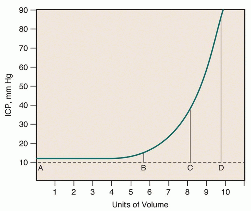
Figure 13-1 ▪ Pressure-volume curve. From point A to just before B, the intracranial pressure (ICP) remains constant, although there is addition of volume (compliance is high). At point B, even though the ICP is within normal limits, compliance begins to change, as evidenced by the slight rise in ICP. From points B to C, the ICP rises with an increase in volume (low compliance). From points C to D, ICP rises significantly with each minute increase in volume (compliance is lost).


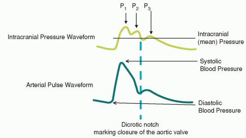
Figure 13-2 ▪ Comparison of arterial and intracranial pressure waveforms.

Factors that influence compliance include the amount of volume increase, the time frame for accommodation of the volume, and the size of the intracranial compartments. Small volume increments made over long periods of time can be accommodated much more easily than a comparable quantity introduced within a short time interval. This concept helps to explain why a slow-growing tumor may become large (roughly equal to the size of a golf ball) without causing an elevation in ICP. The size of the intracranial compartments can vary because of cerebral atrophy, craniectomy, or immaturity of the cranium (i.e., suture lines are not fused). For example, an adult with an acute subdural hematoma, an enlarging lesion (typically over a few days), will develop increased ICP. Of note, in the elderly, some cerebral atrophy occurs with normal aging, thus creating a little more space in the cranial vault. Because of this extra intracranial space, a subdural hematoma in an older person may go unnoticed for days or weeks before there are signs and symptoms of a rise in ICP. However, a critical point is eventually reached beyond which physiologic compensation is exhausted, and there is a dramatic rise in ICP, regardless of how slowly minute-volume increments are added.

Estimating Compliance by Morphologic Changes in Intracranial Pressure Waveform. Cerebral compliance can be estimated clinically by examining morphologic changes in ICP waveforms. This task requires astute observation and an understanding of the ICP wave using a continuous real-time display monitor. The ICP pulse wave arises from arterial pulsations and to a lesser degree from the respiratory cycle. In addition, effects from retrovenous pulsation and choroid plexus pulsations also influence the waveform.2 Observation of any variations in waveform during activities, treatments, or environmental changes allows one to determine the effects of these activities on patients and to protect them from risk, if necessary.

The ICP waveform has three peaks that are of clinical importance. These are aptly named P1, P2, and P3. Other lesser peaks sometimes occur after P3, but their significance is unknown.3 The principal waveforms, in order of appearance, are the following.


  • P1, the percussion wave, which originates from pulsations of the arteries and choroid plexus, is sharply peaked and fairly consistent in amplitude.


  • P2, the tidal wave, is more variable, changes with compliance, and terminates in the dicrotic notch.


  • P3, the dicrotic wave, immediately follows the dicrotic notch.

Clinically, aligning the ICP waveform with an arterial pressure waveform demonstrates the association between systemic and cerebral hemodynamics. See Figure 13-2 for a comparison between ICP and arterial wave forms. Beginning with the ejection of blood from the heart into the brain and provides the arterial influence of the ICP waveform, P1. Throughout the remainder of ventricular systole, the amount of blood entering the brain tapers off and the ICP wave begins to fall.

The second upward trend in the ICP wave is the P2 wave, which is also called the tidal wave. The P2 wave is thought to be due to the reflexive compliance of the surrounding compartmental volumes (brain, venous volume, and CSF) as they affect the P1 wave. The size and shape of the P2 wave and its relationship to P1 (becomes equal to or greater) most directly reflect compliance.4 As compliance decreases, there is less absorption of the percussion wave and P2 will increase. The end of the P2 wave is marked by the presence of the dicrotic notch.

Physiologically, the dicrotic notch is associated with closure of the aortic valve and signals the end of ventricular systole.5 Components of the ICP waveform that occur after the dicrotic notch cannot, therefore, be assumed to relate to arterial influence, but are related to changes in retrograde venous pressures. The dicrotic notch also signals the beginning of P3 and the pressure tapers down to the diastolic position after P3 unless retrograde venous pulsation creates additional waves.

Under stable conditions, the ICP waveform has a very predictable appearance. Changes in the ICP waveform that correspond to different points during the respiratory cycle likely reflect pressure changes in the intrathoracic pressure and decreased venous outflow from the cranium. When ICP is normal with good compliance, the pulse wave formation appears as a descending sawtooth pattern (Fig. 13-3). A rise in ICP with decreasing compliance is reflected as a progressive rise in P2. The P1 and P3 waves rise to a much lesser degree so that the overall pulse wave has a rounded appearance (Fig. 13-4).

Recent research regarding the use of ICP waveform to predict patients at risk for developing increased ICP is contradictory. Fan et al. in 2008 found that P2 elevations were not predictive of disproportionate increases in ICP while earlier studies conducted by Willis and Kirkness found elevations in P2 were predictive of increases in ICP. Table 13-1 highlights ICP changes in various physiological
situations. Because it is possible to observe responses to patient activities, treatments, and environmental factors immediately, it may be possible to use this information to develop an individualized plan of care based on avoidance of individual risk factors. ICP waveform analysis should be a focus of more clinical research to explore its ramifications for integration into clinical practice.


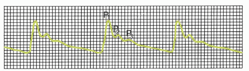
Figure 13-3 ▪ Normal intracranial pressure waveform (ICP = 8 mm Hg).


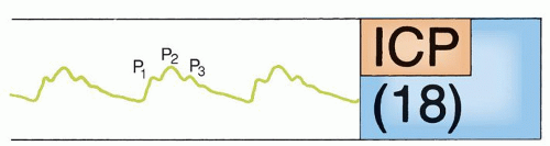
Figure 13-4 ▪ Abnormal intracranial pressure (ICP) waveform (ICP = 18 mm Hg).


Cerebral Hemodynamics

Several concepts related to cerebral hemodynamics are important to an understanding of the pathophysiology of increased ICP.

Cerebral Blood Volume. Cerebral blood volume (CBV) refers to the amount of blood in the brain at a given time. Normally, blood occupies about 10% of the intracranial space. 80% of the blood volume is venous, contained in the lower-pressure venous system. Arterial CBV is affected by the autoregulatory mechanisms that control cerebral blood flow (CBF) by dilating or constricting blood vessels. When cerebral vessels dilate, they have increased capacitance and therefore have an increased blood volume which may lead to an increase in cerebral volume and increase in ICP. A limited compensatory mechanism is operational when ICP begins to rise in which venous blood and CSF are shunted out of the cranium, thus decreasing CBV. However, as the compensatory reserve is exhausted, pressure in the venous system rises, CBV increases, and ICP rises. Depending on the rate of decline in CBF and the duration of ischemia, cerebral infarction can occur.


TABLE 13-1 ICP CHANGES RELATED TO DIFFERING PHYSIOLOGICAL CONDITIONS


CONDITION


ICP CHANGES


Rapidly expanding mass lesion


Increase mean ICP


Increase ICP waveform amplitude


Increase/decrease CSF volume


Increase/decrease mean ICP


Increase/decrease ICP waveform amplitude


Little change in ICP waveform configuration


Severe arterial hypotension


Decrease mean ICP


Decrease ICP waveform amplitude, especially P1


Severe arterial hypertension


Increase mean ICP


Increase ICP waveform amplitude


Severe hypercapnia and hypoxia


Increase mean ICP


Increase ICP waveform amplitude


Rounding of ICP waveform due to increase in later waveform components


Hyperventilation


Decrease mean ICP


Decrease ICP waveform amplitude P2, and to a lesser degree, P3 with little change in P1


Jugular vein compression


Increase mean ICP


Increase ICP waveform amplitude, mainly P2 and P3


From: Kirkness, C. J., Mitchell, P. H., Burr, R. L., March, K. S., & Newell, D. (2000).


Intracranial pressure waveform analysis: Clinical and research implications.


Journal of Neuroscience Nursing. 32, 271-277. Used with permission.


Cerebral Blood Flow. Adequate blood flow and oxygenation are required to maintain normal neural function. At rest, the approximate CBF through the brain is 45 to 65 (average 50) mL/100 g brain per minute; given that a normal brain weighs between 1 and 1.5 kg, this translates into 450 to 1,000 mL/min of blood flow for the whole brain.6 CBF in the cortex is significantly higher than in the white matter due to differences in neural activity and needs. The average CBF in the cortex is 80 to 100 mL/100 g of brain per minute while white matter flow is 20 to 25 mL/100 g of brain per minute. Although the brain is only 2% of body weight, it receives 15% to 20% of total cardiac output and uses 20% of oxygen and 25% of body glucose in the basal state. In normal conditions, the total oxygen consumed by the brain and CBF are almost constant. Local demand for blood can vary depending on differing metabolic needs. Cortical gray matter receives about six times the amount of blood as white matter, and changes in blood flow respond to demands of varying neural activity.7 A basic concept of any hemodynamic system is that the blood flow is directly proportional to the perfusion pressures and inversely proportional to the total resistance of the system. CBF is represented by the following equation, in which MAP = mean arterial pressure, CVP = central venous pressure, CPP = cerebral perfusion pressure, and CVR = cerebrovascular resistance.

CBF = MAP − CVP/CVR or CPP/CVR

Note: Under normal conditions, CVP represents the resistance to venous outflow and CPP = MAP − CVP. Another common formula (CPP = MAP − ICP) incorporates the resistance to flow contributed from ICP.

If the CBF exceeds the amount of blood required for metabolism, a state of hyperemia is said to exist. Hyperemia, also called “luxury perfusion,” is an excess of blood to a part of the body. Commonly, hyperemia is associated with vasodilation of the blood vessels leading to an increased blood volume and increased ICP.


Factors That Modify Cerebral Blood Flow

CBF can be increased or decreased by several extracerebral and intracerebral factors.8

Extracerebral Factors. These factors are primarily related to the cardiovascular system and include systemic blood pressure, cardiac function, and blood viscosity.


  • Blood pressure. The main force that maintains CBF is the pressure difference between the arteries and the veins. In the brain, cerebral venous pressure is low (approximately 5 mm Hg), so that arterial blood pressure is the most important factor in maintaining CBF. Under normal circumstances, intrinsic regulatory mechanisms maintain CBF at a constant level even with systemic arterial blood pressure changes, unless the MAP dips to less than 50 mm Hg.


  • Cardiac function. Systemic arterial blood pressure is dependent on cardiac output and peripheral vasomotor tone (resistance), which are primarily under autonomic control from the vasomotor center of the medulla. Cardiac arrhythmias, altered myocardial function, circulating blood volume, and cardiac disease can affect cardiac output, thus influencing CBF. In addition, various carotid sinus and aortic arch reflexes assist in maintaining a constant blood pressure. Advanced age, atherosclerosis, and certain drugs can alter these reflexes, thus affecting arterial blood pressure and CBF secondarily.


  • Blood viscosity. Blood viscosity is a measure of how thick the blood is. Blood viscosity is generally represented by hematocrit (Hct). A higher Hct correlates with higher viscosity. Fluid replacement therapy and anemia are two primary causes of a decrease in blood viscosity. Anemia may increase blood flow up to 30%, whereas polycythemia may decrease flow by more than 50%. When blood is viscous, vessels dilate and they constrict when blood is dilute.

Intracerebral Factors. The primary intracerebral factors that influence CBF are widespread cerebrovascular artery disease and increased ICP.

Widespread cerebrovascular artery disease can increase cerebrovascular resistance (CVR), resulting in reduced CBF. Processes that rapidly shunt blood from arteries to veins (as in an arteriovenous malformation) result in a condition in which total CBF is increased but local tissue perfusion is decreased.

When increased ICP is present, it is transmitted to the low-pressure venous system, thus increasing cerebral venous pressure and decreasing CBF.


Regulation of Cerebral Blood Flow

Several other intracerebral regulatory mechanisms that can modify CBF include autoregulation, chemical-metabolic regulation, and neurogenic regulation.

Autoregulation. The ability of an organ (such as the brain) to maintain a constant blood flow despite marked changes in arterial perfusion pressure is called autoregulation. Autoregulation is a major homeostatic and protective mechanism occurring in large and small arterioles. Autoregulation is achieved using myogenic, chemical-metabolic, and neurogenic mechanisms. Arterioles contain smooth muscles that respond to stretch receptors and intraluminal pressure, causing vasoconstriction to increase intraluminal pressure and vasodilation to decrease intraluminal pressure. Specifically, autoregulation is primarily a pressure-controlled myogenic mechanism that operates independently, yet synergistically, with other chemical-metabolic and neurogenic autoregulatory mechanisms. Autoregulation provides a constant CBF, maintained within the normal range by adjusting the diameter of blood vessels. Autoregulation operates within limited parameters in healthy people—a mean arterial blood pressure of 50 to 60 to 150 mm Hg results in changes in vascular diameter; as pressure decreases the vessels dilate and as pressure increases vessels constrict. Pressures below 50 to 60 mm Hg result in decrease in CBF and above 150 mm Hg, CBF increases. In patients with chronic hypertension, the curve shifts to the right. Autoregulation generally operates with an ICP of less than 40 mm Hg. Without autoregulation, there is reduced cerebrovascular tone, known as vasomotor paralysis, and the CBF and CBV become passively dependent on changes in blood pressure.

Chemical-Metabolic Regulation. Chemical and metabolic regulation exerts a strong influence on CBF; carbon dioxide, oxygen, and pH are important chemical regulators.


  • Carbon dioxide (CO2), found in the blood and locally in cerebral tissue as an end product of cell metabolism, is a potent agent that influences CBF. Cerebral blood vessels respond indirectly to changes in carbon dioxide (PaCO2) levels that alter pH. Increases in PaCO2 in acidosis lead to vasodilation, thus increasing blood flow. Decreased PaCO2 leads to alkalosis and will cause vasoconstriction and result in a general decrease in blood flow. CBF generally changes by 2% to 3% for each change in PaCO2 within the range of 20 to 80 mm Hg.


  • Oxygen (O2) has an opposite, but less profound effect: reduction in local oxygen (PaO2) produces vasodilation by changes in pH (acidosis), and an increase in local PaO2 produces vasoconstriction. The mechanism by which this is achieved is unclear.


  • H+ ions are also powerful agents that influence CBF. In body fluids, CO2 combines with water to form carbonic acid, with subsequent dissociation of hydrogen ions. Hydrogen ion concentration can also be increased by lactic acid, pyruvic acid, and other acids that result from cell metabolism. Excess hydrogen ions cause cerebral vessels to dilate, which results in a net increase in CBF.


  • pH changes also have an effect on cerebral arterioles; a low pH (acidosis) results in vasodilation and increased CBF, and a high pH (alkalosis) results in vasoconstriction and a decreased CBF. A buildup of the metabolic end products of cell metabolism (e.g., lactic acid, pyruvic acid, carbonic acid) causes localized acidosis. An increase in the concentration of these acids will also increase CBF.

Flow-metabolism coupling is an intrinsic mechanism in which CBF changes in response to changes in metabolic need. If there is an increase in metabolic demand (fever), normally CBF will increase to meet the increased need for cellular nutrients. When flow-metabolism coupling is impaired, two situations can arise: a mismatch in which CBF never meets the demands of the tissue resulting in ischemia or uncoupling where when the brain is at rest, CBF matches the demand, but when demand is increased, CBF is unable to change to meet the demand and ischemia occurs.

Neurogenic Regulation. Neurogenic factors play a lesser role in regulation of CBF than the chemical-metabolic regulators. Neurogenic regulation includes a rich neural network that is extrinsic and intrinsic to the brain.


  • Extrinsic neurogenic control. Sympathetic innervation comes from postganglionic fibers of the superior cervical sympathetic ganglion that innervate the carotid and vertebral arteries and major intracranial branches. Norepinephrine, a vasoconstrictor, is released from the sympathetic fibers. Parasympathetic fibers come from the facial and superficial petrosal nerves to innervate large- and small-diameter cerebral blood vessels. They use acetylcholine as a neurotransmitter, which causes vasodilation.


  • Intrinsic neurogenic control. The intrinsic pathways originate in the brainstem and interneurons in the cerebral cortex. These pathways originate in the brainstem from the locus caeruleus (neurons use norepinephrine to produce microcirculatory vasodilation), raphe nuclei (neurons use serotonin, a vasoconstrictor), and fastigial nuclei of the cerebellum. Cortical interneurons contain both vasoconstrictor and vasodilator substances.

Other Factors. An increase in CBF and CBV can also result from pharmacologic agents such as volatile anesthetic agents (halothane, enflurane, sevoflurane, desflurane, isoflurane, nitrous oxide) and those antihypertensives (sodium nitroprusside, nitroglycerin) that cause vasodilation. Increased CBF and CBV are also associated with rapid eye movement (REM) sleep, arousal, pain, seizures, elevations in body temperature (about 6% per 1°C), and cerebral trauma.

Cerebral Perfusion Pressure. Cerebral perfusion pressure (CPP) is defined as the blood pressure gradient across the brain. An adequate CPP is necessary to maintain an adequate driving force for blood throughout the brain in order to prevent episodes of cerebral ischemia. The CPP found in a normal adult is in the range of ˜80 mm Hg. In the laboratory, ischemia is not seen until the CPP falls below 40 mm Hg.9 In traumatic brain injury (TBI), however, the observations regarding global blood flow and ICP changes may not accurately reflect areas of severe regional ischemia. Therefore, in cerebral trauma, recommendation is to maintain CPP from 50 to
70 mm Hg in adults. In the pediatric population, thresholds are less well defined and most likely depend on the age of the patient. Current recommendations for the pediatric patient are to maintain CPP between 40 and 65 mm Hg.

The decision to maintain a higher threshold for CPP remains controversial. Rosner and Daughton recommend a CPP of 70 to 80 mm Hg for patients with cerebral injury and intracranial hypertension.10 Robertson et al., however, found no difference in 3-month and 6-month outcomes for patients treated to maintain CPP greater than 50 mm Hg versus patients treated to maintain CPP greater than 70 mm Hg and higher incidences of complications such as acute respiratory distress syndrome (ARDS).11 New technology enabling CBF and PbtO2 monitoring may allow that CPP be targeted to a treatment goal rather than an absolute value. One prospective study concluded that episodes of CPP less than 70 mm Hg are associated with a significant decrease in brain tissue oxygenation (PbtO2), whereas this risk is markedly decreased when CPP is maintained greater than 70 mm Hg.12 What is clear is that if CPP is inadequate, ischemia develops; if ischemia is not reversed, infarction results.

CPP is calculated as the difference between the incoming MAP and the opposing ICP. It is represented by the following formula:

CPP = MAP − ICP

CPP is an estimate of the adequacy of cerebral circulation. To calculate CPP, it is first necessary to compute the MAP. This is calculated as follows:

MAP = (systolic − diastolic)/3 + diastolic Example: systolic = 130; diastolic = 82; ICP = 15 MAP = (130 − 82)/3 + 82 = 98 MAP − ICP = CPP 98 − 15 = 83

Cerebrovascular Resistance. CVR is the pressure across the cerebrovascular bed from the arteries to the jugular veins, which is influenced by inflow pressure, outflow pressure, the diameter of the vessels, and ICP. CVR is the resistance created by the cerebral vessels and is controlled by the autoregulatory mechanisms of the brain. The CVR increases with vasoconstriction, decreases with vasodilation, and varies inversely with CBV. Because the cerebral venous system does not have valves as does other veins in the body, any condition that obstructs or compromises venous outflow may cause blood to back up into the intracranial cavity, increase CVR, and therefore, increases CBV and ICP. For example, increasing levels of positive end-expiratory pressure (PEEP) can increase intrathoracic pressure leading to increased jugular venous pressure and result in increased ICP.

Cerebral Metabolism. Under normal circumstances, the brain depends almost exclusively on oxygen and glucose to obtain its energy needs. Neural cells in the brain lack mitochondria and the ability to store energy. Therefore, a constant and critical supply of glucose must be transported to brain cells to produce energy in the form of adenosine triphosphate (ATP), which is synthesized through the glycolytic pathway, Krebs cycle, and respiratory chain (also called the electron transport chain). Both the Krebs cycle and the respiratory chain require oxygen, and this accounts for the brain’s high and critical dependence on oxygen.

Aerobic metabolism is the usual pathway for glucose metabolism. Glucose is metabolized through the previously mentioned pathways to yield 38 moles of ATP per mole of glucose. However, if anaerobic metabolism occurs, Krebs cycle and the respiratory chain cannot be activated because of lack of oxygen. In these circumstances, pyruvate, derived from glycolysis, is metabolized to lactate, yielding only 2 moles of ATP per mole of glucose. Therefore, much less ATP is available to fuel the ATP-dependent sodium-potassium pump of cell membranes.

Studies following TBI suggest that in some patients develop a “hypermetabolism” in the first day in an attempt to meet the cellular needs. Because of an inadequate CBF, the energy is produced by hyperglycolysis, a burning of glucose in the absence of oxygen. When these mechanisms fail, ion channels open leading to an influx of sodium and calcium into the cell. As calcium levels increase intracellularly, mitochondrial function fails and neurons die. In the weeks following the initial injury, metabolic demand may become depressed.


HERNIATION SYNDROMES OF THE BRAIN

Increased ICP that continues to develop will result in herniation. Herniation is defined as the abnormal protrusion of an organ or other body structures through a defect or natural opening in a covering membrane, muscle, or bone. Simply put, brain herniation is when one part of the brain moves beyond its normal border. It is important to note that there are different types of brain herniation. To understand the pathophysiology of these different cerebral mass lesions (Box 13-2), it is most important to understand the principles that govern herniation. The intracranial cavity is divided into several smaller compartments by folds of the fibrous, relatively rigid dura mater. The most important dural folds are as follows.


  • Falx cerebri is a double fold of dura mater that drops into the longitudinal fissure and is partially responsible for dividing the supratentorial space into the left and right hemispheres.


  • Tentorium cerebelli is a double fold of dura mater that forms a tent-like partition (higher in the middle) between the cerebrum and cerebellum. The area above the tentorium is the supratentorial space, and the area below the tentorium is the infratentorial space. To allow the brainstem, blood vessels, and accompanying nerves to pass through the tentorium, there is an oval opening in the tentorium called the tentorial notch or incisura.


  • Falx cerebelli is a double fold of dura mater that separates the cerebellum (in the infratentorial space) into left and right sides.

When cerebral edema or a mass lesion occurs within a compartment, the pressure exerted by the lesion is not evenly distributed. This uneven distribution of pressure results in shifting or herniation of the brain from a compartment of high pressure to one of lesser pressure. The shifting of cerebral structures resulting from pressure is called the mass effect. With mass effect, there is compression and traction of cerebral tissue that results in ischemia. Ischemia is potentially reversible, but without effective treatment, it will lead to infarction, which is irreversible.

In addition, the foramen magnum is the hole at the base of the skull (occipital bone) through which the spinal cord passes. If an elevated ICP resulting from a supratentorial lesion continues to expand unchecked, the uncus of the medial inferior temporal lobe will herniate through the tentorium, with resulting exertion of pressure on the brainstem. This will eventually result in cerebellar tonsillar herniation through the foramen magnum, the only opening in the closed cranial vault. Cerebellar tonsillar herniation is a sure cause of death because of pressure on the vital structures in the medulla.

An expanding mass lesion of the supratentorial space behaves differently than an expanding mass lesion in the infratentorial space. The intracranial pathologic changes radiate downward and away from the supratentorial lesion in a rostral-caudal pattern. (A rostral-caudal pattern means that the deterioration in function proceeds from the head to the tail.) Of particular importance is the clinical presentation of Cushing’s response and of Cushing’s triad. Cushing’s response is an increase in blood pressure with bradycardia while Cushing’s triad is a rise in blood pressure, widening pulse pressure, bradycardia, and irregular respirations. These symptoms are seen with posterior fossa lesions or when the brainstem is compressed as part of downward progression of herniation.17


Supratentorial Herniation

Progressive supratentorial lesions develop clinical signs and symptoms in a sequence of ocular, motor, cardiovascular, and respiratory function. This pattern indicates the predictable continuum of rostral-caudal failure that proceeds from the diencephalon to the midbrain, followed by the pons, and finally medullary function. The pattern of deterioration is predictable with a significant hemorrhage, an abscess ruptures into the ventricles, or a contraindicated lumbar puncture rapidly changes the intracranial dynamics, resulting in compression of the medulla.

Three major patterns of herniation, described by Plum and Posner in their classic work, identify syndromes caused by expanding supratentorial lesions: (1) cingulate herniation, (2) central transtentorial herniation, and (3) uncal transtentorial herniation17 (Fig. 13-5). A developing supratentorial lesion results in two clinically significant syndromes, a central syndrome and an uncal syndrome. These two syndromes are the major clinical presentations encountered in practice. Clinically, they are two distinct patterns early in their course, but both merge into a singular pattern once the pathophysiology begins to involve the midbrain level and below (brainstem structures).


Cingulate Herniation

An expanding lesion in one cerebral hemisphere can cause pressure medially so that the cingulate gyrus is forced under the falx cerebri, displacing it toward the opposite side. This displacement of the falx cerebri can compress the local blood supply and cerebral tissue, which causes edema and ischemia, which further increase the degree of ICP elevation. Cingulate herniation (also called a subfalcine herniation) is common, but little is known about its clinical signs and symptoms, except in those instances when the blood supply of the anterior cerebral artery is compromised as a result of the subfalcine shift.


Central Transtentorial Herniation (Central Syndrome)

Central herniation is the rostral-caudal downward displacement of cent of the cerebral hemispheres as a result of


  • a lesion located on the central neural axis (diencephalon).


  • an extracerebral lesion located around the central apex of the cranium (midline).


  • bilateral lesions in the hemispheres.


  • unilateral cingulate herniation.

The lesion produces a downward displacement of the cerebral hemispheres, basal ganglia, diencephalon, and midbrain through the tentorial incisura. The diencephalon can be compressed tightly against the midbrain with such force that edema and hemorrhage result. Compression of the posterior cerebral artery may be seen on cerebral arteriogram. The early symptoms of the central syndrome include the following.


TABLE 13-2 FACTORS ASSOCIATED WITH INCREASED INTRACRANIAL PRESSURE


FACTORS AND DESCRIPTION


MECHANISMS


Hypercapnia


PCO2 ≥ 45 mm Hg


Elevated CO2 results in increased CBF, which leads to increased CBV and ICP.


Excessive levels of CO2 in the blood Potent cerebral vasodilator


Hypercapnia results from underventilation (decreased ventilatory volume or decreased ventilatory rate) of a patient in such circumstances as follows.


  • Sleep apnea


  • Pulmonary diseases/conditions (e.g., atelectasis, pneumonia, COPD, neurogenic pulmonary edema, ARDS)


  • Oversedation


  • Shallow respirations, as seen with anxiety reactions, severe pain, undersedation, or ventilatory asynchrony


  • Pressure on brainstem respiratory centers


  • Improperly calibrated ventilator (e.g., rate, sensitivity)


Hypoxemia


PaO2 < 50 mm Hg


Decreased O2 in the blood Has much less effect as a vasodilator compared to CO2


Decreased O2 does not increase cerebral vasodilation until it is ˜50 mm Hg or less.


Hypoxemia results from the following.


  • Insufficient concentration of O2 administered during O2 therapy


  • Insufficient ventilation during and after suctioning


  • Inadequate ventilation during intubation


  • Partial or complete airway obstruction


  • Increased oxygen consumption


Respiratory Procedures


Suctioning


PEEP


Asynchrony of respiratory rate when Ambu bag is used


Intubation


Suctioning decreases O2, increases CO2, and partially obstructs the airway with a catheter.


PEEP increases intrathoracic pressure, which leads to increased central venous pressure, cerebral venous pressure, and ICP.


Asynchronous use of the Ambu bag causes a response that is similar to the PEEP response.


Same as suctioning.


Vasodilating Drugs


Anesthetic agents (halothane, enflurane, isoflurane, nitrous oxide)


Some antihypertensives (nitroglycerin, Nipride)


Some histamines


Vasodilation causes increased CBF, resulting in increased ICP.


Some Body Positions


Trendelenburg’s position (always contraindicated in neuroscience patients)


Prone position (increased intra-abdominal and intrathoracic pressures; also, neck flexion impedes venous drainage)


Extreme hip flexion (increased intra-abdominal pressure)


Hip flexion on a pendulous abdomen (increased intra-abdominal pressure)


Angulation of the neck; neck flexion, even from a small, improperly positioned pillow, or improper lateral positioning when turned (which impedes venous return from the brain)


Turning the patient laterally if the head of the bed is up and the knees are flexed on the abdomen (increased intra-abdominal pressure)


Obstruction of venous return from the brain increases CBV, which results in increased ICP.


The venous cerebrovascular system has no valves; thus, an increase in intra-abdominal, intrathoracic, or neck pressure is communicated as increased pressure throughout the open venous system, thus impeding drainage from the brain and increasing ICP.


It has been an accepted practice to elevate the head of the bed 30 degrees to facilitate drainage from the brain; current research is inconclusive as to what is the best degree of head elevation for promoting venous drainage from the brain.


Pressure on Neck


Snug “track tape,” soft collar, or other constricting material


These impede venous drainage from the brain.


Isometric Muscle Contractions


Increased muscle tension without lengthening of the muscle


Examples: pushing against the bed with one’s feet, as in pushing oneself up in bed; pulling on an extremity restraint; shivering; decortication; decerebration; other rigidity


Isometric muscle contractions increase systemic blood pressure and contribute to further elevation of increased ICP in the patient who is on the borderline of brain compliance or who already has increased ICP.


Passive range of motion (PROM) exercises do not involve isometric contractions because the length of the muscle does change during contraction. Therefore, PROM exercises should be included in the plan of care.


Chlorpromazine (Thorazine) has been used to control shivering; pancuronium bromide (Pavulon) and baclofen (Lioresal) have been used for decerebration in the patient at risk for ICP spikes.


Valsalva’s Maneuver


Exhalation against a closed epiglottis. Examples: straining at stool, moving in bed, sneezing


Increased intra-abdominal or increased intrathoracic pressure impedes venous return from the brain, thereby increasing ICP.


If the patient’s ICP is already elevated or brain compliance is borderline, spikes in ICP may occur.


Coughing


Increases intra-abdominal and intrathoracic pressure as a result of muscle visceral contractions


Increased intra-abdominal and intrathoracic pressures impede venous drainage from the brain.


Also, the increased pressure is transmitted through the spinal subarachnoid space to the intracranial subarachnoid space and through the veins that communicate with the dural venous sinuses and intracranial subarachnoid space. The venous return from the cranial vault is impeded, resulting in increased ICP.


Noxious Stimuli


For example, invasive procedures, such as lumbar puncture, or painful nursing procedures, such as removal of tape from skin


Activation of the sympathetic nervous system is probably the major cause of increased blood pressure, increased CBF, and increased ICP, particularly in the patient who already has increased ICP.


Activities That Increase Cerebral Metabolism


Arousal from sleep


REM phase of sleep


Seizure activity


Hyperthermia


A focal or generalized increase in cerebral metabolism results from these activities. There is regional or generalized increased CBF, which is reflected in increased CBV, which causes an increase in ICP.


Clustering of Activities


In a patient with already increased ICP, clustering of patient care activities (e.g., bathing, turning) and other activities known to increase ICP can cause dangerous elevations in ICP and plateau waves in the patient at risk. The impact of nursing activities may be compounded after having blood drawn or undergoing an invasive procedure.


Note that suctioning is notorious for increasing ICP in the patient at risk.


The compounding effect of activities causes an increase in blood pressure, CBF, and ICP; elevations of ICP can be high enough to cause plateau waves and cerebral ischemia.


Spacing of procedures allows the patient’s ICP to return to a safe baseline level. Observing the effects and the return to baseline on an ICP monitor provides a guide for delivering safe care to the patient at risk.


CBF, cerebral blood flow; CBV, cerebral blood volume; ICP, intracranial pressure; COPD, chronic obstructive pulmonary disease; ARDS, acute respiratory distress syndrome; PEEP, positive end-expiratory pressure; PROM, passive range of motion; REM, rapid eye movement.


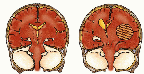
Figure 13-5 ▪ Cross-section of a normal brain (left) and a brain with intracranial shifts from supratentorial lesions (right). (1) Herniation of the cingulate gyrus under the falx. (2) Herniation of the temporal lobe into the tentorial notch. (3) Downward displacement of the brainstem through the notch. (From: Plum, F., & Posner, J. (1972). Diagnosis of stupor and coma. (2nd ed.). Contemporary neurology series. Philadelphia: F.A. Davis.)


  • Deterioration in the level of consciousness (LOC) (confusion and restlessness)


  • Bilateral small, reactive pupils (in early diencephalic stage)


  • Gradual loss of upward (vertical) gaze


  • Contralateral monoplegia or hemiparesis to hemiplegia


TABLE 13-3 PROGRESSION OF SIGNS AND SYMPTOMS OF THE CENTRAL SYNDROMEa


ASSESSMENT


EARLY DIENCEPHALON


LATE DIENCEPHALON


MIDBRAIN UPPER PONS


LOWER PONS/UPPER MEDULLA


MEDULLA


Outcome


Potentially Reversible


Poor Prognosis-Irreversible


Level of consciousness


Pattern varies from decreased alertness and behavior with difficulty to concentrate


Some become agitated; others become drowsy


Stupor to coma


Deep coma


Deep coma


Deep coma


Ocular


Pupillary size and reaction to light


Extraocular movement


Oculocephalic (OC) and oculovestibular (OV) response


Small (1-3 mm), reactive, but contraction difficult to see; examine using bright light


Oculocephalic (OC) and oculovestibular (OV) response


Conjugate or slightly dysconjugate at rest; may note roving eyes


OC and OV intact


Small (1-3 mm), reactive, but range of contraction slight; difficult to see


Brisk lateral OC; impaired upward conjugate on vertical OC


Full lateral OV response


Midpoint (3-5 mm), nonreactive


Often irregularly shaped


Impaired OC


OV difficult to elicit; dysconjugate with limited horizontal movement


Midpoint (3-5 mm), nonreactive


Often irregularly shaped


Absent OC and OV


Dilated and nonreactive


Absent OC and OV


Motor


Function


Pre-existing hemiparesis or hemiplegia develop to bilateral signs in early diencephalic stage


Hemiparesis or hemiplegia worsens and paratonic rigidity develops homolateral to lesionb


Purposeful response to painful stimuli


Decortication first contralateral to primary lesion in response to painful stimuli, then bilateral


May see a pattern of decortication ipsilateral and decerebration contralateral to primary lesion


Decortication to bilateral decerebration in response to painful stimuli or sometimes spontaneously


Flaccid at rest


Occasional flexor response of lower extremities to painful stimuli


Flaccid at rest


Occasional flexor response of lower extremities to painful stimuli


Reflexes


Babinski’s sign absent


Bilateral Babinski’s sign less vigorous ipsilateral to lesion. Grasp reflex present


Bilateral Babinski’s sign present


Bilateral Babinski’s sign present


Respirations


Deep sighs, yawns, and occasional pauses


Cheyne-Stokes syndrome


Gradual change to sustained hyperventilation


Normal pattern of respirations, but more rapid (20-40/min) and shallow


Apneustic and cluster breathing


Slow and irregular rate and amplitude (ataxic respirations); interrupted by deep sighs, gasps, and periods of apnea


Apnea


Other


Diabetes insipidus


Fluctuation in temperature; hyperthermia


Often hyperthermia


Cushing’s triad


Often hyperthermia


Cushing’s triad


aAn important point to keep in mind when observing a patient with a possible herniation syndrome is that there is predictable order to the development of signs and symptoms. The neurological deterioration in both central and uncal syndromes proceeds in an orderly rostral-caudal direction; the diencephalon, midbrain, pons, and finally, the medulla are affected from the increasing pressure. Signs and symptoms characteristic of each area can be identified. Notice that the last stages of central and uncal herniation are the same.

b Often there is an original hemispheric lesion that results in contralateral hemiparesis or hemiplegia. With diencephalic involvement, hemiparesis or hemiplegia worsens and extremities ipsilateral to the lesion develop paratonic resistance (an intermittent abnormal increase in resistance to passive movement in a comatose patient).


Table based on work of Plum, F., & Posner, J. B. (1982). The diagnosis of stupor and coma (3rd ed.). Philadelphia: F. A. Davis.


The progression of signs and symptoms with continued pressure is detailed in Table 13-3. There is clinical importance to the signs and symptoms caused by the diencephalic stage of progression. If the supratentorial process can be alleviated before midbrain signs and symptoms occur, there is a good possibility of complete
recovery. However, after the area of pathophysiology has expanded beyond the diencephalon and into the brainstem, the process is generally irreversible and prognosis poor. The underlying pathophysiology is that ischemia and compression, both reversible conditions, are the basis for the signs and symptoms of diencephalic involvement. Once the midbrain becomes involved, infarction has begun and the condition is most likely irreversible.17


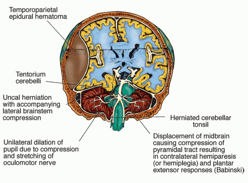
Figure 13-6 ▪ Cross-section of the brain showing herniation of part of the temporal lobe through the tentorium as a result of a temporoparietal epidural hematoma. (From: Kentzel, K. C. (1997). Advanced concepts in clinical nursing (2nd ed.). Philadelphia: J. B. Lippincott.)


Uncal Transtentorial Herniation (Uncal Syndrome)

The most common cerebral herniation syndrome is the uncal syndrome. An expanding lesion of the uncus of the hippocampal gyrus of the inferior, medial temporal lobe herniates through the incisura of the tentorium. The diencephalon and midbrain are compressed and displaced to the opposite side by the uncal herniation. With this lateral displacement, the cerebral peduncle (contralateral to the primary uncal herniation) may be compressed against the firm, unyielding edge of the tentorium incisura, producing Kernohan’s notch. This finding is important because it results in hemiparesis ipsilateral to the expanding supratentorial lesion (Fig. 13-6).18

Evaluating changes in pupillary findings and LOC is an important tool in differentiating central and uncal herniation syndromes. In the case of uncal herniation, the diencephalon, associated with consciousness, may not be the first anatomic area affected, so that impaired consciousness is not consistently an early sign of impending uncal herniation. The oculomotor nerve (cranial nerve [CN] III) and the posterior cerebral artery on the same side of the expanding temporal lobe lesion are frequently caught between the overhanging edematous uncus and the free edge of the tentorium. The entrapment of the oculomotor nerve results in ipsilateral pupillary dilation. Unilateral pupillary dilation is the earliest and consistent sign of uncal herniation. The progression of signs and symptoms is outlined in Table 13-4. The signs of uncal herniation include the following.


  • Gradual ipsilateral (to primary lesions) pupillary dilation, sluggish pupillary reaction to light (earliest sign), and possible development of an ovoid pupil (CN III)


  • Paralysis of the oculomotor extraocular muscles (CN IV, VI)


  • Restlessness, then LOC deteriorating from stupor to coma (frontal lobe and diencephalon)


  • Contralateral hemiparesis or hemiplegia (motor strip of frontal lobe and cerebral peduncles)


  • Most often, progression to decerebrate posturing; note that decorticate posturing is unusual (red nucleus lesion)


  • Positive Babinski’s signs (upper motor neuron lesion)


  • Respiratory changes (e.g., hyperventilation) (pontine lesion)


  • Finally, dilated, fixed pupils; flaccidity; and respiratory arrest (brainstem infarction)

As noted during early development of the uncal syndrome, the unilateral dilating, sluggish pupil is usually the only sign noted for several hours before other signs occur. There are no abnormal changes in extraocular movement, motor function, or respirations. Any motor deficits present are those specific to the supratentorial lesion. However, after any signs of herniation or brainstem compression appear, deterioration may proceed rapidly in a timeline of only a few hours in which a fully conscious patient deteriorates to deep coma. This means that unless uncal herniation is recognized early and interventions are effective, after midbrain (upper portion of the brainstem) involvement is noted, the patient progresses as in central herniation.


Effects of Supratentorial Herniation

Any supratentorial herniation syndrome can initiate vascular and obstructive complications that can further exaggerate the neurological deterioration. Compression of the aqueducts of the ventricular system can cause CSF circulation to be interrupted. As a result, major spikes in ICP and obstructive hydrocephalus can develop. Cingulate herniation can compress both arterial and venous vessels (portions of the anterior cerebral artery and the great cerebral vein), causing exacerbation of already present ischemia and edema.

With uncal herniation, the herniated tissue through the tentorial incisura compresses the posterior cerebral artery, resulting in partial occipital lobe infarction and edema. Brainstem edema, ischemia, and hemorrhage can develop from the diencephalon to the pons-medulla area secondary to the downward displacement from central herniation.

The result of progressive, unresolved downward displacement from any of the supratentorial herniation syndromes is brainstem herniation, in which the medulla herniates into the foramen magnum. Brain death is immediate and is attributable to medullary compression, ischemia, and infarction. The medulla controls vital functions such as respiration, blood pressure, cardiac function, and vasomotor tone, all of which are absolutely necessary to sustain life.


TABLE 13-4 PROGRESSION OF SIGNS AND SYMPTOMS OF THE UNCAL SYNDROMEa


ASSESSMENT


EARLY DIENCEPHALON


LATE DIENCEPHALON


MIDBRAIN UPPER PONS


LOWER PONS/UPPER MEDULLA


MEDULLA


Outcome


Potentially Reversible


Reverse Poor Prognosis-Irreversible


Level of consciousness


May not be impaired initially


After deterioration begins, quick progression to deep stupor and coma


Deep coma


Deep coma


Deep coma


Ocular


Pupillary size, reaction to light, and shape


Unilateral dilating pupil ipsilateral to primary lesion


Sluggish response Round to ovoid


Fully dilated pupil ipsilateral to lesion


Nonreactive


Round to ovoid


Other pupil dilates


Two possibilities for pupil contralateral to lesion:


Fully dilated and fixed or


Enlarged to midpoint (5-6 mm) and fixed


Bilateral midpoint (5-6 mm) pupils


Often irregularly shaped


Bilateral fully dilated and nonreactive


Extraocular movement


Full movement


Paralysis of oculomotor soon after dilated pupil


Oculocephalic (OC) and oculovestibular (OV) response


OC and OV intact or contralateral eye of OV on cold calorics, may not move medially


OC and OV sluggish, then absent


OC and OV absent


OC and OV absent


OC and OV absent


Motor


Function


No abnormalities may be present; if present, related to primary supratentorial lesion


  • Contralateral intermittent abnormal increase in resistance to passive movement


  • Ipsilateral hemiplegia


  • Decerebrate posturing of limbs (decortication is uncommon)


Bilateral decerebration


  • Flaccid at rest


  • Occasional flexor response of lower extremities to painful stimuli


  • Flaccid at rest


  • Occasional flexor response of lower extremities to painful stimuli


Reflexes


Babinski’s sign


Bilateral positive


Babinski’s sign


Bilateral positive


Babinski’s sign


Bilateral positive


Babinski’s sign


Respirations


Normal pattern and rate


Most often hyperventilation (20-40/min), rarely Cheyne-Stokes reaction


Hyperventilation (20-40/min)


Normal pattern of respirations, but more rapid (20-40/min) and shallow


Apneustic and cluster breathing


Slow and irregular in rate and amplitude (ataxic respirations); interrupted by deep sighs, gasps, and periods of apnea


Apnea


Other


Possible ptosis ipsilateral to primary lesion


Often hyperthermia


Cushing’s triad


Often hyperthermia


Cushing’s triad


aAn important point to keep in mind when observing a patient with a possible herniation syndrome is that there is predictable order to the development of signs and symptoms. Neurological deterioration in both central and uncal syndromes proceeds in an orderly rostral-caudal direction; the diencephalon, midbrain, pons, and, finally, medulla are affected from the increasing pressure. Signs and symptoms characteristic of each area can be identified. Notice that the last stages of central and uncal herniation are the same.


Table based on work of Plum, F., & Posner, J. B. (1982). The diagnosis of stupor and coma (3rd ed.). Philadelphia: F. A. Davis.


Infratentorial Herniation

Lesions of the infratentorial compartment contributing to herniation are much less frequent than those involving the supratentorial region. The infratentorial compartment includes the brainstem and cerebellum. The three possible effects of an expanding lesion in the infratentorial compartment include the following.


  • Direct compression of the brainstem, cerebellum, or their vascular supply


  • Upward transtentorial herniation of the brainstem and cerebellum through the tentorial incisura, resulting in maximal pressure on the midbrain


  • Downward herniation of one or both cerebellar tonsils through the foramen magnum to the cervical spine with compression of the medulla, an immediate cause of death (Fig. 13-7)

An expanding lesion causes compression and ischemia to selected structures. The increased pressure interferes with the normal function of the involved tissue and causes edema, ischemia, infarction, and necrosis if the process is not reversed. As the lesion continues to enlarge, the only egress from the infratentorial compartment is through the tentorial incisura (above) to the supratentorial compartment or through the larger orifice, the foramen magnum (below), into the cervical spine. In either case, neurological demise is rapid with high mortality.


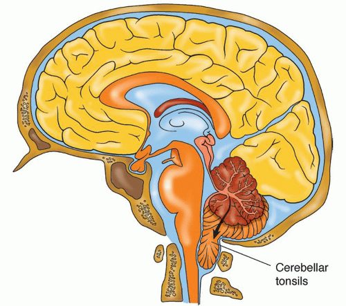
Figure 13-7 ▪ Herniation of the cerebellar tonsils into the foramen magnum is the final outcome of increased intracranial pressure. Respiratory centers within the medulla oblongata are compressed, and apnea leads to cardiac arrest and death.

The brainstem structures, particularly the medulla oblongata, contain centers for vital functions. If medullary compression develops, immediate death occurs as a result of respiratory and cardiac arrest. Expanding infratentorial lesions can also encroach on a portion of the ventricular system (third or fourth ventricle), resulting in acute hydrocephalus. The signs and symptoms noted with an infratentorial herniation vary widely depending on the brainstem area most involved. It is unclear whether posterior fossa lesions causing upward transtentorial herniation produce a consistent syndrome.17 The most prominent signs associated with upward herniation include the following.


  • Immediate onset of deep coma


  • Conjugate downward deviation of the eye to failure of upward voluntary or reflex movement (pretectal compression)


  • Small, equal, fixed pupils (pontine compression) to unequal, midpoint fixed pupils


  • Decerebration


  • Abnormal respiratory patterns (e.g., slow rate with intermittent deep sighs or ataxia)


  • Vital sign abnormalities (Cushing’s signs)

In summary, herniation syndromes are life-threatening occurrences that can progress rapidly. Early recognition of signs and symptoms (Chart 13-1) is important for prompt intervention to prevent neurological demise.


CHART 13-1 Signs and Symptoms of Impending Herniation


  • Decreased level of consciousness (coma)


  • Pupillary abnormalities


  • Motor dysfunction (hemiplegia, decortication, or decerebration)


  • Impaired brainstem reflexes (corneal, gag, swallowing)


  • “Cushing’s triad” of alterations in vital signs (bradycardia, hypertension, and respiratory irregularity)


SIGNS AND SYMPTOMS OF INCREASED INTRACRANIAL PRESSURE


Perspective

Increased ICP is a syndrome with multiple patterns (central syndrome, uncal syndrome), not a specific disease entity. A large percentage of neuroscience patients are at risk for developing increased ICP. Nursing assessment of neurological signs is directed at detecting early signs and symptoms of increased ICP when nursing and medical interventions are still effective. The performance and interpretation of the neurological exam by knowledgeable clinicians is a most sensitive indicator of neurologic change. The astute clinician must continually evaluate a comparison between the baseline neurological assessment and ongoing assessments to interpret the evolving relationship of these changes to changes in ICP. When late signs appear (e.g., brainstem signs of changes in vital signs or respiratory pattern), it may be too late for effective interventions to reverse cerebral deterioration, herniation, or even death.

As discussed previously, several cerebral compensatory mechanisms provide adequate CBF, CPP, and substrates for cerebral metabolism. These compensatory mechanisms act even when there is evidence of increased ICP. Cushing’s response is a compensatory response that attempts to provide adequate CPP in the presence of rising ICP. The signs associated with a Cushing’s response include the following.


  • A rising systolic pressure widening pulse pressure


  • Bradycardia


  • Irregular respirations

These signs profile late brainstem dysfunction resulting from rising ICP and correlate with decreasing brain compliance.

Cushing’s triad (also called Cushing’s sign) has subtle, yet important differences from Cushing’s response. Whereas Cushing’s response is a compensatory response, Cushing’s triad represents a loss of compensatory mechanisms and is a presentation of brainstem dysfunction, which correlates with low or loss of brain compliance. Cushing’s triad, which is a late finding and may not be present in all cases of herniation, includes the following.

Only gold members can continue reading. Log In or Register to continue

Stay updated, free articles. Join our Telegram channel

Jul 14, 2016 | Posted by in NURSING | Comments Off on Intracranial Hypertension: Theory and Management of Increased Intracranial Pressure

Full access? Get Clinical Tree

Get Clinical Tree app for offline access