Estrogens and progestins: basic pharmacology and noncontraceptive applications

CHAPTER 61


Estrogens and progestins: basic pharmacology and noncontraceptive applications


Estrogens and progestins are hormones with multiple actions. They promote female maturation, and help regulate the ongoing activity of female reproductive organs. In addition, they affect bone mineralization and lipid metabolism. The principal endogenous estrogen is estradiol. The principal endogenous progestational hormone is progesterone. Both hormones are produced by the ovaries. During pregnancy, large amounts are produced by the placenta. In addition, small amounts of estrogens and progestins are produced in peripheral tissues.


Clinical applications of the female sex hormones fall into two major categories: contraceptive and noncontraceptive applications. In this chapter, we focus on noncontraceptive uses. Contraception is discussed in Chapter 62. The principal noncontraceptive application of estrogens and progestins is menopausal hormone therapy (HT): estrogens are given to manage hot flushes and other menopausal symptoms; progestins are given to oppose estrogen-mediated stimulation of the endometrium.


The menstrual cycle


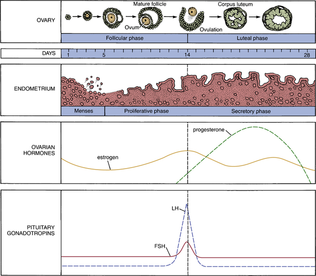
Because much of the clinical pharmacology of the estrogens and progestins is related to their actions during the menstrual cycle, understanding the menstrual cycle is central to understanding these hormones. Accordingly, we begin by reviewing the menstrual cycle. The anatomic and hormonal changes that take place during the cycle are summarized in Figure 61–1. As indicated, the first half of the cycle (days 1 through 14) is called the follicular phase, and the second half is called the luteal phase. One full cycle typically takes 28 days.


Ovarian and uterine events.

The menstrual cycle consists of a coordinated series of ovarian and uterine events. In the ovary, the following sequence occurs: (1) several ovarian follicles ripen; (2) one of the ripe follicles ruptures, causing ovulation; (3) the ruptured follicle evolves into a corpus luteum; and (4) if fertilization does not occur, the corpus luteum atrophies. As these ovarian events are taking place, parallel events take place in the uterus: (1) while ovarian follicles ripen, the endometrium prepares for nidation (implantation of a fertilized ovum) by increasing in thickness and vascularity; (2) following ovulation, the uterus continues its preparation by increasing secretory activity; and (3) if nidation fails to occur, the thickened endometrium breaks down, causing menstruation, and the cycle begins anew.


The roles of estrogens and progesterone.

The uterine changes that occur during the cycle are brought about under the influence of estrogens and progesterone produced by the ovaries. During the first half of the cycle, estrogens are secreted by the maturing ovarian follicles. As suggested by Figure 61–1, these estrogens act on the uterus to cause proliferation of the endometrium. At midcycle, one of the ovarian follicles ruptures and then evolves into a corpus luteum. For most of the second half of the cycle, estrogens and progesterone are produced by the newly formed corpus luteum. These hormones maintain the endometrium in its hypertrophied state. At the end of the cycle, the corpus luteum atrophies, causing production of estrogens and progesterone to decline. In response to the diminished supply of ovarian hormones, the endometrium breaks down.


The role of pituitary hormones.

Two anterior pituitary hormones—follicle-stimulating hormone (FSH) and luteinizing hormone (LH)—play central roles in regulating the menstrual cycle. During the first half of the cycle, FSH acts on the developing ovarian follicles, causing them to mature and secrete estrogens. The resultant rise in estrogen levels exerts a negative feedback influence on the pituitary, thereby suppressing further FSH release. At midcycle, LH levels rise abruptly (see Fig. 61–1). This LH surge causes the dominant follicle to swell rapidly, burst, and release its ovum. Following ovulation, the ruptured follicle becomes a corpus luteum and, under the influence of LH, begins to secrete progesterone.


From the foregoing, it is clear that precisely timed alterations in the secretion of FSH and LH are responsible for coordinating the structural and secretory changes that occur throughout the menstrual cycle. The mechanisms that regulate secretion of FSH and LH are complex and beyond the scope of this book.


Estrogens


Biosynthesis and elimination


Females.

In premenopausal women, the ovary is the principal source of estrogen. During the follicular phase of the menstrual cycle, estrogens are synthesized by ovarian follicles; during the luteal phase, estrogens are synthesized by the corpus luteum. The major estrogen produced by the ovaries is estradiol. In the periphery, some of the estradiol secreted by the ovaries is converted into estrone and estriol, hormones that are less potent than estradiol itself. Estrogens are eliminated by a combination of hepatic metabolism and urinary excretion.


During pregnancy, large quantities of estrogens are produced by the placenta. Excretion of these hormones results in high levels of estrogens in the urine. (The urine of pregnant mares is extremely rich in estrogens and serves as a commercial source of these hormones.)


Males.

Estrogen production is not limited to females. In the human male, small amounts of testosterone are converted into estradiol and estrone by the testes. Enzymatic conversion of testosterone in peripheral tissues (eg, liver, fat, skeletal muscle) results in additional estrogen production.


Physiologic and pharmacologic effects


Effects on primary and secondary sex characteristics of females

Estrogens support the development and maintenance of the female reproductive tract and secondary sex characteristics. These hormones are required for the growth and maturation of the uterus, vagina, fallopian tubes, and breasts. In addition, estrogens direct pigmentation of the nipples and genitalia.


Estrogens have a profound influence on physiologic processes related to reproduction. During the follicular phase of the menstrual cycle, estrogens promote (1) ductal growth in the breast, (2) thickening and cornification of the vaginal epithelium, (3) proliferation of the uterine epithelium, and (4) copious secretion of thickened mucus from endocervical glands. In addition, estrogens increase vaginal acidity (by promoting local deposition of glycogen, which is then acted upon by lactobacilli and corynebacteria to produce lactic acid). At the end of the menstrual cycle, a decline in estrogen levels can bring on menstruation. However, it is the fall in progesterone levels at the end of the cycle that normally causes breakdown of the endometrium and resultant menstrual bleeding. Following menstruation, estrogens promote endometrial restoration.


During pregnancy, the placenta produces estrogen in large amounts. This estrogen stimulates uterine blood flow and growth of uterine muscle. In addition, it acts on the breast to continue ductal proliferation. However, final transformation of the breast for milk production requires the combined influence of estrogen, progesterone, and human placental lactogen.


Metabolic actions

Estrogens can affect various nonreproductive tissues. Important among these are bone, blood vessels, the heart, liver, and central nervous system (CNS).


Bone.

Estrogens have a positive effect on bone mass. Under normal conditions, bone undergoes continuous remodeling, a process in which bone mineral is resorbed and deposited in equal amounts. The principal effect of estrogens on the process is to block bone resorption, although estrogens may also promote mineral deposition.


During puberty, the long bones grow rapidly under the combined influence of growth hormone, adrenal androgens, and low levels of ovarian estrogens. When estrogen levels grow high enough, they promote epiphyseal closure, and thereby bring linear growth to a stop.


After menopause, more bone is resorbed than is deposited, and hence bone mass declines. Why does this occur? Because the braking influence of estrogen on bone resorption is gone. Although estrogen therapy can help preserve bone mass, other drugs—raloxifene, bisphosphonates, calcitonin, and teriparatide—are safer, and hence are preferred (see Chapter 75).


Clinical pharmacology


Adverse effects

The principal concerns with estrogen therapy are the potential for endometrial hyperplasia, endometrial cancer, breast cancer, and cardiovascular events. Most other adverse effects are more of a nuisance than a concern.


Endometrial hyperplasia and carcinoma.

Prolonged use of estrogens alone by postmenopausal women is associated with an increased risk of endometrial carcinoma. However, when estrogens are used in combination with a progestin, there is little or no risk of uterine cancer. Why? When used alone, estrogens act on the endometrium to cause proliferation and hyperplasia. In a few cases, hyperplasia progresses to carcinoma. Concurrent use of a progestin greatly reduces cancer risk by antagonizing estrogen-mediated endometrial proliferation and by reversing hyperplasia. Accordingly, whenever estrogen is given to postmenopausal women who have an intact uterus, a progestin should be given as well. During the first 3 to 6 months of treatment, about 30% of women experience benign, irregular bleeding. If bleeding persists, or if it starts after prolonged therapy, the possibility of endometrial carcinoma should be evaluated.


Breast cancer.

Does estrogen increase the risk of breast cancer? Yes—but primarily in postmenopausal women who are using estrogen combined with a progestin. In younger women, and in women using estrogen without a progestin, the risk, if any, is very low. Results of the Women’s Health Initiative (WHI) indicate that treatment of postmenopausal women with estrogen plus a progestin produces a small increase in the risk of breast cancer—but treatment with estrogen alone carries little or no risk (see below under Menopausal Hormone Therapy). In contrast, the Women’s Contraceptive and Reproductive Experience study has shown that, for most younger women, use of estrogen plus a progestin for contraception does not increase breast cancer risk (see Chapter 62). How can we explain these contradictory results? The best explanation is that risk is a function of age—being relatively high in older women and very low in younger women. In either population—old or young—the risk of breast cancer from estrogen alone has not been firmly established. Of note, after the results of the WHI were published, there was a dramatic decline in postmenopausal estrogen use, followed by a significant decline in the incidence of breast cancer. This observation reinforces the conclusion that postmenopausal hormone therapy increases breast cancer risk.


How does estrogen increase breast cancer risk? Estrogen does not cause breast cancer—it only promotes the growth of a cancer that already exists, and then only if the cancer is estrogen receptor–positive. Accordingly, estrogen-dependent breast cancer must be ruled out before initiating estrogen treatment. Furthermore, because all women are at potential risk for breast cancer, and because estrogens may slightly increase that risk, women taking estrogens should have a yearly breast exam by a health professional. For women older than 40, an annual mammogram is recommended.


Cardiovascular events.

In postmenopausal women, estrogen, used either alone or combined with a progestin, increases the risk of venous thromboembolism (VTE) and stroke. In addition, estrogen alone increases the risk of coronary heart disease and myocardial infarction, but only in women over age 60. The risk of VTE is greatest during the first years of treatment, and is greater with oral dosing than with transdermal or intravaginal dosing. These effects are discussed further under Menopausal Hormone Therapy.


In premenopausal women, use of combination estrogen/progestin oral contraceptives increases the risk of VTE. Although the risk is dose dependent, even low-dose formulations cause an estimated 15 to 30 cases per 100,000 woman-years of use. The cardiovascular risk associated with oral contraceptives is discussed further in Chapter 62.


Nausea.

Nausea is the most frequent undesired response to the estrogens. Fortunately, nausea diminishes with continued use, and is rarely so severe as to necessitate treatment cessation. Nausea can be minimized by administering estrogens with food, initiating therapy at low doses, and, when appropriate, using a topical estrogen formulation (eg, gel, spray, patch, vaginal ring).


Therapeutic uses

Estrogens have contraceptive and noncontraceptive applications. In this chapter, discussion is limited to the noncontraceptive applications. Use of estrogens for contraception is discussed in Chapter 62.


Acne.

Estrogens, in the form of oral contraceptives, can help control acne. Treatment is limited to females at least 15 years old who want contraception. The use of estrogen for acne is discussed in Chapter 105.


Routes of administration


Transdermal.


Transdermal estradiol is available in four formulations:


The emulsion is applied once daily to the top of both thighs and the back of both calves. The spray is applied once daily to the forearm, never to the breasts or pubic area. The gel is applied once daily to one arm, from the shoulder to the wrist. The patches are applied to the skin of the trunk (but not the breasts), allowing estrogen to be absorbed through the skin and then directly into the bloodstream. Rates of estrogen absorption with transdermal formulations range from 14 to 60 mcg/24 hr, depending on the product employed.


Compared with oral formulations, the transdermal formulations have four advantages:


Selective estrogen receptor modulators (SERMs)


SERMs are drugs that activate estrogen receptors in some tissues and block them in others. These drugs were developed in an effort to provide the benefits of estrogen (eg, protection against osteoporosis, maintenance of the urogenital tract, reduction of LDL cholesterol) while avoiding its drawbacks (eg, promotion of breast cancer, uterine cancer, and thromboembolism). Three SERMs are available: tamoxifen [Nolvadex], toremifene [Fareston], and raloxifene [Evista]. None of these offers all of the benefits of estrogen, and none avoids all of the drawbacks.


Tamoxifen was the first SERM to be widely used. By blocking estrogen receptors, tamoxifen (and its active metabolite, endoxifen) can inhibit cell growth in the breast. As a result, the drug is used extensively to prevent and treat breast cancer. Unfortunately, blockade of estrogen receptors also produces hot flushes. By activating estrogen receptors, tamoxifen protects against osteoporosis and has a favorable effect on serum lipids. However, receptor activation also increases the risk of endometrial cancer and thromboembolism. The pharmacology of tamoxifen and toremifene (a close relative of tamoxifen) is discussed at length in Chapter 103.


Raloxifene is very similar to tamoxifen. The principal difference is that raloxifene does not activate estrogen receptors in the endometrium, and hence does not pose a risk of uterine cancer. Like tamoxifen, raloxifene protects against breast cancer and osteoporosis, promotes thromboembolism, and induces hot flushes. Raloxifene is approved only for prevention and treatment of osteoporosis, and for prevention of breast cancer in high-risk women. Raloxifene is discussed at length in Chapter 75.


Progestins


Progestins are compounds that have actions like those of progesterone, the principal endogenous progestational hormone. As their name implies, the progestins act prior to gestation to prepare the uterus for implantation of a fertilized ovum. In addition, progestins help maintain the uterus throughout pregnancy.


Biosynthesis


Progesterone is produced by the ovaries and the placenta. Ovarian production occurs during the second half of the menstrual cycle. During this period, progesterone is synthesized by the corpus luteum, in response to LH released from the anterior pituitary. If implantation of a fertilized ovum does not occur, progesterone production by the corpus luteum ceases, and menstrual flow begins. However, if implantation does take place, the developing trophoblast will produce its own luteotropic hormone—human chorionic gonadotropin (hCG)—that will stimulate the corpus luteum to continue making progesterone. For the first 7 weeks of gestation, the placenta depends entirely on progesterone from the corpus luteum. However, between weeks 7 and 10, production of progesterone is shared between the corpus luteum and placenta. After 10 weeks of gestation, progesterone made by the placenta is sufficient to support pregnancy, and hence ovarian progesterone production declines. Placental synthesis of progesterone and estrogen continues throughout the pregnancy.


Physiologic and pharmacologic effects


Effects during the menstrual cycle.

Progesterone secreted during the second half of the menstrual cycle converts the endometrium from a proliferative state into a secretory state. If implantation does not occur, progesterone production by the corpus luteum declines. The resultant fall in progesterone levels is the principal stimulus for the onset of menstruation.


Effects during pregnancy.

As noted, progesterone levels increase during pregnancy. These high levels suppress contraction of uterine smooth muscle, and thereby help sustain pregnancy. Unfortunately, progesterone also suppresses contraction of GI smooth muscle, which leads to prolonged transit time and constipation. In the breast, progesterone promotes growth and proliferation of alveolar tubules (acini), the structures that produce milk. Metabolic effects include suppression of arterial pCO2, altered serum bicarbonate content, and elevation of serum pH. Lastly, progesterone may help suppress the maternal immune system, thereby preventing immune attack on the fetus.


Jul 24, 2016 | Posted by in NURSING | Comments Off on Estrogens and progestins: basic pharmacology and noncontraceptive applications

Full access? Get Clinical Tree

Get Clinical Tree app for offline access