Drugs for multiple sclerosis

CHAPTER 23


Drugs for multiple sclerosis


Multiple sclerosis (MS) is a chronic, inflammatory, autoimmune disorder that damages the myelin sheath of neurons in the central nervous system (CNS), causing a wide variety of sensory, motor, and cognitive deficits. Initially, most patients experience periods of acute clinical exacerbations (relapses) alternating with periods of complete or partial recovery (remissions). Over time, symptoms usually grow progressively worse—although the course of the disease is unpredictable and highly variable. Among young adults, MS causes more disability than any other neurologic disease. Nonetheless, most patients manage to lead fairly normal lives, and life expectancy is only slightly reduced. Multiple sclerosis affects about 400,000 people in the United States and 2.5 million worldwide.


Drug therapy of MS changed dramatically in 1993, the year the first disease-modifying agent was approved. Prior to this time, treatment was purely symptomatic. We had no drugs that could alter the disease process. By using disease-modifying drugs, we can now slow the progression of MS, decrease the frequency and intensity of relapses, and delay permanent neurologic loss. As a result, we can significantly improve prognosis, especially if treatment is started early.


Overview of MS and its treatment


Pathophysiology


What initiates the autoimmune process?

No one knows. The most likely candidates are genetics, environmental factors, and microbial pathogens. We suspect a genetic link for two reasons. First, the risk of MS for first-degree relatives of someone with the disease is 10 to 20 times higher than the risk for people in the general population. Second, the risk of MS differs for members of different races. For example, the incidence is highest among Caucasians (especially those of northern European descent), much lower among Asians, and nearly zero among Inuits (the indigenous people of the Arctic). We suspect environmental factors because the risk is not the same in all places: In the United States, MS is more common in northern states than in southern states; around the globe, MS is most common in countries that have a moderately cool climate, whether in the northern or southern hemisphere; and, as we move from the equator toward the poles, the incidence of MS increases. Microbial pathogens suspected of initiating autoimmunity include Epstein-Barr virus, human herpesvirus 6, and Chlamydia pneumoniae. The bottom line? Multiple sclerosis appears to be a disease that develops in genetically vulnerable people following exposure to an environmental or microbial factor that initiates autoimmune activity.


Signs and symptoms


People with MS can experience a wide variety of signs and symptoms. Depending on where CNS demyelination occurs, a patient may experience paresthesias (numbness, tingling, pins and needles), muscle or motor problems (weakness, clumsiness, ataxia, spasms, spasticity, tremors, cramps), visual impairment (blurred vision, double vision, blindness), bladder and bowel symptoms (incontinence, urinary urgency, urinary hesitancy, constipation), sexual dysfunction, disabling fatigue, emotional lability, depression, cognitive impairment, slurred speech, dysphagia, dizziness, vertigo, neuropathic pain, and more. The intensity of these symptoms is determined by the size of the region of demyelination. To quantify the impact of MS symptoms, most clinicians employ the Kurtzke Expanded Disability Status Scale (EDSS), an instrument that measures the impact of MS on nine different functional systems (eg, visual, sensory, cerebellar). The results are tabulated and reported on a scale from 0 to 10, with 0 representing no disability and 10 representing death. An EDSS of 4 or greater indicates difficulties with ambulation. Symptoms of MS are discussed further under Drugs Used to Manage MS Symptoms.


MS subtypes


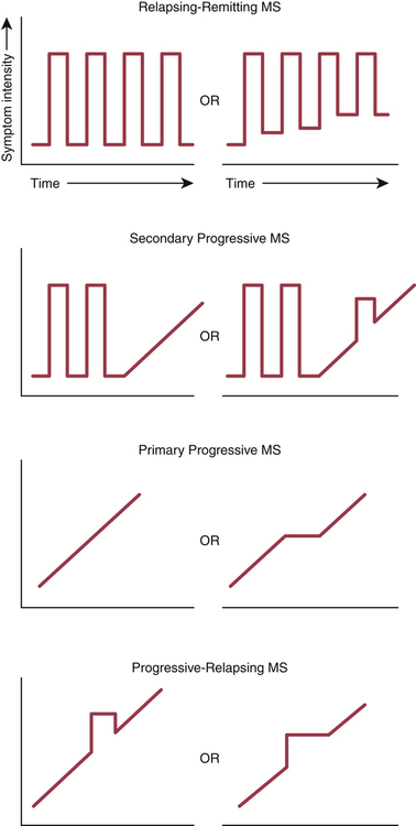
There are four subtypes of MS—relapsing-remitting, secondary progressive, primary progressive, and progressive-relapsing—defined by the clinical course the disease follows. Symptom patterns that characterize the MS subtypes are depicted in Figure 23–1.


Diagnosis


Diagnosis of MS is based on clinical presentation supplemented with laboratory data. As a rule, we cannot diagnose MS on the basis of symptoms or signs or laboratory tests alone. Rather, for most patients, all three kinds of information are needed. In addition, because the signs, symptoms, and test results that suggest MS can also suggest other disorders, a positive diagnosis cannot be made until all other possibilities have been ruled out. Diagnosis of MS is confounded by interpatient variability in symptoms and the course the disease follows.


In 1965, the following diagnostic criteria were introduced:


Since these criteria were introduced, additional diagnostic tools have become available. Important among these are magnetic resonance imaging (MRI), cerebrospinal fluid (CSF) tests, and measurement of the visual evoked potential (VEP). All three tests can help confirm a suspected diagnosis of MS, but they cannot, by themselves, provide definitive proof of the disease. Properties of these tests are as follows:


• MRI is the most sensitive way to image the brain. Sensitivity can be made even greater with gadolinium, an IV contrast agent. MRI is especially good for detecting areas of demyelination. However, it is important to note that, in some patients who have clinically definite MS, an MRI scan may fail to detect any lesions. Hence, a negative scan does not necessarily rule out MS. Conversely, because other disorders can produce a positive scan, a positive scan, by itself, does not prove the presence of MS, although it can help confirm a suspected case.


• Tests of CSF are used to assess immune activity within the CNS. Two tests are employed: measurement of immunoglobulin G (IgG) levels and measurement of oligoclonal IgG bands (OCBs), which indicate intrathecal production of antibodies. More than 90% of patients with MS have OCBs. However, as with MRI scans, other disorders can also produce positive test results—and negative results may be seen in some patients who do have MS. Accordingly, CSF analysis, by itself, can neither confirm nor rule out MS.


• The VEP test measures how quickly the brain responds to a visual stimulus, and hence indirectly measures conduction velocity in the optic nerve. In patients with MS, the VEP is delayed, indicating conduction velocity is slowed (owing to demyelination of the optic nerve). However, as with MRI scans and CSF tests, the VEP test is not specific for MS: Other disorders can produce positive results, and some patients with MS may get negative results.


Diagnostic criteria for MS were revised by the McDonald committee in 2001, revised again in 2005, and revised yet again in 2010 (Table 23–1). The latest criteria are still founded on the patient’s clinical presentation, but also incorporate information from MRI scans and CSF tests. These criteria were adopted to permit the earliest possible diagnosis of MS, and hence permit the earliest possible implementation of disease-modifying therapy.


TABLE 23–1 


2010 Revised McDonald Criteria for Diagnosis of MS


Number of Attacks Clinical Evidence of Lesions Additional Data Needed for a Diagnosis of MS
2 or more Objective clinical evidence of 2 or more lesions or objective clinical evidence of 1 lesion with reasonable historic evidence of a prior attack None: Clinical evidence alone will suffice.
2 or more Objective clinical evidence of 1 lesion Proof of dissemination in space shown by either MRI (ie, at least 2 lesions visible on the MRI in CNS regions typically affected by MS) or await another clinical attack implicating a different CNS site.
1 Objective clinical evidence of 2 or more lesions Proof of dissemination in time shown by either MRI (eg, a new lesion is visible on a follow-up MRI) or await a second clinical attack
1 Objective clinical evidence of 1 lesion (clinically isolated syndrome) Proof of dissemination in space shown by either MRI or await a second clinical attack implicating a different site
plus
Proof of dissemination in time shown by either MRI or await a second clinical attack
0 (Insidious neurologic progression suggestive of MS but with no clear clinical attack)   One year of disease progression (retrospectively or prospectively determined)
plus
Two out of three of the following:


image


*CSF = cerebrospinal fluid. A positive CSF test shows either oligoclonal bands different from those in serum or a raised IgG index.


Adapted from Polman CH, Reingold SC, Banwell B, et al: Diagnostic criteria for multiple sclerosis: 2010 revisions to the McDonald Criteria. Ann Neurol 69:292–302, 2011.


Drug therapy overview


In patients with MS, drugs are employed to (1) modify the disease process, (2) treat an acute relapse, and (3) manage symptoms. We have no drugs that can cure MS.


Disease-modifying therapy

Disease-modifying drugs can decrease the frequency and severity of relapses, reduce development of brain lesions, decrease future disability, and help maintain quality of life. In addition, they may prevent permanent damage to axons. However, it is important to note that, although these drugs can slow disease progression, they do not cure MS. Also, they do not work for all patients. Those with relapsing-remitting MS benefit most.


There are two main groups of disease-modifying drugs: immunomodulators and immunosuppressants (Table 23–2). The immunomodulators—interferon beta, glatiramer acetate, natalizumab, and fingolimod—are safer than mitoxantrone (the major immunosuppressant in use), and hence are generally preferred.


Relapsing-remitting MS.

All patients with relapsing-remitting MS—regardless of age, frequency of attacks, or level of disability—should receive one of the immunomodulators:


Treatment should begin as soon as possible after relapsing-remitting MS has been diagnosed. Why? Because early treatment can help prevent axonal injury, and may thereby prevent permanent neurologic deficits.


Treatment should continue indefinitely. The principal reasons for stopping would be toxicity or a clear lack of effect. Unfortunately, if disease-modifying therapy is stopped, disease progression may return to the pretreatment rate.


If treatment with an immunomodulator fails to prevent severe relapses or disease progression, treatment with mitoxantrone (an immunosuppressant) should be considered. However, keep in mind that mitoxantrone can cause serious toxicity (eg, myelosuppression, heart damage), and hence should be reserved for patients who truly need it.


Treating an acute episode (relapse)

A short course of a high-dose IV glucocorticoid (eg, 500 mg to 1 gm of methylprednisolone daily for 3 to 5 days) is the preferred treatment of an acute relapse. Glucocorticoids suppress inflammation and can thereby reduce the severity and duration of a clinical attack. As discussed in Chapter 72, these drugs are very safe when used short term, elevation of blood glucose being the principal concern. By contrast, long-term exposure can cause osteoporosis and other serious adverse effects. Accordingly, frequent use (more than 3 times a year) or prolonged use (longer than 3 weeks at a time) should be avoided.


Acute relapse may also be treated with IV gamma globulin. This option can be especially helpful in patients intolerant of or unresponsive to glucocorticoids. Results have been good.


Disease-modifying drugs I: immunomodulators


Seven immunomodulators are available: glatiramer acetate [Copaxone], natalizumab [Tysabri], fingolimod [Gilenya] and four preparations of interferon beta [Avonex, Rebif, Betaseron, Extavia]. With the exception of natalizumab, all of these drugs are recommended as first-line therapy for patients with relapsing-remitting MS and for patients with secondary progressive MS who still experience acute exacerbations. Natalizumab is reserved for patients with relapsing-remitting MS who have not responded to at least one of the other four drugs. Why? Because, very rarely, natalizumab has been associated with a potentially fatal infection of the brain.


All of the first-line immunomodulators—glatiramer, fingolimod, and the interferon beta preparations—have nearly equal efficacy, decreasing the relapse rate by about 30%. Because benefits are very similar, selection among these drugs is based primarily on patient and prescriber preference. If a particular drug is intolerable or ineffective, a different one should be tried. Natalizumab is more effective than the first-line drugs, decreasing the relapse rate by 68%, but is also more dangerous. As noted, although the immunomodulators can modify the course of MS, they do not cure the disease.


With the exception of fingolimod, all of the first-line immunomodulators are administered by self-injection (IM or SQ). Fingolimod is administered PO. Natalizumab, a second-line drug, is administered by IV infusion (at an approved infusion center). All seven drugs are expensive: One year of treatment costs between $35,000 and $48,000. Properties of these drugs are summarized in Table 23–2.


Interferon beta preparations


Description and mechanism

Interferon beta is a naturally occurring glycoprotein with antiviral, antiproliferative, and immunomodulatory actions. Natural interferon beta is produced in response to viral invasion and other biologic inducers. In patients with MS, it is believed to help in two ways. First, it inhibits the migration of proinflammatory leukocytes across the blood-brain barrier, thereby preventing these cells from reaching neurons of the CNS. Second, it suppresses T helper cell activity.


Two forms of interferon beta are used clinically: interferon beta-1a [Avonex, Rebif] and interferon beta-1b [Betaseron, Extavia]. Both forms are manufactured using recombinant DNA technology. Interferon beta-1a contains 166 amino acids plus glycoproteins, and is identical to natural human interferon beta with respect to amino acid content. Interferon beta-1b contains only 165 amino acids and has no glycoproteins, and hence differs somewhat from the natural compound. The two preparations of interferon beta-1a—Avonex and Rebif—differ slightly in their glycoprotein content, and are administered by different routes (see below). The two preparations of interferon beta-1b—Betaseron and Extavia—are identical. In fact, they are both manufactured in the same plant.


Adverse effects and drug interactions

Interferon beta is generally well tolerated, although side effects are common.


Drug interactions.

Exercise caution when combining interferon beta with other drugs that can suppress the bone marrow or cause liver injury.


Preparations, dosage, and administration


Jul 24, 2016 | Posted by in NURSING | Comments Off on Drugs for multiple sclerosis

Full access? Get Clinical Tree

Get Clinical Tree app for offline access